Activity-guided Purification of N-benzyl-N-methyldecan-1-amine from Garlic and Its Antitumor Activity against CT-26 Colorectal Carcinoma in BALB/C Mice
Rajasekar Seetharaman1,2†, Seong Mi Choi1†, Lu Guo1, Zheng Wei Cui1, Duuriimaa Otgonbayar1, Ju Ha Park1, Young-Seok Kwon3, Jung Ho Kwak3, Young Hee Kwon4, Ji Hyun Min4, Jum Soon Kang1 and Young Whan Choi1*
1Department of Horticultural Bioscience, Pusan National University, Miryang 627-706, Korea
2Global Institute of Stem Cell Therapy and Research (GIOSTAR) Inc. Pvt. Ltd., Ahmedabad, Gujrat, India
3Onion and Garlic Lab, Vegetable Research Division, National Institute of Horticultural & Herbal Science, Muan 55365, Korea
4Chungcheongbuk-do Agricultural Research & Extension Services, Cheongju 28130, Korea Received July 24, 2019 /Revised October 14, 2019 /Accepted October 15, 2019
A components of garlic (Allium sativum) have anti-proliferative effects against various types of cancer.
We aimed to investigate the capacity of garlic compounds to anti-tumor on a various cancer cell lines.
Fractionation of garlic extract, guided by antiproliferative activity against human gastric cancer (AGS) cells, has resulted in the isolation of N-benzyl-N-methyldecan-1-amine (NBNMA). We investigated the effect of newly isolated NBNMA from garlic cloves on the inhibition of the growth of CT-26, AGS, HepG2, HCT-116, MCF7, B16F10, and Sarcoma-180 cells for in vitro and CT-26 colon carcinoma cells in vivo. NBNMA exhibited an antiproliferative effect in CT-26 cells by apoptotic cell death. NBNMA exhibited down-regulation of anti-apoptotic Bcl-2 proteins and up-regulation of apoptotic Bad protein expression in western blot analyses. In addition, NBNMA meagre activated caspase 3 and caspase 9, initiator caspases of the extrinsic and intrinsic pathways of apoptosis. NBNMA treatment at a dose of 10 mg/kg for 21 days in experimental mice implanted with tumors resulted in significant reduction of the tumor weight (43%). NBNMA exhibited both in vitro and in vivo anticancer activity. These re- sults indicate that NBNMA has promising potential to become a novel anticancer agent from garlic cloves for the treatment of colon carcinoma cancer.
Key words : Activity-guided purification, anti-cancer, colorectal cancer, garlic, N-benzyl-N-methyldecan- 1-amine
†Authors contributed equally.
*Corresponding author
*Tel : +82-55-350-5522, Fax : +82-55-350-5529
*E-mail : [email protected]
This is an Open-Access article distributed under the terms of the Creative Commons Attribution Non-Commercial License (http://creativecommons.org/licenses/by-nc/3.0) which permits unrestricted non-commercial use, distribution, and reproduction in any medium, provided the original work is properly cited.
Journal of Life Science 2019 Vol. 29. No. 10. 1062~1070 DOI : https://doi.org/10.5352/JLS.2019.29.10.1062
Introduction
Garlic (Allium sativum L.) has been used as a spice and an ingredient in folk medicine since ancient times [23].
Studies on garlic and its derivatives have revealed a range of potential drugs for treating ailments that include throm- bosis [5, 6], cardioprotective effects [11, 21, 25, 27, 30], cancer [8, 10, 34], oxidative stress [4, 13, 33] and inflammatory asso- ciated diseases [24]. A number of bioactive compounds have been reported from garlic; of these, Allicin is formed due to the hydrolysis of alliin when the garlic tissue is damaged.
To evaluate the quality of garlic and garlic products, it is
important to consider all the precursors and the biological active substances present [22, 28]. Among them ajoene has been shown to inhibits platelet aggregation by altering the platelet membrane via an interaction with sulphydryl groups [3, 6], anti-microbial [12, 18], and potent anti-cancer activity against variety of cancer cells [35]. In human non-leukaemia malignant cells, several organosulphur garlic compounds including allicin and its corresponding sulfide as well as ajoene have inhibited the proliferation and in- duced apoptosis of human breast, bladder, colorectal, hep- atic, prostate cancer, lymphoma and skin tumor cell lines [7, 19, 26, 32]. Colorectal cancer (CRC) has been the world wide fourth most commonly diagnosed cancer in men and third in women. In recent days the incidence of CRC has increased. Meanwhile, in Korea, incidence rates have risen markedly over the past few years. From 2003 to 2005, CRC was the third most commonly diagnosed cancer in Korea with 12.0% of all cancer diagnoses [17]. Hence the present study was focused on in vivo anti-tumor effect of pure
N-benzyl-N-methyldecan-1-amine; NBNMA compound from garlic cloves on CT-26 murine colorectal carcinoma.
Materials and Methods
Plant materials and isolation of the pure compound Garlic cloves were purchased directly from the Danyang Food Co. (Danyang, Korea) in January, 2009. The isolation of N-benzyl-N-methyldecan-1-amine was previousely re- ported Jeong et al. [14].
Chemicals and cell line
HPLC grade solvents including hexane, chloroform, di- chloromethane, methanol, acetonitrile and water (Fishers Scientific Korea, Seoul, Korea) were used for the extraction, column chromatography and mobile phases of HPLC. Fetal bovine serum (FBS), RPMI-1640 medium and anti-biotics were purchased from Gibco (Grand Island, NY, USA), and dimethylsulfoxide (DMSO), 3-(4,5-dimethylthiazol-2)-2, 5-di- phenyl-2H-tetrazoliumbromide (MTT) and 5-fluorouracil were purchased from Sigma (St. Louis, MO, USA). The cell lines CT-26, AGS, HepG2, HCT-116, MCF7, B16F10, and Sarcoma- 180 cells were obtained from Korean Cell Line Bank in Seoul, South Korea, maintained in RPMI-1640 medium supplemented with 10% FBS, anti-biotics (100U/ml penicillin G, 100 mg/ml streptomycin), and maintained in an incubator at 37℃ with a humidified atmosphere at 5% CO2 concentration.
Cell viability assay
The effects of NBNMA on the viability of CT-26 cells were evaluated by MTT assay. Cells were seeded into the wells of a 96 well plate at a density of 1×10⁴ cells/well and then allowed to adhere for 24 hr at 37℃ in a 5% CO2 atmosphere.
After incubation, the cells were treated with desired concen- trations of NBNMA (1.19. 2.39, 4.78, 9.56, 14.35 and 19.13 μM) or DMSO as vehicle control for 24 hr. After the addition of 50 ml of tetrazolium dye, 3-(4, 5-dimethylthiazol-2)-2 and 5-diphenyl-2H-tetrazoliumbromide (MTT; 2 mg/ml in PBS), the cells were incubated at 37℃ for 4 hr. The formazan de- posits that formed were dissolved in DMSO and the absorb- ance of each well was measured at 540 nm in an Epoch mi- croplate reader (BioTek instruments, Winooski, VT, USA).
Triplicate experiments were performed at each concentration and the results were presented as mean ± S.D. The percent- age of untreated cell viability was used as the control to calculate relative cell viability. The inhibitory concentration,
IC50, was calculated as the concentration that resulted in 50%
inhibition of cell proliferation.
Western blot analysis
The CT-26 cells were grown at a density of 1×10⁶ cells in 100 mm culture dishes for 24 hr and then treated with various concentration of NBNMA. After 24 hr treatment, both live and dead cells were collected and washed twice with PBS. Protein was extracted from the cells and the re- sults were isolated using a Qproteome mammalian protein preparation kit (Qiagen, Hilden, Germany). The total protein content in the cell lysate was quantified using a 2D Quant kit (Amersham Biosciences, Amersham, UK). The con- ditioned media (30 mg of protein/sample) was run on a Western blot using 13% SDS-PAGE and then transferred to a PVDF membrane (Millipore Corporation, Billerica, MA, USA). The membrane was then blocked with 5% skimmed milk in Tris buffered saline containing 0.1% Tween 20 (T-TBS). After washing with T-TBS, the membrane was pro- bed with rabbit anti-mouse primary antibodies from the Bcl-2 family of proteins (Bcl-2, Bcl-xL, Bid, Bad and Bax anti- bodies), caspase proteins (pro-caspase 3, pro-caspase 8 and pro-caspase 9 antibodies), poly (ADP-ribose) polymerase (PARP) and internal standard β-actin (Millipore Corporation, Billerica, MA, USA), and then incubated over- night at 4℃. Subsequently, the membrane was incubated at room temperature with horseradish peroxidase conjugated goat anti-rabbits secondary antibody (Millipore Corporation, Billerica, MA, USA) for 2 hr. The resulting protein signals were detected using an ECL Western blotting kit (Thermo scientific, Rockford, IL, USA).
Caspase assay
NBNMA treated and nontreated cell lines (1×10⁶ cells) were lysed in lysis buffer (1% Triton X-100, 0.32 M sucrose, 5 mM EDTA, 10 mM Tris–HCl at pH 8.0, 2 mM dithio- threitol, 1 mM PMSF, 1 mg/ml aprotinin and 1 mg/ml leu- peptin) for 30 min at 4℃, followed by centrifugation at 10,000 g for 30 min. For determination of caspase activity, 50 ml of caspase specific reaction mixtures were used. The substrates DEVD-pNA, IETD-pNA and LEHD-pNA for cas- pase 3, 8 and 9, respectively (200 mM), were mixed in- dividually with cytosolic extracts (30 mg of total protein) from the cell lines and incubated for 1 hr at 37℃. After in- cubation, the fluorescence of the extracts was determined (excitation wavelength, 400 nm; emission wavelength, 505
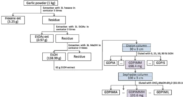
Fig. 1. Activity-guided purification scheme of GDPIMMIH from the garlic powder.
nm) using a fluorescence plate reader (BioTek instruments, Winooski, VT, USA).
DNA fragmentation assay
The cells were treated with different concentrations of NBNMA and lysed on ice in a buffer containing 10 mM Tris –HCl (pH 7.4), 150 mM NaCl, 5 mM EDTA, and 0.5% Triton X-100 for 30 min. The lysates were vortexed and cleared by centrifugation at 10,000 g for 20 min. The fragmented DNA in the supernatant was extracted using an equal volume of neutral phenol: chloroform: isoamylalcohol (25:24:1, v/v/v) and analyzed electrophoretically on 1.0% agarose gel con- taining ethidium bromide (EtBr) (Sigma, St. Louis, MO, USA)
Animals
BALB/C female mice, 6-7 weeks old were purchased from Samtako (Osan-shi, Kyunggi-do, South Korea) and housed in polycarbonate cages under laboratory conditions with a 12 hr day-night cycle, room temperature of 22±2℃ and hu- midity of 50-60%. They were fed standard pelleted rats chow and had free access to water during the experiment. The animal experiments were approved by the Pusan National University, Institutional Animal Care and Use Committee (PNU-2015-0808).
Tumor induction and drug treatment
CT-26 mouse colon carcinoma cells were collected from
cell cultures by trypsinization and then injected subcuta- neously (2×10⁶ cells) into the right flank region of BALB/C mice. After 24 hr of tumor induction, the mice were orally administrated with 3.83 and 19.13 μM/kg of NBNMA at ev- ery three days interval over a 16 day treatment period.
Tumor control mice were treated with the vehicle PBS.
Whereas 5-fluorouracil (5-FU) as the positive control was in- traperitoneally injected at a single time dose with 384.38 μ M/kg. The tumor volume was calculated using the formula, tumor volume (mm2) = AB2/2. The length (A) and width (B) of the tumor from each mouse was measured every 3 days. The tumor inhibition ratio was calculated by the for- mula: inhibition ratio (%) = [(X-Y)/X] ×100, whereby X was the average tumor weight of the negative control and Y was the average tumor weight of the treated group. The animals were sacrificed at 20 days after treatment, then tumors were removed and immediately weighed.
Statistical analysis
Results were provided as mean ± SD for each condition and statistically analysed by the Student–Newman–Keuls test. P<0.05 was considered statistically significant.
Results
Purification of anti-proliferative compounds by Bio- guided fractionation
The purification scheme for the isolation of anti-pro-
Fig. 2. Percent inhibition of fractions from garlic powder on AGS cell line. AGS cell were incubated for 24 hr after treatment with 20 μg/ml fractions. Percent inhibition was assayed by MTT assay. Data are expressed as the mean ± SD.
N CH3
CH3 2 1
3 4
5 6
7 8
9
10 11 12 13 14 15 16 17 18
19
Fig. 3. The structure of GDPIMMIH was elucidated N-benzyl-N-methyldecan-1-amine (NBNMA) by 1D and 2D NMR spectral data.
liferative compound from the garlic powder shown in Fig 1. Preliminary investigations of hexane (GDH), ethyl acetate (GDE) and ethyl alcohol (GDP) extracts were tested for its in vitro anti-proliferative activity. The GDP extract (13 g) was chromatographed on a Diaion HP20 Resin column with a step gradient (0, 25, 50 and 90%) of EtOH in water and meth- anol (MeOH) to obtain 21 fractions (GAPIA-GDPIS). In order to search the inhibitory anti-proliferative compounds from GDP, we examined the cytotoxic effects of 21 fractions over a 24 hr treatment period using an MTT assay. Of the differ- ent extracts, GDPIMM showed better anti-proliferative activ- ity against AGS cancer cells. Hence, we focused on GDPIMM to further search anti-proliferative compound (Fig. 1). Frac- tion GDPIMM (221.3 mg) was rechromatographed on a Sephadex LH20 column with CHCl3:MeOH:dH2O (65:35:10) to obtained 11 fractions (GDPIMMIA-IK) and measured its MTT assay. Of the 11 fractions, GDPIMMIH (38.2 mg) was purified and found to significantly inhibit cell growth in a 20 μg/ml (Fig. 2). The structure of GDPIMMIH was de- termined with 1D and 2D NMR and MS spectral data and elucidated as N-benzyl-N-methyldecan-1-amine (NBNMA) (Jin-Woo Jeong et al., 2014) (Fig. 3).
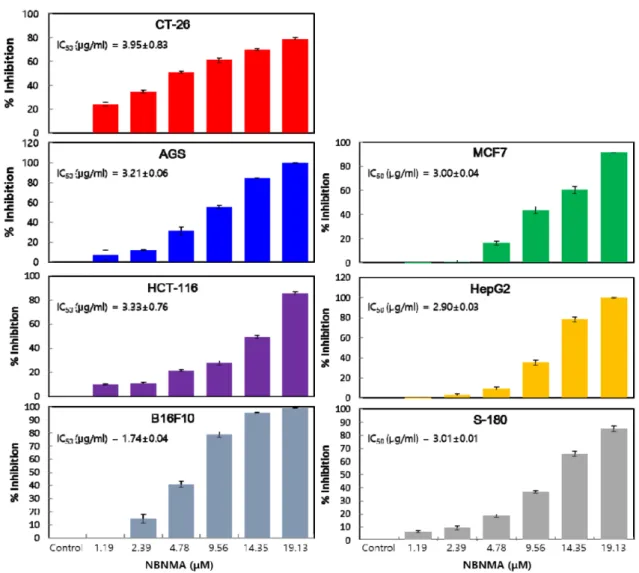
Anti-proliferative effect of NBNMA
Preliminary investigations of NBNMA were made for its in vitro anti-proliferative activity. In order to study the in- hibitory effect of NBNMA on CT-26, AGS, HepG2, HCT-116,
MCF7, B16F10, and Sarcoma-180 cells growth, we examined the cytotoxic effects of serial concentrations of NBNMA over a 24 hr treatment period using an MTT assay (Fig. 4).
NBNMA was found to significantly inhibit cell growth in a dose dependent manner. Interestingly, NBNMA appeared to be a more potent inhibitor of CT-26 cells in particular, with IC50 values of 15.11 μM. Treatment of CT-26 cells with NBNMA for 24 hr at increasing concentrations resulted in the inhibition of cell proliferation in the range of 14%–80%
at concentrations of 1.19-19.13 μM.
NBNMA regulates the expression of cell apoptosis regulating proteins
In order to determine if the inhibitory effect of NBNMA on cell proliferation was due to apoptosis, an immunoblot was carried out using Bcl-2 and caspase family proteins. The results showed that NBNMA could either induce or inhibit the expression of apoptotic regulating proteins. When com- pared with control cells, the CT-26 cells treated with differ- ent concentration of NBNMA for 24 hr achieved a meagre reduction of anti-apoptotic levels Bcl-2 protein. However, the level of protein apoptotic Bad members, gradually in- creased in a dose dependent manner (Fig. 5). Generally, the apoptosis observed in this study was caspase-dependant.
Cell lysates from CT-26 cells that had been dosed with NBNMA for 24 hr were probed with specific antibodies in order to evaluate the level of caspase 3, and 9 proenzymes,
Fig. 4. Inhibitory effect of NBNMA on CT-26, AGS, MCF7, HCT-116, HepG2, B16F10 and S-180 carcinoma cells. 1×10⁴ cells/well were seeded in a 96 well plate and incubated for 24 hr, followed by treatment either with DMSO control or NBNMA at different concentrations. The resulting cell viabilities were detected by an MTT assay, and the results are expressed as a percentage of control ± SD and represent the average of three separate experiments.
indicators of caspase activation. The NBNMA was activated caspase 3 at 19.13 and 38.26 μM and caspase 9 at 38.26 μM (Fig. 5). The levels of caspase 3 and 9 elevated during NBNMA treatment in a high concentrations with Western blot (Fig. 5).
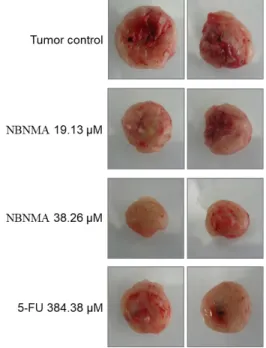
NBNMA inhibits the growth of CT-26 colon carcinoma in BALB/C mice
To determine the effect of NBNMA on colon carcinoma growth in vivo, CT-26 tumor cells were subcutaneously in- oculated into syngeneic BALB/C mice. 24 hr after tumor in- oculation the animals were systematically treated either with vehicle, NBNMA or 5-FU as positive control. The tumor vol- umes of the control, NBNMA and 5-FU treated groups were provided in Fig. 6-Fig.8. Of the different concentrations test-
ed, NBNMA doses of 5 19.13 μM/kg body weight on a 16 days oral administration exhibited 43.16±8.54% prevention of solid tumor growth. At the end treatment period, the tu- mor weight (0.89±0.13 g) and tumor volume (1,586±167mm3) in mice treated with 3.83 μM/kg NBNMA group was sig- nificantly lower when compared with the control (p<0.01).
Notably, NBNMA at a dose of 19.13 μM/kg inhibited tumor proliferation more effectively than the positive control 5-FU (30.51±5.77%).
DNA fragmentation
Further experiments were conducted to determine if the inhibitory effect of NBNMA on solid tumor growth was the result of apoptotic cell death. We also analyzed whether DNA fragmentation, another hallmark of apoptosis, was in-
Fig. 5. Immunoblotting analysis of Bcl-2, Bad, caspase-3 and caspase-9 in control and NBNMA treatment CT-26 cells. Immunoblot expression of Bcl-2, Bad, caspase-3 and caspase-9 in control and NBNMA groups were treated with CT-26 cancer cells at different concentration for 24 hr. Data are shown as the means ± SD of 3 independent experiments. * indicates p<0.05 and
**indicates p<0.01 vs. the control group.
Fig. 6. Effect of NBNMA on tumor growth rate of mice with CT-26 solid tumor. Significant inhibition of tumor growth at 23.63±8.07 and 49.84±7.55% were recorded in the dos- ing groups of NBNMA at 3.83 and 19.13 μM/kg for 16 days after treatment, respectively. Whereas the positive control 5-FU (384.38 μM/kg) inhibit the tumor growth rate with 36.78±5.16%.
Fig. 7. Effect of NBNMA and 5-fluorouracil (5-FU) on CT-26 solid tumor in mice at 20 days after treatment. Signifi- cant inhibition of tumor weight at values of 43% and 28% were recorded in the dosing groups for NBNMA at 19.13, and 3.83 μM/kg, respectively. Meantime, the positive control 5-FU inhibits 30% of tumor weight.
duced by NBNMA treatment of CT-26 tumor on BALB/C mouse. Following agarose gel electrophoresis of tumors treated with NBNMA 3.83 and 19.13 μM/kg body weight for 21 days, a typical ladder pattern of inter-nucleosomal fragmentation was observed (Fig. 9).
Discussion
The N-benzyl-N-methyldecan-1-amine (NBNMA) is the
first report of the occurrence of a natural source. The newly isolated NBNMA from garlic cloves was also tested for their in vitro cytotoxicity towards CT-26, AGS, HepG2, HCT-116,
Fig. 8. Average weight of CT-26 solid tumor at 20 days after treatment. Each value represents the mean ± SD, n=10 (p values **<0.01, *<0.05 vs. untreated control).
Fig. 9. Apoptotic DNA pattern of agarose gel electrophoretic of NBNMA treatment on CT-26 colon carcinoma cells induced in BZLB/C mice. Lane 1; CT-26 cells–induced I mice, Lane 2 ; Administration of NBNMA 3.83 μM/kg body weight, Lane 3 ; Administration of NBNMA 19.13/
kg μM body weight, , Lane 4 ; Administration of positive control 5-FU 384.38 μM/kg body weight (B) To analyze the DNA fragmentation, the genomic DNA was ex- tracted, electrophoresed in a 1.0% agarose gel and then visualized by EtBr staining.
MCF7, B16F10, and Sarcoma-180 cells. We also tested for their in vitro anti-cancer potential in a panel of solid tumor cells (CT-26). The NBNMA exerted significant growth in- hibitory effects on xenografted CT-26 solid tumor in BALB/
C mouse. NBNMA is potential cancer treatment agents showing anti-tumor activity against a variety of cancer cells and triggering multiple cell death pathways [14, 15]. Howev- er, first studies are available on the anti-proliferative activity of NBNMA against CT-26, AGS, HepG2, HCT-116, MCF7, B16F10, and Sarcoma-180 cells, and the mechanism by which it suppresses carcinogenesis remains unclear. An under-
standing of the underlying mechanism of induction of apop- tosis by NBNMA will benefit the development of chemo- preventives and chemotherapeutics for treating colon carci- noma. This study evaluated the anti-proliferative activity of NBNMA on CT-26, AGS, HepG2, HCT-116, MCF7, B16F10, and Sarcoma-180 cells using for in vitro and CT-26 for in vivo tests, with a special focus on the effect of NBNMA on apoptosis. Cell proliferation is governed by apoptosis, and the ability of cancer chemotherapeutic agents to induce apoptosis is an important in determining their therapeutic relevance [9, 29]. A cytotoxicity assay showed that NBNMA significantly inhibited the growth of CT-26 cells in a dose dependent manner. We demonstrated that NBNMA in- hibited cell growth and induced apoptosis in CT-26 cells in vitro and in vivo. These biochemical events were possibly associated with activation of pro-apoptotic protein Bad, sup- pression of anti-apoptotic Bcl-2, and activation of the caspase cascade. Apoptosis is a complex biological process involving many pathways and has been known to be a promising strat- egy for cancer prevention. Bax is a Bcl-2 family protein with proapoptotic activity, which has been shown to trigger cyto- chrome c release from mitochondria and Bax is present in the cytosol and weakly associated with mitochondria [2].
Consisting of both anti-apoptotic member Bcl-2 and pro- apoptotic member Bad, as they play a vital role in the mi- tochondrial apoptosis pathway [2]. As an anti-apoptotic pro- tein, Bcl-2 confers a negative control in the pathway of the cellular suicide machinery, whereas Bad competes with Bcl-2 to promote cell death [31]. In order to investigate the mecha- nism underlying apoptosis induced by NBNMA in CT-26 cells, we tested the expression of Bcl-2 family proteins and Bad. From the results NBNMA showed meagre reduction in Bcl-2 expression, and increased the level of Bad protein expression in a concentration dependent manner. This result affirmed that NBNMA induces apoptosis in CT-26 cells through the governance of apoptosis regulating proteins.
Previous reports showed that chemopreventive compounds induced apoptosis in cancer cells by the activation of caspase 3 [16]. Similar results were observed in this study as NBNMA upregulated the caspase 3 and 9 pro-enzymes, and elevated caspase 3 and 9 at a high concentrations in CT-26 cells. Caspase 3 plays a pivotal role in the execution of apop- tosis and its activation occurs by proteolytic cleavage [1, 20].
From the results, it was concluded that NBNMA induces apoptosis in CT-26 cells by the activation of caspase 3 and caspase 9 at high concentrations above 19.13 μM and 38.26
μM, respectively. Evidence of cytotoxicity in in vitro system is a property possessed by most clinically used anti-tumor agents. Furthermore, the NBNMA also demonstrated benefi- cial effects in a clinically relevant CT-26 murine melanoma model.
In conclusion, NBNMA produces significant anti-cancer effects, in both in vitro and in vivo conditions, by inducing apoptosis of CT-26 cells through the down regulation of Bcl-2 family proteins and the up regulation of Bad ex- pression. Of particular note, NBNMA can be used as a po- tent agent in the treatment of cancer.
Acknowledgment
This work was carried out with the support of "Cooper- ative Research Program for Agriculture Science & Technol- ogy Development (Project No. N01201804120010)" Rural Development Administration, Republic of Korea.
References
1. Adams, J. M. and Cory, S. 1998. The Bcl-2 protein family:
arbiters of cell survival. Science (New York, NY) 281, 1322- 1326.
2. Antonsson, B., Montessuit, S., Sanchez, B. and Martinou, J.
C. 2001. Bax is present as a high molecular weight oligom- er/complex in the mitochondrial membrane of apoptotic cells. J. Biol. Chem. 276, 11615-11623.
3. Apitz-Castro, R., Badimon, J. J. and Badimon, L. 1992. Effect of ajoene, the major antiplatelet compound from garlic, on platelet thrombus formation. Thromb. Res. 68, 145-155.
4. Argüello-García, R., Medina-Campos, O. N., Pérez-Hernán- dez, N., Pedraza-Chaverrí, J. and Ortega-Pierres, G. 2010.
Hypochlorous acid scavenging activities of thioallyl com- pounds from garlic. J. Agri. Food Chem. 58, 11226-11233.
5. Ariga, T., Seki, T., Ando, K., Teramoto, S. and Nishimura, H. 1996. Antiplatelet principle found in the essential oil of garlic (Allium sativum) and its inhibition mechanism. In Developments in Food Engineering; Yano, T., Matsuno, R., Nakamura, K., Eds.; Blackie Academic and Professional:
Tokyo, Japan, 1994; pp 1056-1058.
6. Block, E., Ahmad, S., Catalfamo, J. L., Jain, M. K. and Apitz- Castro, R. 1986. Antithrombotic organosulfur compounds from garlic-structural, mechanistic, and synthetic studies. J.
Am. Chem. Soc. 108, 7045-7055.
7. Dirsch, V. M., Gerbes, A. L. and Vollmar, A. M. 1998. Ajoene, a compound of garlic, induces apoptosis in human promye- loleukemic cells, accompanied by generation of reactive oxy- gen species and activation of nuclear factor kappaB. Mol.
Pharmacol. 53, 402-407.
8. Feng, W., Teo, X. Y., Novera, W., Ramanujulu, P. M., Liang, D., Huang, D., Moore, P. K., Deng, L. W. and Dymock, B.
W. 2015. Discovery of new H2S-releasing phosphordi- thioates and 2,3-dihydro-2-phenyl-2-sulfanylenebenzo[d][1, 3,2]oxazaphospholes with improved antiproliferative activ- ity. J. Med. Chem. 58, 6456-6480.
9. Fogarty, C. E. and Bergmann, A. 2017. Killers creating new life: caspases drive apoptosis-induced proliferation in tissue repair and disease. Cell Death Differ. 24, 1390-1400.
10. Gemici, B., Elsheikh, W., Feitosa, K. B., Costa, S. K. P., Muscara, M. N. and Wallace, J. L. 2015. H2S-releasing drugs:
Anti-inflammatory, cytoprotective and chemopreventative potential. Nitric Oxide 46, 25-31.
11. Guo, W., Cheng, Z. Y. and Zhu, Y. Z. 2013. Hydrogen sul- fide and translational medicine. Acta Pharmacologica Sinica 34, 1284-1291.
12. Heo, G. J, Oh, D. Y., Yi, S. W. and Shin, G. W. 2016.
Antimicrobial activity of Korean garlic extract against fish pathogenic bacteria isolated from cultured Olive Flounder, Paralichthys olivaceus in Korea. J. Prev. Vet. Med. 40, 7-11.
13. Ho, C. Y., Cheng, Y. T., Chau, C. F. and Yen, G. C. 2012.
Effect of diallyl sulfide on in vitro and in vivo Nrf2-mediated pulmonic antioxidant enzyme expression via activation ERK/p38 signaling pathway. J. Agri. Food Chem. 60, 100-107.
14. Jeong, J. W., Park, S., Park, C., Chang, Y. C., Moon, D. O., Kim, S. O., Kim, G. Y., Cha, H. J., Kim, H. S., Choi, Y. W., Kim, W. J., Yoo, Y. H. and Choi, Y. H. 2014. N-ben- zyl-N-methyldecan-1-amine, a phenylamine derivative iso- lated from garlic cloves, induces G2/M phase arrest and apoptosis in U937 human leukemia cells. Oncol. Rep. 32, 373-381.
15. Kaowinn, S., Kaewpiboon, C., Kim, J. E., Lee, M. R., Hwang, D. Y., Choi, Y. W., Kim, H. W., Park, J. K., Song, K. M., Lee, N. H., Maeng, J. S. and Chung, Y. H. 2018. N-Benzyl-N- methyl-dodecan-1-amine, a novel compound from garlic, exerts anti-cancer effects on human A549 lung cancer cells overexpressing cancer upregulated gene (CUG). Eur. J.
Pharmacol. 841, 19-27.
16. Keum, Y., S, Kim, J., Lee, K. H., Park, K. K., Surh, Y. J., Lee, J. M., Lee, S. S., Yoon, J. H., Joo, S. Y., Cha, I. H. and Yook, J. I. 2002. Induction of apoptosis and caspase-3 activa- tion by chemopreventive [6]-paradol and structurally re- lated compounds in KB cells. Cancer Lett. 177, 41-47.
17. Kim, D. H. 2009. Risk factors of colorectal cancer. J. Kor.
Soc. Coloproctol. 25, 356-362.
18. Ledezma, E. and Apitz-Castro, R. 2006. Ajoene the main ac- tive compound of garlic (Allium sativum): a new antifungal agent. Rev. Iberoam. Micol. 23, 75-80.
19. Nishikawa, T., Yamada, N., Hattori, A., Fukuda, H. and Fujino, T. 2002. Inhibition by ajoene of skin-tumor promo- tion in mice. Biosci. Biotechnol. Biochem. 66, 2221-2223.
20. Nunez, G., Benedict, M. A., Hu, Y. and Inohara, N. 1998.
Caspases: the proteases of the apoptotic pathway. Oncogene 17, 3237-3245.
21. Ou, H. C., Tzang, B. S., Chang, M. H., Liu, C. T., Liu, H.
W., Lii, C. K., Bau, D. T., Chao, P. M. and Kuo, W. W. 2010.
Cardiac contractile dysfunction and apoptosis in streptozo- tocin-induced diabetic rats are ameliorated by garlic oil
초록:활성추적분리법에 의해서 순수분리한 마늘 N-benzyl-N-methyldecan-1-amine이 CT-26 세 포주 이식 BALB/C mice의 항암효과
라자세카 시타르만1,2†․최성미1†․궈루1․추이정웨이1․두리마 오타곤바야르1․박주하1․권영석3․곽정호3․ 권영희4․민지현4․강점순1․최영환1*
(1부산대학교 원예생명과학과, 2GIOSTAR 인도, 3국립원예특작과학원 채소과, 4충청남도 농업기술센터)
마늘(Allium sativum)의 주요 생리활성 성분들은 다양한 종류의 암에 대해 항암효과가 보고되고 있다. 본 연구 에서는 활성추적분리방법(activity-guided purification)을 이용하여 마늘의 항암성분을 발굴하고자 하였다. 마늘 에탄올 추출물을 칼럼크로마토그래피로 얻은 각각의 분획물에 대해서 AGS세포의 증식 억제율을 검증하여 가장 효과가 좋은 분획물로부터 물질을 순수분리하여 구조를 동정한 결과 N-benzyl-N-methyldecan-1-amine (NBNMA) 로 밝혀졌다. NBNMA의 암생장 억제효능을 검증하기 위해서 CT-26, AGS, HepG2, HCT-116, MCF7, B16F10 및 Sarcoma-180 세포에 대한 in vitro 효과와 CT-26 결장암 세포를 마우스에 이식한 다음 in vivo 효과를 조사하였다.
NBNMA는 Bcl-2의 down-regulation과 Bad의 up-regulation을 유도하여 CT-26 세포의 세포사멸 촉진시켰다. 또 한, NBNMA는 세포사멸의 외적 및 내적 경로에서 caspases 억제자인 caspase 3과 caspase 9의 활성을 약간 증가 시켰다. CT-26세포를 이식한 쥐에 19.13μM/kg의 NBNMA를 21일 동안 경구투여한 결과 암종의 크기가 43% 감소 하였다. NBNMA는 in vitro 및 in vivo에서 항암 효과를 나타내었는데, 이러한 결과는 마늘로부터 순수분리한 NBNMA가 대장암치료를 위한 항암제 후보물질로서 활용 가능성이 있을 것으로 기대된다.
supplementation. J. Agric. Food Chem. 58, 10347-10355.
22. Pârvu, M., Pârvu, A. E., Rosca-Casian, O., Vlase, L. and Groza, G. 2010. Antifungal activity of Allium obliquum. J.
Med. Plants Res. 4, 138-141.
23. Rivlin, R. S. 2001. Historical perspective on the use of garlic.
J. Nutr. 131, 951S-954S.
24. Schäfer, G. and Kaschula, C. H. 2014. The immunomodula- tion and anti-inflammatory effects of garlic organosulfur compounds in cancer chemoprevention. Anticancer Agents Med. Chem. 14, 233-240.
25. Shen, Y., Shen, Z., Luo, S., Guo, W. and Zhu, Y. Z. 2015.
The cardioprotective effects of hydrogen sulfide in heart dis- eases: From molecular mechanisms to therapeutic potential.
Oxid. Med. Cell. Longev. 2015, 925167. http://dx.doi.org/
10.1155/2015/925167
26. Sigounas, G., Hooker, J., Anagnostou, A. and Steiner, M.
1997. S-allylmercaptocysteine inhibits cell proliferation and reduces the viability of erythroleukemia, breast, and pros- tate cancer cell lines. Nutr. Cancer 27, 186-191.
27. Snijder, P. M., Frenay, A. R., De Boer, R. A., Pasch, A., Hillebrands, J. L., Leuvenink, H. G. D. and Van Goor, H.
2015. Exogenous administration of thiosulfate, a donor of hydrogen sulfide, attenuates angiotensin II-induced hyper- tensive heart disease in rats. British J. Pharmacol. 172, 1494- 1504.
28. Stajner, D., Canadanovic-Brunet, J. and Pavlovic, A. 2004.
Allium schoenoprasum L., as a natural antioxidant. Phytother.
Res. 18, 522-524.
29. Tseng, L. J., Jao, Y. T. and Mo, L. R. 2002. Preoperative stag- ing of colorectal cancer with a balloon-sheathed miniprove.
Endoscopy 34, 564-568.
30. Vazquez-Prieto, M. A., González, R. E., Renna, N. F., Gal- marini, C. R. and Miatello, R. M. 2010. Aqueous garlic ex- tracts prevent oxidative stress and vascular remodeling in an experimental model of metabolic syndrome. J. Agric. Food Chem. 58, 6630-6635.
31. Williams, J. G. and Smith, J. P. 1993. Alcohol and other drug use among adolescents: Family and peer influences. J. Subst.
Abuse 5, 289-294.
32. Yang, J. Y., Della-Fera, M. A., Nelson-Dooley, C. and Baile, C. A. 2006. Molecular mechanisms of apoptosis induced by ajoene in 3T3-L1 adipocytes. Obesity (Silver Spring) 14, 388- 397.
33. Yin, M. C., Hwang, S. W. and Chan, K. C. 2002. Nonenzy- matic antioxidant activity of four organosulfur compounds derived from garlic. J. Agri. Food Chem. 50, 6143-6147.
34. Zhao, L., Wang, Y., Yan, Q., Lv, W., Zhang, Y. and He, S.
2015. Exogenous hydrogen sulfide exhibits anti-cancer ef- fects though p38 MAPK signaling pathway in C6 glioma cells. Biol. Chem. 396, 1247-1253.
35. Zong, J. and Martirosyan, D. M. 2018. Anticancer effects of garlic and garlic-derived bioactive compounds and its po- tential status as functional food. Bioactive Compd. Health Dis.
1, 16-35.