• 검색 결과가 없습니다.

A verification on the physical effectiveness of therapeutic horseback riding exercise: Focused on the EMG analysis

N/A
N/A
Protected

Academic year: 2021

Share "A verification on the physical effectiveness of therapeutic horseback riding exercise: Focused on the EMG analysis"

Copied!
8
0
0

로드 중.... (전체 텍스트 보기)

전체 글

(1)

ISSN 2288-1069 (Online)

http://dx.doi.org/10.12925/jkocs.2018.35.2.317

A verification on the physical effectiveness of therapeutic horseback riding exercise: Focused on the EMG analysis

You-Sin Kim․Jae-Young Yang․Namju Lee

Department of Leisure Sports, School of Sports Science, Jungwon University, Goesan-gun, Chungbuk, 28024, Republic of Korea

Department of Physical Education, Graduate School, Jungwon University, Goesan-gun, Chungbuk, 28024, Republic of Korea

Department of Leisure Sports and Sports Health Medicine, School of Sports Science, Jungwon University, Goesan-gun, Chungbuk, 28024, Republic of Korea (Received April 24, 2018; Revised June 18, 2018; Accepted June 19, 2018)

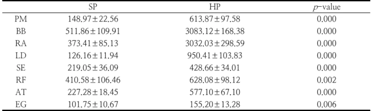
Abstract : Various studies related to therapeutic horseback riding have been reported to be positive for the therapeutic effect of patients with cerebral palsy; however, most of the previous studies focused on to muscle development with training period related to the physical effects of therapeutic horseback riding. To identify the causes and phenomena of muscular activation of the body through actual therapeutic horseback riding exercise and to promote the excellence of physical effects of therapeutic horseback riding. This study was a nonrandomized prospective positive-controlled trial design. Twelve teenaged males with cerebral palsy were selected who had experienced riding exercise for 8-12 months. This study measured 8 muscle activities of the pectoralis major muscle (PM), biceps brachii (BB), rectus abdominis muscle (RA), latissimus dorsi muscle (LD), spinal erector muscle (SE), rectus femoris muscle (RF), anterior tibial muscle (AT), and external gastrocnemius muscle (EG) by using electromyography (EMG). Muscle activity was significantly higher in horse riding position than sitting on the common chair in all muscles (PM, BB, RA, LD, SE, RF, AT, and EG). The activity of the body muscles according to the difference of horse walking method (walk: WA; sitting trot: ST;

and riding trot: RT) of therapeutic horse riding showed the highest muscle activity in the PM muscle at ST, and the highest activity at BB, RA, LD, SE, and AT muscles at ST and RT, and showed the highest muscle activity in RF and EG muscle at RT. The results of this study suggest that intervention for the treatment of cerebral palsy patients can use therapeutic riding exercise as a rehabilitation method.

Keywords : Therapeutic horseback riding, Rehabilitation, Walk, Trot, EMG



Corresponding author

(E-mail: namju1210@gmail.com)

(2)

1. Introduction

Therapeutic horseback riding is one therapeutic method among rehabilitation sports for the disabled and it induces overcoming emotional, social, and physical disability for the better life [1]. Therapeutic horseback riding plays a role in the overall balance and harmony in the human body rather than treating only one part of the disorder and especially, therapeutic horseback riding is widely used as a treatment and management tool in cerebral palsy, learning disability, amputation, autism, spinal cord injury, neurological disorders, and emotional disorders [2].

Cerebral palsy is a type of physical lesion that occurs in the developing fetal period or early childhood brain tissue and has a permanent limitation on the development of movement such as walking disorder, movement disorder, and sensory disorder [3]. It is a chronic disease that causes physical disability in various parts of the body movement [4], and requires physical and occupational therapy during lifetimes [5]. It also causes other health problems such as sight, hearing, speech, and language disorders and the most usual form is rigid paralysis and 70-80% of rigid paralysis patients are classified as hemiplegia, quadriplegia, and paraplegia [6].

In 21

st

century, as a sporting method for improving performance of patients with cerebral palsy, therapeutic horseback riding is being actively promoted to stabilize the posture and to stimulate a part of the body using horse [7-10] and previous studies reports that the treatment effect of patients with cerebral palsy is positive [8,11]. One previous therapeutic horseback riding study [12]

reported that patients with cerebral palsy, who conducted therapeutic horseback riding for 1 time per week for 18 weeks, showed decreased muscle tone and thus therapeutic horseback riding has been reported to be effective in relaxing abnormal stiff muscles. Silva, et al

[13] also mentioned that therapeutic horseback riding usually gives riders to experience the pressure of the sensory nerves, which is similar to the pattern of movement that occurs in the pelvis when a person is walking, so that the body temperature and movement of the horses delivered at this time improve the circulation of the passenger and helps to relax the rigid muscles.

Various studies related to therapeutic horseback riding have been reported to be positive for the therapeutic effect of patients with cerebral palsy; however, most of the previous studies focused on to muscle development with training period related to the physical effects of therapeutic horseback riding and were only related with the identification of the cause of muscle development on how much muscle of the body was mobilized during actual therapeutic horseback riding exercise [14,15]. Therefore, the purpose of this study was to identify the causes and phenomena of muscular activation of the body through actual therapeutic horseback riding exercise and to promote the excellence of physical effects of therapeutic horseback riding in patients with cerebral palsy.

2. Materials and Method 2.1. Participants

This study was a non-randomized

prospective positive-controlled trial to

investigate the physical effectiveness of

therapeutic horseback riding exercise. In this

study, 12 teenaged males with dyskinetic

cerebral palsy participated in the experiment

after receiving consent to participate in the

experiment by their caregivers and their

physicians. The characteristics of this study

participants are shown in Table 1. This study

participants were selected as cerebral palsy

patients who had experienced riding exercise

for 8-12 months, and their functional

classification was classified as athetoid (slow

(3)

N Age

(yrs) Body Mass

(kg) Height

(cm) BMI

(kg/m

2

) Career

(mon) functional

classification GMFCS 12 16.83±0.37 65.42±0.77 168.42±0.69 23.07±0.30 9.58±0.23 athetoid level 2 Values are mean ± SE

GMFCS, gross motor functional classification system Table 1. Physical characteristics of participants

torsional motion type) among dyskinetic (movement ideal) of global involvement, and the gross motor functional classification system (GMFCS) level was limited to GMFCS level 2.

Informed consent from each participant with parental agreement and physicians’ consent were obtained before conducting this study.

2.2. Experimental approach to the problem Experiments to collect the data required for this study were conducted at the “T” horse riding area in Cheongju city. In consideration of the validity of the measurements at the time of the experiment, the horse for this study was limited to the same species of Shetland pony.

Before starting the experiment, participants were informed about the anatomical position, sitting position (SP), horse riding position (HP), walk (WA), sitting trot (ST), and riding trot (RT) and all participants fully practiced those with the help of a guardian, a horse leader, and two side walkers to ensure smooth movement. The mean speed for each method

was 100-110m/min for WA and

220-240m/min for trot (ST and RT) [16], and 12 steps were performed at one time, and the values ​​of 6 motion cycles excluding the first 3 steps and the last 3 steps were extracted for each horse walking method [17, 18]. With 5 tests for each horse walking method, the measurement results of 30 strides (6 strides × 5 times) were analyzed.

For the analysis of electromyography (EMG), a surface electrode was fixed on the muscle surface to be measured. To eliminate the measurement error, the hair protruding from the muscular part was removed with a razor

and the medical alcohol (Hitech bio Pharm, Busan, Korea) and then the surface electrode was attached. Based on the previous study [19], all surface electrodes were attached on pectoralis major muscle (PM), biceps brachii (BB), rectus abdominis muscle (RA), latissimus dorsi muscle (LD), spinal erector muscle (SE), rectus femoris muscle (RF), anterior tibial muscle (AT), and external gastrocnemius muscles (EG), which was representative muscles of right trunk, upper limb, and lower extremity.

The experimental procedure of EMG according to the therapeutic horseback riding method was applied by counterbalanced design.

One Life Cam VX-5000 digital video camera (Microsoft Corporation, WA, USA) was used to synchronize all data with each anatomical position, SP, HP, image data, and EMG data.

For EMG measurement, myo MUSCLE 3.8.2

program (Noraxon USA Inc., AZ, USA) was

used. The surface electrode was connected to a

Model 542 DTS EMG sensor (Noraxon USA

Inc., AZ, USA) with a small surface electrode

T 246 H (Bio-Protech Inc., Wonju, Korea)

with a diameter of 20 mm. The electrodes

were placed in a 2cm distance and the

electrodes were connected to the TeleMyo ™

Desktop Direct Transmission System (Noraxon

USA Inc., AZ, USA). The EMG signal

processing method sets the bandwidth to

10-350 Hz and the sampling rate to 1,024

Hz. EMG signals were rectified and then

smoothed with a root mean square (RMS) of

200 ms.

(4)

SP HP p -value

PM 148.97±22.56 613.87±97.58 0.000

BB 511.86±109.91 3083.12±168.38 0.000

RA 373.41±85.13 3032.03±298.59 0.000

LD 126.16±11.94 950.41±103.83 0.000

SE 219.05±36.09 428.66±34.01 0.000

RF 410.58±106.46 628.08±98.12 0.002

AT 227.28±18.45 577.10±67.10 0.000

EG 101.75±10.67 155.20±13.28 0.006

Values are mean ± SE

%RVC, percentage of reference voluntary contraction; SP, sitting position; HP, horse riding position; PM, pectoralis major muscle; BB, biceps brachii; RA, rectus abdominis muscle; LD, latissimus dorsi muscle; SE, spinal erector muscles; RF, rectus femoris muscle; AT, anterior tibial muscle; EG, external gastrocnemius muscle

Table 2. Comparison of average rectified variable muscle activity during SP and HP (Unit: %RVC) 2.3. Procedures

In this study, the standardization process was performed to compare EMG signals between subjects and between the muscles. The EMG signal standardizing method assumes that the muscle contraction of the specific motion is standard voluntary contraction (RVC) and the% RVC method was used [19]. The baseline RVC was defined as the duration of each subject's anatomical position for 10 seconds, and the data value for 10 seconds was subjected to RMS, and the average EMG signal level during the middle 4 seconds except for the first and last 3 seconds were used to standardize the EMG signals of each muscle with % RVC.

2.4. Statistical Analysis

All data was expressed as the mean and standard error (mean ± SEs) and calculated by analyzing the experimental results with IBM SPSS Statistics 23 (IBM Corporation, NY, USA). Paired t-test was used to compare the difference of muscle activity in SP and HP.

One-Way RM (repeated-measures) ANOVA was used to compare the difference of muscle activity according to various walking method of the horse (WA, ST, and RT) and if there

was a statistically significant difference, Bonferroni method was used as a post analysis method. Statistical significance was set at p <.05.

3. Results

Table 2 shows the results of muscle activity of 8 muscles in comparison with SP and HP.

Measurement of muscle activity in PM, BB, RA, LD, SE, RF, AT, and EG resulted that HP showed higher muscle activity than SP in all the muscles and each results are as follows; (t=-5.30, p =0.000), (t=-9.38, p =0.000), (t=-11.92, p =0.000), (t=-8.66, p =0.000), (t=-23.17, p =0.000), (t=-3.91, p =0.002), (t=-5.33, p =0.000), and (t=-3.21, p =0.006).

During therapeutic horseback riding, 8

muscle activity changes of the WA, ST, and

RT are shown in Table 3. The ST showed the

highest activity in PM than RT and WA, and

the statistical result was (F

(2,118)

=22.70,

p =0.000). At ST and RT, the highest muscle

activity was found in BB, RA, LD, SE, and

AT than in WA and each results are as

follows; (F

(2,118)

=6.21, p=0.004), (F

(2,118)

=14.02,

(5)

WA ST RT p-value Post-hoc PM 1041.57±133.30 2771.03±230.03 2024.74±169.56 0.000 WA<RT<ST

BB 10235.23±1129.53 20563.06±1878.15 17957.36±3022.67 0.004 WA<RT, ST RA 3710.50±659.93 10118.22±943.20 10076.68±1259.15 0.000 WA<RT, ST LD 2341.47±302.42 8750.24±1401.11 6692.57±756.31 0.000 WA<RT, ST SE 1720.60±273.59 6115.22±1305.74 9645.75±3373.14 0.036 WA<ST, RT RF 719.96±122.13 3018.49±199.09 5008.63±317.18 0.000 WA<ST<RT AT 2147.16±300.41 4331.27±763.76 4135.56±471.03 0.012 WA<RT, ST

EG 359.96±78.98 636.22±74.97 1258.02±122.22 0.000 WA, ST<RT

Values are mean ± SE

%RVC, percentage of reference voluntary contraction; SP, sitting position; HP, horse riding position; PM, pectoralis major muscle; BB, biceps brachii; RA, rectus abdominis muscle; LD, latissimus dorsi muscle; SE, spinal erector muscles; RF, rectus femoris muscle; AT, anterior tibial muscle; EG, external gastrocnemius muscle

Table. 3. Comparison of average rectified variable muscle activity during WA, ST, and RT (Unit: %RVC)

p =0.000), (F

(2,118)

=12.23, p =0.000), (F

(2,118)

=3.59, p =0.036), and (F

(2,118)

=4.89, p =0.012). Lastly, at RT, RF, and EG showed the highest muscle activity than in ST and WA and each results are (F

(2,118)

=89.06, p =0.000) and (F

(2,118)

=23.69, p =0.000).

4. Discussion

Major findings of this study are as follows;

1) muscle activity was significantly higher in HP than SP in all muscles (PM, BB, RA, LD, SE, RF, AT, and EG), 2) The activity of the body muscles according to the difference of horse walking method (WA, ST, and RT) of therapeutic horseback riding showed the highest muscle activity in the PM muscle at ST, and the highest activity at BB, RA, LD, SE, and AT muscles at ST and RT, and showed the highest muscle activity in RF and EG muscle at RT.

Cerebral palsy is accompanied by various paralysis of the body caused by brain lesions, so it is necessary to cope with the problems such as reduction of joint motion range,

paralysis due to muscle tension, walking ability disorder, and motor dysfunction [12].

Particularly, due to the nature of cerebral

palsy, it is very important to intervene to

relieve the developmental delay because it

affects the physical development and has

difficulties in maintaining the daily life. This is

because it can be a management to minimize

the inconvenience of daily life even if it is not

a fundamental treatment, and the key to

rehabilitation is to maximize the individual's

potential [20]. As one of these intervention

methods, therapeutic horseback riding exercise

has been reported to show many effects on

patients with cerebral palsy [21,22]. In the

case of studies on the excellence of physical

characteristics by performing therapeutic

horseback riding exercise for patients with

cerebral palsy, Alfredson, et al. [23] reported

that the group that performed rehabilitation

riding exercise increased bone mass due to

weight load and strength of hamstrings and

quadriceps femoris was significantly increased

as a result of examining the strength of 20

adolescent female professional riders who

exercised for 7 hours a week and 20

(6)

adolescent females who did not exercise.

McGibbon, et al. [11] investigated the effect of exercise on the group of rehabilitation riding exercise group and the group that stretched in the round barrel for the same time. They used EMG and 47 children with spastic cerebral palsy were participated. Results showed that the group of children who underwent a rehabilitation riding exercise program had higher muscle strength.

Hodges and Richardson [24] mentioned that the activity of the trunk muscles plays a major role in the movement of the pelvis and the movement of the lower limb, and is an essential factor in maintaining the lumbar stability. This study compared muscle activity at SP and HP and found that PM (412.07 %), BB (602.33 %), RA (811.98 %), LD (753.33

%), SE (195.69 %), RF (152.97 %), AT (253.91 %), and EG (152.53 %) showed significantly higher muscle activity in HP compared to SP. During therapeutic horseback riding, PM (266.04 %), BB (200.90 %), RA (272.69 %), LD (373.70 %), SE (560.60 %), RF (695.68 %), AT (201.72 %), and EG (349.48 %) showed significantly higher muscle activity in ST and RT compared to WA.

These findings proved that physical exercise is excellent even if only the HP is performed rather than the SP and trot (ST or RT) is more effective than WA during therapeutic horseback riding exercise. These results may be consistent with the goal of rehabilitation riding therapy, which is to optimize the strength of the body, by decreasing the stiffness pattern of patients with cerebral palsy by maintaining proper posture while sitting on the horse. In addition, this study also proved that the actual rehabilitation riding therapy approach, which has been avoided clinically in the treatment of patients with cerebral palsy, is an effective treatment method.

A study for the effect of rehabilitation riding therapy through EMG on the symmetry in

muscle activity of the trunk and upper legs [25], it reported that children aged 4-12 years with rigid cerebral palsy had significantly higher symmetry in muscle activity in WA compared to HP. Similarly, this study result ssupports higher muscle activity of PM (169.67

%), BB (331.97 %), RA (122.37 %), LD (246.36 %), SE (401.39 %), RF (114.62 %), AT (372.06 %), and EG (231.93%) in WA compared to HP. This suggests that therapeutic horseback riding exercise is effective in improving general physical exercise function of cerebral palsy patients.

5. Conclusion

HP and trot (ST and RT) had a significant effect on the muscle activity of patients with cerebral palsy, and it was found that the physical effect of the therapeutic horseback riding was positive for the improvement of muscle strength of cerebral palsy patients. If patients with cerebral palsy can successfully perform WA, the basic course of therapeutic horseback riding exercise, we recommend the trot (ST and RT) method, which is more helpful in improving physical strength. The results of this study suggest that the method of intervention for treatment of cerebral palsy patients can use therapeutic horseback riding exercise as a rehabilitation method. In future research, it would be expected that the physical effects of therapeutic horseback riding exercise should be studied according to several types of disability, gender, and age.

Acknowledgment

This research did not receive any specific

grant from funding agencies in the public,

commercial, or not-for-profit-sectors.

(7)

References

1. M. Lessick, R. Shinaver, K. Post, J.

Rivera, B. Lemon, "Therapeutic horse riding: Exploring this alternative therapy for women with disabilities". AWHONN Lifelines. Vol.8, pp. 48–53, (2004).

2. Z. Nqwena, R. Naidoo, "The effect of therapeutic horseback riding on heart rate variability of children with disabilities". Afr J Disabil, Vol.5, No.1 pp. 1 - 8, (2016).

3. M. Bax, M. Goldstein, P. Rosenbaum, A.

Leviton, N. Paneth, B. Dan, B. Jacobsson, D. Damiano, "Proposed definition and classification of cerebral palsy". Dev Med Child Neurol, Vol.47, No.8 571-576, (2005).

4. K. Nelson, B. Russman, “Cerebral Palsy”

In K. Swaiman, Pediatric neurology. St Louis: Mosby Inc (2002).

5. N. I. Ionatamishvili, D. M. Tsverava, M.

Sh. Loriia, E. G. Sheshaberidze, M. M.

Rukhadze, "Riding Therapy as a Method of Rehabilitation of Children with Cerebral Palsy". Fiziol Cheloveka, Vol30, No.5 pp.

69-74, (2004).

6. J. A. Blackman, A. Healy, E. S. Ruppert,

"Participation by pediatricians in early intervention: impetus from Public Law"

pp. 99-457, Pediatrics, Vol.89, No.1 pp.

98-102, (1992).

7. R. Cherng, H. F. Liao, H. Leung, A.

Hwang, "The effectiveness of therapeutic horseback riding in children with spastic cerebral palsy". Adapt Phys Activ Q, Vol.21, No.2 pp. 103-121, (2004).

8. G. S. Liptak, "Complementary and alternative therapies for cerebral palsy".

Ment Retard Dev Disabil Res Rev, Vol.11, No.2 pp. 156-163, (2005).

9. M. C. Bergene, "Therapeutic horseback riding in individuals with disabilities. Effect on Hip Range of Motion". Senior Research Project. Marian College of Fond du Lac, WI , (2006).

10. F. Beinotti, N. Correia, G. Christofoletti,

G. Borges, "Use of hippotherapy in gait training for hemiparetic post-stroke". Arq Neuropsiquiatr, Vol.68, No.6 pp. 908-913, (2010).

11. N. H. McGibbon, W. Benda, B. R.

Duncan, D. Silkwood-Sherer, "Immediate and long-term effects of hippotherapy on symmetry of adductor muscle activity and functional ability in children with spastic cerebral palsy". Arch Phys Med Rehabil, Vol.90, No.6 pp. 966-974, (2009).

12. J. A. Sterba, B. T. Roger, A. P. France, D.

A. Vokes, "Horseback riding in children with cerebral palsy: effect on gross motor function". Dev Med Child Neurol, Vol.44, No.5 pp. 301-308, (2002).

13. M. B. Silva e Borges, M. J. Werneck, L.

da Silva Mde, L. Gandolfi, R. Pratesi,

"Therapeutic effects of a horse riding simulator in children with cerebral palsy".

Arq Neuropsiquiatr, Vol.69, pp. 799-804, (2011).

14. G. Asselin, J. H. Penning, S. Ramanujam, R. Neri, C. Ward, "Therapeutic horse back riding of a spinal cord injured veteran: a case study". Rehabil Nurs, Vol.37, No.6 pp. 270-276, (2012).

15. S. Muñoz-Lasa, G. Ferriero, R. Valero, F.

Gomez-Muñiz, A. Rabini, F. Varela,

"Effect of therapeutic horseback riding on balance and gait of people with multiple sclerosis". G Ital Med Lav Ergon, Vol.33, No.4 pp. 462-467, (2011).

16. P. Sarah, E. Samantha, D. Zoe, Blackwell Publishing Company, pp. 96, (2009).

17. B. Fruehwirth, C. Peham, M. Scheidl, H.

Schobesberger, "Evaluation of pressure distribution under an English saddle at walk, trot and canter". Equine Vet J, Vol.36, No.8 pp. 754-757, (2004).

18. B. Winkelmayr, C. Peham, B. Frühwirth,

T. Licka, M. Scheidl, "Evaluation of the

force acting on the back of the horse with

an English saddle and a side saddle at

walk, trot and canter". Equine Vet J,

Vol.38, No. S36, pp 406-410, (2006).

(8)

19. J. R. Cram, G. S. Kasman, J. Holtz.

Electrode placement. In: Cram JR, Kasman GS, Holtz J. Introduction to surface electromyography. Gaithersburg, MD.

Aspen Publishers, (1998).

20. L. Powell, J. Barlow, A. Cheshire, "The training and support programme for parents of children with cerebral palsy: a process evaluation". Complement Ther Clin Pract, Vol.12, No.3 pp. 192-199, (2006).

21. J. A. Sterba, "Does horseback riding therapy or therapist-directed hippotherapy rehabilitate children with cerebral palsy"?

Dev Med Child Neurol, Vol.49, No.1 pp.

68-73, (2007).

22. J. Y. Kwon, H. J. Chang, J. Y. Lee, Y.

Ha, P. K. Lee, Y. H. Kim, "Effects of hippotherapy on gait parameters in children with bilateral spastic cerebral palsy". Arch Phys Med Rehabil, Vol.92, No.5 pp. 774-779, (2011).

23. H. Alfredson, G. Hedberg, E. Bergström, P. Nordström, R. Lorentzon, "High thigh muscle strength but not bone mass in young horseback-riding females". Calcif Tissue Int, Vol.62, No.6 pp. 497-501, (1998).

24. P. W. Hodges, C. A. Richardson,

"Contraction of the abdominal muscle associated with movement of the lower limb". Phys Ther, Vol.77, No.2 pp.

132-142, (1997).

25. W. Benda, N. H. McGibbon, K. L. Grant,

"Improvements in muscle symmetry in children with cerebral palsy after equine-assisted therapy (Hippotherapy)". J Altern Complement Med, Vol.9, No.6 pp.

817-825, (1998).

수치

Table  2.  Comparison  of  average  rectified  variable  muscle  activity  during  SP  and  HP  (Unit:  %RVC)2.3

참조

관련 문서

The purpose of this study is to analyze the thesis that Taekwondo poomsae has positive effects on Physical Fitness of Students with Intellectual

Many studies using the assumed mode method have been found for the free vibration analysis of stiffened plate with known elastic boundary conditions.. However

The purpose of this study was to examine the effects of a swimming program for 12 weeks on health-related physical fitness and growth hormone in

The effect of Combined Exercise program on Body composition, Physical fitness and thickness of subcutaneous fat in. Obese

Taken together, by applying physical strength exercise program to the elite table tennis players we can conclude that physical strength exercise program has positive effects

Various studies have been performed to improve properties of material, electrical and mechanical properties of railway train according to the increase of the speed in the

Through an empirical analysis of employability of persons with cerebral palsy this study aims to examine the factors affecting their employability and

In future studies focused on antioxidant effect and active oxygen reduction in resistance exercise using thera-band will be conducted in-depth research that