• 검색 결과가 없습니다.

In vitro antioxidant, anti-hyperglycemic, anti-cholinesterase, and inhibition of nitric oxide production activities of methanol and hot water extracts of Russula rosacea mushroom

N/A
N/A
Protected

Academic year: 2021

Share "In vitro antioxidant, anti-hyperglycemic, anti-cholinesterase, and inhibition of nitric oxide production activities of methanol and hot water extracts of Russula rosacea mushroom"

Copied!
10
0
0

로드 중.... (전체 텍스트 보기)

전체 글

(1)

1

This is an Open-Access article distributed under the terms of the Creative Commons Attribution Non-Commercial License (http://

creativecommons.org/licenses/by-nc/3.0) which permits unrestricted non-commercial use, distribution, and reproduction in any medium, provided the original work is properly cited.

J. Mushrooms 2015 March, 13(1):1-10 http://dx.doi.org/10.14480/JM.2015.13.1.1 Print ISSN 1738-0294, Online ISSN 2288-8853

© The Korean Society of Mushroom Science

*Corresponding author E-mail : [email protected]

Tel : +82-32-835-4617, Fax : +82-32-835-0763 Received March 9, 2015

Revised March 23, 2015 Accepted March 25, 2015

In vitro antioxidant, anti-hyperglycemic, anti-cholinesterase, and inhibition of nitric oxide production activities of methanol and hot water extracts of Russula rosacea mushroom

Ki Nam Yoon and Tae Soo Lee

1,

*

Department of Clinical Laboratory Science, Ansan University, 155 Ansan Dae-hak-ro, Sangrok-gu, Ansan, 426-701, Korea

1

Division of Life Sciences, Incheon National University, (Songdo-dong) 119 Academy-ro, Yeonsu-gu, Incheon 406-772, Korea

ABSTRACT: Russula rosacea, a mycorrhizal fungus, has been used for edible and medicinal purposes. This study was conducted to evaluate the in vitro antioxidant, anti-hyperglycemic, anti-cholinesterase, and nitric oxide inhibitory effects of the fruiting bodies from R. rosacea extracted with methanol, and hot water. The 1,1-diphenyl-2-picryl hydrazyl (DPPH) radical scavenging activities of the methanol and hot water extracts (2.0 mg/ml) of R. rosacea were comparable with BHT, the positive control. The chelating effects of the mushroom and hot water extracts were significantly higher than that of BHT. The reducing power of methanol and hot water extract (6 mg/ml) were significantly lower than that of BHT. Seven phenolic compounds were detected from acetonitrile and hydrochloric acid solvent extract of the mushroom. alpha-amylase and alpha-glucosidase inhibitory activities of methanol and hot water extracts were lower than that of acarbose, the positive control. The acetylcholinesterase and butyrylcholinesterase inhibitory effects were moderate compared with galanthamine, the standard drug. Nitric oxide (NO) production in lipopolysaccharide (LPS) induced RAW 264.7 cells were inhibited significantly by the mushroom extracts in a concentration dependent manner. Therefore, we demonstrated that fruiting bodies of R. rosacea possess in vitro antioxidant, anti-hyperglycemic, anti-cholinesterase, and NO production inhibitory activities. The experimental results suggest that the fruiting bodies of R. rosacea are good natural antioxidant, anti-hyperglycemic, anti-cholinesterase, and anti-inflammatory sources.

KEYWORDS: Anti-cholinesterase, Anti-hyperglycemic, Anti-inflammation, Antioxidant, Russula rosacea

Introduction

Traditionally, mushrooms have a long history of use in the prevention and treatment of various diseases such as hepatitis, hypertension, hypercholesterolemia and cancers (Wasser and Weis, 1999) Recent studies have shown that mushroom extracts contain active components that suppress the pathological processes of many diseases such as diabetes, dementia and inflammatory diseases

(Ying et al, 1987).

Oxidation is essential biological processes for producing energy fuel in living creatures. Free radicals and related reactive oxygen species (ROS) which are produced in vivo, sometimes result in tissue damages and cell death.

Damage caused by free radicals might be related to diseases including atherosclerosis, hyperlipidemia, and diabetes. Although almost all organisms possess antioxidant defense and repair systems that protect them against oxidative damages, these systems are not sufficient to prevent the damage entirely (Lobo et al, 2010). However, foods sources which containing antioxidant ingredients including flavonoid and polyphenolic compounds may be useful to protect the human body from oxidative damage (Mayne, 2003).

The α-amylase and α-glucosidase are major enzymes

responsible for the hydrolysis of starch and carbohydrates

to mono-saccharide. For diabetes patients, inhibition of

these enzymes are important for the regulation of

postprandial glucose levels. Numerous scientific researchers

have demonstrated that mushrooms possessed medical

(2)

properties, including a hypeglycemic inhibitory effect (Perera and Li, 2011). Therefore, mushrooms may be a potential source for the dietary supplement in prevention or treatment of diabetes.

Acetylcholinesterase (AChE) is an enzyme hydrolyzes acetylcholine (ACh), the neurotransmitter found at cholinergic brain synapses (Quinn, 1987). An AChE inhibitor is a chemical that inhibits AChE from breaking down acetylcholine, thereby increasing the action of ACh. AChE inhibitors can increase cholinergic transmission by preventing the hydrolysis of released ACh, thus making more ACh available at the cholinergic synapse (Benzi and Morreti, 1998).

This makes AChE inhibitors the most effective means to treat the cognitive symptoms of Alzheimer's disease (Kalauni et al, 2002). AChE inhibitors such as physostigmine, tacrine, donepezil, rivastigmine, and galanthamine are the only currently available drugs for the treatment of AD. However, these drugs are known to have limitations for clinical use due to their short half-lives and side-effects (Sung et al, 2002). As such, the search for potent natural products that selectively inhibit AChE activity is necessary.

Russula rosacea, a mycorrhizal mushroom, belongs to the family Russulaceae, order Agaricales, Basidiomycota The fruiting bodies of R. rosacea, has been used for edible and medicinal purposes in Asian countries for centuries, however only a few studies on physiologically beneficial activities have been conducted (Park and Lee, 2003). Therefore, the objective of this study was to evaluate the in vitro antioxidant, anti-hyperglycemic, and anti-cholinesterase activities of methanol, and hot water extracts from the fruiting bodies of R. rosacea. The constituents of phenolic compounds of fruiting bodies of the mushroom were also analyzed.

Materials and Methods

Chemicals and reagents

Dibutyl hydroxy toluene (BHT), potassium ferricyanide, trichloroacetic acid, ferrous chloride, ferric chloride, ferrozine, Folin-Ciocalteu reagent, methanol, 3,4-dihydroxy- L-phenylalanine, dimethyl sulfoxide (DMSO), tris base, glacial acetic acid, trichloroacetic acid and galantamine, were obtained from Sigma-Aldrich (St. Louis, MO, USA). All chemicals and solvents used for the experiment and high performance liquid chromatography (HPLC) were analytical grade.

Mushroom and extraction

Fresh and mature fruiting bodies of R. rosacea, collected from Seoraksan National Park, were hot air- dried at 40

o

C for 48 h and finely pulverized. 10 grams of powdered sample was extracted with 200 ml of 80%

methanol with stirring at 150 rpm for 24 h at 25

o

C to obtain the methanol extracts. The mixture was filtered through two layers of Whatman No. 1 filter paper (Whatman, Maidstone, UK). The same quantity of sample was boiled at 100

o

C for 3 h with 200 ml deionized distilled water to obtain the hot water extract.

The mixture was cooled to room temperature and filtered through Whatman No. 1 filter paper. The residues were then extracted with two additional 200 ml aliquots of methanol, and deionized water, as described above. The combined extracts were evaporated with a rotary evaporator at 40

o

C, and the remaining solvent was removed with a freeze-drier. Yields of the methanol and hot water extracts of R. rosacea were 16.32% and 18.35% (w/w), respectively.

DPPH radical scavenging

DPPH free radical scavenging activity was measured by evaluating hydrogen atoms or electron donation ability of the extracts and some pure compounds were measured as bleaching of the purple colored DPPH methanol solution (Cuendet et al., 1997). 4 ml of various concentrations (0.125-2.0 mg/ml) of the extracts in methanol was added to 1.0 ml of DPPH radical solution in methanol (final DPPH concentration, 0.2 mM). The mixture was shaken vigorously and allowed to stand for 30 min, and the absorbance of the solution was measured at 517 nm using a spectrophotometer.

Inhibition of the DPPH free radical in percent (I %) was calculated as:

I % = [(A

control

−A

sample

)/A

control

]×100

Where, A

control

is the absorbance of the control reaction (containing all reagents except the test compound), and

A

sample

is the absorbance of the test compound. BHT was

used as positive control.

Chelating effects on ferrous ions

The chelating effect was determined according to the

method of Dinis et al (1994). Briefly, 2 ml of various

concentrations (0.063-1.0 mg/ml) of the extracts in

methanol was added to a solution of 2 mM FeCl

2

(0.05

ml). The reaction was initiated by adding 5 mM

(3)

ferrozine (0.2 ml). Total volume was adjusted to 5 ml with methanol, and the mixture was shaken vigorously and incubated at room temperature for 10 min. The absorbance of the solution was measured by spectrophotometer at 562 nm. The inhibition percentage of the ferrozine-Fe

2+

complex formation was calculated as:

Ferrous ions chelating effect (%) = [(A

control

−A

sample

)/A

control

]×100

Where, A

control

is the absorbance of the control (control contained FeCl

2

and ferrozine; complex formation molecules), and A

sample

is the absorbance of the test compound. BHT was used as positive control.

Reducing power

Reducing power was determined according to the method of Gulcin et al (2003). Each extract (0.5-6.0 mg/

ml) in methanol (2.5 ml) was mixed with 2.5 ml 200 mM sodium phosphate buffer (pH 6.6) and 2.5 ml 1%

potassium ferricyanide, and the mixture was incubated at 50

o

C for 20 min. Then, 2.5 ml 10% trichloroacetic acid was added, and the mixture was centrifuged at 200×g (6K 15; Sigma, Munich, Germany) for 10 min. The upper layer (2.5 ml) was mixed with 2.5 ml deionized water and 0.5 ml 0.1% ferric chloride. Finally, the absorbance was measured at 700 nm against a blank.

BHT was used as positive control.

HPLC analysis of phenolic compounds

20 standard phenolic compounds, including gallic acid, homogentisic acid, protocatechuic acid, (+)-catechin, chlorogenic acid, ( −)epicatecin, (−)-epigallocatecin gallate, caffeic acid, vanillin, rutin hydrate, p-coumaric acid, ferulic acid, naringin, myricetin, resveratrol, quercetin, naringenin, kaempferol, formononetin, and biochanin-A were purchased from Sigma-Aldrich (St. Louis, MO, USA). The standard stock solutions (25, 50, 75 and 100 ppm) were prepared in methanol. Sample compounds were identified based on retention times of authentic standards and were quantified by comparing their peak areas with those of the standard curves.

Sample preparation for the phenolic compound analysis was followed by Alam et al (2011). Two grams of dry mushroom powder were mixed with 10 ml of acetonitrile and 2 ml of hydrochloric acid and stirred for 2 h at room temperature. The extract was filtered through No. 42 Whatman filter paper. The extract was

freeze-dried below -70

o

C, and the residues were redissolved in 10 ml of 80% aqueous methanol (HPLC grade), and filtered through a 0.45 μm nylon membrane filter (Titan, Rockwood, TN, USA). Then 10 μl filtrate was loaded onto an Alliance

®

HPLC system 2695 (Waters, Milford, MA 01757, USA) equipped with a quaternary solvent pump and an automatic injector.

Separation was achieved on a XSELECT CSH

reverse phase C-18 column (150 mm ×4.6 mm ×3.5 μm) (Waters, Ireland) maintained at 40

o

C. The mobile phase was consisted of a gradient mixture of a solvent A (0.85%

phosphoric acid solution) and solvent B (acetonitrile), with a flow rate of 0.5 ml/min. The gradient was started with 100% of solvent A and adjusted for 93% of solvent A and 7% of solvent B in 5 min; 91% of solvent A and 9% solvent B in 10 min; 85% of solvent A and 15% of solvent B in 15 min; 78% of solvent A and 22% of solvent B in 30 min; 75% of solvent A and 25% of solvent B in 40 min; 62% of solvent A and 38% of solvent B in 45 min; and 100% of solvent B in 60 min. Run time was 65 min. Detection of phenolic compounds was performed with Waters 2988 photodiode array detector at 280 nm as the preferred wavelength.

Data acquisition and processing were carried out using the Waters Empower

TM

2 software (Milford, MA 01757, USA).

α-amylase inhibition

The α-amylase inhibitory activity was performed by the previous method by Su et al (2013) with minor modifications. 200 μl of various extracts (0.125-2.0 mg/

ml) prepared in 20 mM phosphate buffer (pH 6.9), were mixed with 200 μL of porcine pancreatic α-amylase (0.5 mg/ml), and pre-incubated at 25

o

C for 10 min. Then, 200 μl of 1% starch solution was added and incubated at 25

o

C for 30 min. The reaction was stop by the addition of 1.0 ml dinitrosalicylic acid reagent (1.0 g of 3.5-dinitrosalicylic acid in 20 ml of 2 M NaOH + 50 ml distilled water + 30 g potassium sodium tartrate tetrahydrate).

The content was dissolved in distilled water and made

up to 100 ml. The test tubes were incubated in a boiling

water bath for 5 min and then cooled at room

temperature. The reaction mixture was then measured at

540 nm with spectrophotometer. Acarbose was used for

the positive control. The percentage inhibition was

calculated as, % inhibition=[(A

control

−A

sample

)/A

control

] ×100,

where A is the absorbance reading. Results are expressed

as means±SD of triplicate measurements.

(4)

α-glucosidase inhibition

The α-glucosidase inhibition assay was carried out by the method by Su et al (2013) with some modifications.

100 μl of various extracts (0.125-2.0 mg/ml) prepared in 100 mM phosphate buffer (pH 6.9) and α-glucosidase from Saccharomyces cerevisiae (100 μl of 1.0 U/ml) were mixed and incubated at 37

o

C for 10 min. Then, 100 μl of 5 mM p-nitrophenyl-α-D-glucopyranoside (pNPG) was added and the reaction mixture was incubated at 37

o

C for 10 min and stopped by adding 10 ml of 0.1 M sodium carbonate. The α-glucosidase inhibition was determined by measuring the yellow colored para-nitrophenyl released from pNPG at 405 nm with spectrophotometer. Acarbose was used for the positive control. The percentage inhibition was calculated as, % inhibition=[(A

control

−A

sample

)/A

control

]×100, where A is the absorbance reading. Results are expressed as means±SD of triplicate measurements.

Anti-acetylcholinesterase activity

AChE inhibitory activity was determined by slightly modified method of Ellman et al (1961). Electric eel AChE (Type-VI-S, Sigma, C3389-500UN) was used as an enzyme source, while acetylthiocholine iodide was employed as a substrate of the reaction. 5,5-dithio-bis(2- nitrobenzoic) acid (DTNB) was used for the measurement of the AChE activity. Briefly, 120 μl of 100 mM sodium phosphate buffer (pH 8.0), 30 μl of sample solution dissolved in methanol at different concentrations of the mushroom (0.063-1.0 mg/ml) and 30 μl of AChE (3 U/

ml) were mixed and incubated for 30 min at 25

o

C, and 10 μl of 0.5 mM DTNB were added. The reaction was then initiated by the adding 10 μl of acetylthiocholine iodide (0.71 mM). The hydrolysis of acetylthiocholine iodide was monitored at a wavelength of 412 nm, utilizing a 96-well microplate reader (SpectraMax 340PC, Sunnyvale, California, USA). Percentage of inhibition of AChE was calculated by comparison of reaction rates of samples relative to blank sample using the formula (E- S)/E ×100, where E is the activity of enzyme without test sample, and S is the activity of enzyme with test sample. Galanthamine was used for the positive control.

Anti-butyrylcholinesterase activity

Butyrylcholinesterase inhibitory activity was measured by modified method of Ellman et al (1961). Horse serum BChE (Sigma, C1057-1KU) was used as an enzyme source, while butyrylcholine iodide was

employed as a substrate of the reaction. DTNB was used for the measurement of the butyrylcholinesterase activity. Briefly, 120 μl of 100 mM sodium phosphate buffer (pH 8.0), 30 μl of sample solution dissolved in methanol at different concentrations of the mushroom extract (0.063-1.0 mg/ml) and 30 μl of BChE (0.35 U/ml) were mixed and incubated at 25

o

C for 30 min, and 10 l of 0.5 mM DTNB were added. The reaction was then initiated by the adding 10 μl of butyrylcholine iodide (0.2 mM). The hydrolysis of butyrylcholine iodide was monitored at a wavelength of 412 nm, utilizing a 96-well microplate reader (SpectraMax 340PC, Sunnyvale, California, USA). Percentage of inhibition of BChE was calculated by comparison of reaction rates of samples relative to blank sample using the formula (E −S)/E ×100, where E is the activity of enzyme without test sample, and S is the activity of enzyme with test sample.

Nitric oxide (NO) inhibitory production

RAW 264.7 macrophage cells were seeded into 96- well plates with 5 ×10

5

cells/well and allowed to overnight. Then, medium was removed and replaced with 0.2 ml of fresh medium and incubated for 1 h.

Then, LPS (1 μg/ml) was supplemented to the medium and incubated in the presence or absence of the mushroom extracts for 24 h. The supernatant of culture medium was collected and 50 μl were used for NO determination. The NO accumulated in culture medium was measured by Griess assay (Abas et al. 2006).

Briefly, 50 μl of cell culture medium were mixed with an equal volume of Griess reagent (equal volumes of 1% (w/v) sulfanilamide in 2.5% (v/v) phosphoric acid and 0.1% (w/v) naphthylethylenediamine dihydrochloride), incubated at room temperature for 10 min, and then the absorbance was measured at 540 nm, using a microplate reader (SpectraMax 340PC, Sunnyvale, California, USA).

The amount of NO present in the samples was measured with the known concentrations of sodium nitrite as standard curve.

Statistical Analysis

Data are expressed as means ±standard deviations of

three replicate determinations and were analyzed by

SPSS V.13 (SPSS Inc., Chicago, Il, USA). One way

ANOVA (Analysis of Variance) and Duncan’s new

multiple range test were used to determine the

differences among the means. Results were considered

significant if p-values ≤ 0.05.

(5)

Results and Discussion

DPPH radical scavenging activity

The DPPH radical scavenging activities of the methanol, and hot water extracts from the fruiting bodies of R. rosacea on DPPH radicals increased with increasing concentration. At 0.125-2.0 mg/ml, the scavenging effects of the methanol, and hot water extracts on the DPPH radical ranged from 12.66-80.17%, and 11.98- 87.85%, respectively (Fig. 1). However, BHT at 0.125- 2.0 mg/ml, the positive control showed excellent scavenging abilities of 96.19-96.97%. The scavenging activity of the hot water extract was a little higher than that of methanol extract in the range of concentration at 0.5-2.0 mg/ml. The highest scavenging activity (87.85%) at 2.0 mg/ml was found in the hot water extract, but the value was significantly lower (p<0.001) than that of BHT, the positive control. These results indicate that methanol extract of the mushroom possessed good activity, whereas the hot water extracts showed moderate activity, at the concentrations tested. Cheung et al (2003) reported that DPPH scavenging effects of methanol and hot water extracts from fruiting bodies of Lentinula edodes were 3.39-29.4%, and 38.3-40.04% at 1.5-9.0 mg/ml, respectively, whereas scavenging effects of methanol and hot water extracts from fruiting bodies of Volvariella volvacea were 3.39-29.4%, and 38.3- 40.04%, respectively at the same concentration. It seemed that the scavenging effects of methanol and hot water extracts from fruiting bodies of R. rosacea were more effective than those of mushrooms mentioned above.

Chelating effects on ferrous ions

The metal chelating activities of the methanol, and hot water extracts at five different concentrations (0.125- 2.0 mg/ml) from R. rosacea fruiting bodies on ferrous ions were investigated. The chelating effects of the methanol and hot water extracts at 0.125-2.0 mg/ml ranged from 29.90- 79.90%, and 17.63-87.63%, respectively (Fig. 2). The chelating effects increased with increasing extract concentrations. The strongest chelating effect (87.63%) was obtained from the hot water extract at 2.0 mg/ml. At this concentration, a little lower chelating effect was obtained from the methanol extract (79.90%), whereas the chelating ability of BHT (71.17%) were significantly lower (p<0.001) than those of the methanol and hot water extracts.

Mau et al (2002) reported that the ferrous ion chelating activities of methanol extracts from Ganoderma lucidum, G. tsugae, and Coriolus versicolor were 55.5, 44.8, and 13.2% at 2.4 mg/ml, respectively. The chelating ability of methanol extracts from Tremella fusiformis, Auricularia fuscosuccinea, and Auricularia mesenterica were 77.45, 73.37, and 53.63% at 2.0 mg/ml, respectively (Mau et al, 2001). The methanol and hot water extracts evaluated here showed significantly higher chelating effects on ferrous ions than that of the standard, BHT at the 0.125-2.0 mg/ml. It seemed that chelating ability of R. rosacea on ferrous ions was more effective than those of mentioned above. Chelating agents may serve as secondary antioxidants because they reduce the redox potential thereby stabilizing the oxidized form of metal ions. The high chelating activities of the methanol and hot water extracts found in fruiting bodies of R. rosacea would be beneficial for promoting good health.

Fig. 1. DPPH radical scavenging activities of methanol and hot water extracts from fruiting bodies of Russula rosacea against 1,1-diphenyl-2-picrylhydrazyl. The values are expressed as means ±SD (n=3). Fr.MeOH, fractions extracted with 80% methanol; Fr.HW, fractions extracted with hot water; BHT, butylated hydroxytoluene. ***p ≤0.001; **p ≤0.01 vs. group treated with BHT only.

Fig. 2. Chelating effects of methanol and hot water extracts

from fruiting bodies of Russula rosacea. The values are

expressed as means ±SD (n=3). Fr.MeOH, fractions extracted

with 80% methanol; Fr.HW, fractions extracted with hot

water; BHT, butylated hydroxytoluene. ***p ≤0.001; **p ≤0.01

vs. group treated with BHT only.

(6)

Reducing power

The reducing power of methanol and hot water extracts from R. rosacea were analyzed and compared with reductive capability of BHT, the standard compound. At 0.125-2.0 mg/ml, the reducing power of the methanol extract (1.13-1.62) and hot water extract (1.11-1.42) showed significantly lower (p<0.001) reducing power compared with BHT (2.47-2.73).

These results revealed that the reducing power of the methanol and hot water extracts of R. rosacea increased with increasing extract concentrations, whereas reducing capability of BHT increased very slowly in a concentration dependent manner. Menaga et al (2013) reported that methanol extract of Pleurotus florida fruiting bodies showed reducing power of 0.91 at 0.5 mg/ml, which was lower than reducing power of our methanol and hot water extracts of 1.29 and 1.21 at the same concentration, respectively. The reducing power of a cold water extract of Hypsizygus marmoreus was 0.99 at 5 mg/ml, whereas those of ethanol, and hot water extracts were 0.27, and 0.36 at the same concentration, respectively (Lee et al, 2007).

Our results apparently showed that the reducing power of R. rosacea was higher than those of P. florida and H. marmoreus.

HPLC analysis of phenolic compounds

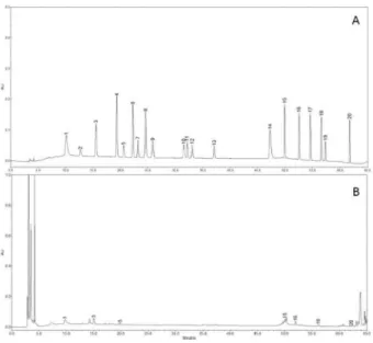
To determine the phenolic compound contents from fruiting bodies of R. rosacea, HPLC was employed for the analysis. A total of 7 phenolic compounds were detected and phenolic compound concentration was 27.25 μg/g. (Fig. 4). The phenolic compounds detected

from the mushroom extract were gallic acid, protocatechuic acid, chlorogenic acid, resveratrol, quercetin, kaempferol, and biochanin-A (Fig. 4B). The highest and lowest concentrations of phenolic compounds were resveratrol (7.38 μg/g) and biochanin-A (0.21 μg/g). These findings were comparable to those of previous studies on mushrooms in which mushroom species contain different types of phenolic compounds ranging from 3 to 15, and gallic acid is the most common phenolic compound found in mushrooms (Kim et al, 2008). Phenolic compounds are classified as simple phenols, phenolic acids, and polyphenols. Flavonoids are a group of polyphenolic compounds with well-known health beneficial properties such as free radical scavenging, hydrolytic inhibition, and anti-inflammatory activities so on (Frankel, 1997). Several reports have shown a close correlation with antioxidant activity and phenolic compound concentration (Duan et al, 2007; Gryglewski et al, 1987; Zhao et al, 2006). Thus, phenolic compound content could be used as an indicator of antioxidant capacity. Mushroom extracts having high levels of phenolic compounds exhibit extensive free radical- scavenging activities as hydrogen donors or electron- donating agents, as well as metal ion-chelating properties.

Fig. 3. Reducing power of methanol and hot water extract from fruiting bodies of Russula rosacea. The values are expressed as means ±SD (n=3). Fr.MeOH, fractions extracted with 80% methanol; Fr.HW, fractions extracted with hot water; BHT, butylated hydroxytoluene. ***p ≤0.001; **p ≤0.01 vs. group treated with BHT only.

Fig. 4. HPLC analysis of phenolic compounds. (A) standard compounds; (B) Russula rosacea; 1, gallic acid; 2, homogentisic acid; 3, protocatechuic acid; 4, (+)-catechin; 5, chlorogenic acid; 6, (-)-epicatechin; 7, (-)-epigallocatechin gallate; 8, caffeic acid; 9, vanillin; 10, rutin hydrate; 11, p- coumaric acid; 12, ferulic acid; 13, naringin; 14, myricetin;

15, resveratrol; 16, quercetin; 17, naringenin; 18, kaempferol;

19, formonoentin; 20, biochanin-A

(7)

α-amylase inhibition

The α-amylase inhibitory activity of methanol and hot water extracts of R. rosacea fruiting bodies were tested and compared with acarbose, the positive control. At five different concentrations (0.125-2.0 mg/ml), the inhibitory effects of methanol and hot water extracts were 5.20-56.27% and 6.92-43.05%, respectively, whereas that of acarbose, the standard drug was 40.61-89.91%, which was significantly higher (p<0.001) than that of R.

rosacea extracts (Fig. 5). These results showed that α- amylase inhibition activities of methanol and hot water extracts of R. rosacea were moderate and concentration dependent manner. Nguyen et al (2014) found that methanol extract of Coprinellus micaceus fruiting bodies showed α-amylase inhibition ability of 33.33% at 2.0 mg/ml, which was lower than that of methanol extract of R. rosacea (56.27%) at the same concentration.

However α-amylase inhibitory effect of hot water extract of R. rosacea was lower (43.05%) than that of C. micaceus (67.28%) at the identical concentration tested.

α-glucosidase inhibition

The α-glucosidase inhibitory effects of the methanol, and hot water extracts ranged from 30.06-71.91%, and 32.44-74.07% at the 0.125-2.0 mg/ml, respectively (Fig.

6). However, acarbose, the positive control showed significantly higher inhibition abilities of 32.27-81.81%

p<0.001) at the same concentrations tested. The α- glucosidase inhibition activity of the hot water extract was slightly higher than that of methanol extract at the range of 1.0-2.0 mg/ml. The highest inhibitory activity was

found at 2.0 mg/ml in the hot water extract (74.07%), but the value was significantly lower (p<0.001) than that of acarbose. Nguyen et al (2014) reported that methanol and hot water extracts of Coprinellus micaceus fruiting bodies showed α-glucosidase inhibition effect of 62.26%

and 67.29% at 2.0 mg/ml, which was lower than those of our methanol and hot water extracts of R. rosacea (71.91% and 74.07%) at the same concentration. These results indicated that methanol and hot water extract of the mushroom possessed moderate α-glucosidase inhibitory activities compared with acarbose. It is likely that this mushroom extracts can be used for lowering high postprandial blood sugar levels of diabetic type II patints and can also be employed in preventing obesity.

Anti-acetylcholinesterase activity

AChE also called acetylhyrolase catalyze acetylcholine, one of neurotransmitter found in the cholinergic brain synapses, where AChE serves to terminate synaptic transmission. The activities of AChE sometimes lead to several neurological disorders such as Alzheimer’s disease, senile dementia, and ataxia (Kalauni et al, 2002). The AChE inhibitory activity of methanol, and hot water extracts from R. rosacea fruiting bodies were investigated. At 0.063-1.0 mg/ml, the inhibition effects of methanol and hot water extracts ranged from 35.1- 66.8%, and 32.7-65.5%, respectively (Fig. 7). The AChE activities were inhibited by methanol and hot water in a concentration dependent manner. The AChE inhibitory effect obtained from the methanol and hot water extracts were statistically similar in all concentration tested, whereas the inhibitory activity of galanthamine (97.80%), Fig. 5. α-amylase inhibitory activity of methanol and hot

water extracts from fruiting bodies of Russula rosacea. The values are expressed as means ±SD (n=3). Fr.MeOH, fractions extracted with 80% methanol; Fr.HW, fractions extracted with hot water. ***p≤0.001; **p≤0.01 vs. group treated with acarbose only.

Fig. 6. α-glucosidase inhibitory activity of methanol and hot

water extracts from fruiting bodies of Russula rosacea. The

values are expressed as means ±SD. Fr.MeOH, fractions

extracted with 80% methanol; Fr.HW, fractions extracted

with hot water. ***p≤0.001; **p≤0.01 vs. group treated with

acarbose only.

(8)

the positive control, was significantly higher (p<0.001) than those of methanol and hot water extracts. It was reported that phenolic acids and flavonoid derivatives including gallic acid, chlorogenic acid, quercetin, caffeic acid, ferulic acid, ellagic acid, catechin, rutin and luteoin-7-rutinoside were strong inhibitors of AChE enzyme (Orhan et al, 2007; Nagarani, et al, 2014). The experimental results (Fig. 4) also suggested that fruiting body of R. rosacea contained 7 different phenolic compounds including gallic acid (3.35 μg/g), chlorogenic acid (3.13 μg/g) and quercetin (1.99 μg/g) have good inhibitory potential toward AChE enzyme, which were in good agreements with those of phenolic acid and flavonoid compounds mentioned above. Therefore, it could be concluded that the moderately good AChE inhibitory activity found in the methanol and hot water extracts might be due to polyphenolic compounds contained in the mushroom fruiting bodies.

Anti-butyrylcholinesterase activity

The butyrylcholinesterase inhibitory effects of methanol and hot water extracts from R. rosacea were analyzed and compared with galanthamine. At 0.063-1.0 mg/ml, the BChE inhibitory activities of methanol extract (45.67-75.57%) and hot water extract (33.66-74.08%) were significantly lower than that of galanthamine (Fig.

8). The AChE inhibitory activity of methanol and hot water extracts from R. rosacea were increased with increasing extract concentrations. Orhan et al (2007) screened 4 phenolic acids and 10 flavonoid derivatives for BChE inhibitory activities. Among them, chlorogenic acid, gallic acid, quercetin, genistein, leteolin-7-O-

galactoside, naringin, silibinin, and silymarin have shown good BChE inhibitory activities. In our experiment, 3 phenolic compounds such as gallic acid, chlorogenic acid and quercetin were found in the fruiting bodies of R. rosacea (Fig. 4). These results could explain the moderate inhibitory effects of the methanol and hot water extracts of the mushroom on BChE.

NO inhibitory production

The accumulated concentration of NO in the culture medium was measured by the Griess assay (Abas et al, 2006). After treatment with LPS on RAW 264.7 cells for 24 h, concentration of NO increased about 5.55-fold (7.72-42.83 mM), whereas the NO production of cells treated with various concentrations of methanol, and hot water extracts of fruiting bodies of R. rosacea were significantly inhibited at the concentrations from 0.25 to 1.0 mg/ml concentration in a dose dependent manner (Fig. 9). The NO production on RAW 264.7 cells treated only with methanol extract of 1.0 mg/ml was 23.32 μM, which was significantly lower than 42.83 μM of LPS only treated group. The LPS-induced RAW 264.7 cells treated with hot water extract of 1 mg/ml also significantly decreased the production of NO by 69.58% compared with LPS only treated group (Fig.

4B). From the results, it is concluded that inhibitory activity of NO production in methanol extract on LPS- induced RAW 264.7 cells was better than hot water extract tested.

No cytotoxicity (data not shown) was observed at the concentrations tested as determined by MTT test Fig. 7. Acetylcholinesterase inhibitory activity of methanol

and hot water extracts from fruiting bodies of Russula rosacea. The values are expressed as means ±SD (n=4).

Fr.MeOH, fractions extracted with 80% methanol; Fr.HW, fractions extracted with hot water. ***p≤0.001 vs. group treated with galanthamine only.

Fig. 8. Butyrylcholinesterase inhibitory activity of methanol and hot water extracts from fruiting bodies of Russula rosacea. The values are expressed as means ±SD (n=4).

Fr.MeOH, fractions extracted with 80% methanol; Fr.HW,

fractions extracted with hot water. ***p≤0.001 vs. group

treated with galanthamine only.

(9)

(Mosmann, 1983). From these results, it is expected that the inhibition of NO production by the mushroom extracts in RAW 264.7 cells would be caused by decreased iNOS protein expression. Park and Park (2005) reported that ethanol extracts of cultured mycelia and fruiting body of Cordyceps militaris suppressed production of NO and iNOS protein in LPS-stimulated RAW 264.7 macrophages in a concentration dependent manner. Furthermore, ethanol extract of G. lucidum fruiting bodies also inhibited significantly NO production in LPS-activated RAW 264.7 cells (Song et al, 2004). Hur et al (2012) reported that ethanol extract of fruiting bodies from Russula virescens showed good NO inhibitory effects on LPS-stimulated RAW 264.7 cells at 0.5-5.0 mg/ml concentration. It seemed that inhibitory effect of NO production in LPS-induced RAW 264.7 cells by the fruiting body extracts of R. rosacea was similar to those of C. militaris, G. lucidum and R.

virescens.

Conclusions

In conclusion, the antioxidant, anti-hyperglycemic, anti- cholinesterase, and NO production inhibitory activities of methanol, and hot water extracts from R. rosacea fruiting bodies were found, and 7 phenolic compounds were also detected. The results suggested that R.

rosacea fruiting bodies possess good natural antioxidant, anti-hyperglycemic, anti-cholinesterase, and NO production inhibitory sources.

Acknowledgements

This study was supported by research grant from Ansan University in 2015.

References

Abas F, Lajis NH, Israf DA, Khozirah S, Kalsom YU. 2006.

Antioxidant and nitric oxide inhibition activities of selected Malay traditional vegetables. Food Chem. 95(4):

566-573.

Alam N, Yoon KN, Lee KR, Lee JS, Lee TS. 2011.

Phenolic compounds concentration and appraisal of antioxidant and antioxidant and antityrosinase activities from the fruiting bodies of Pleurotus eryngii. Adv.

Environ. Biol. 5(6):1104-1113.

Benzi G, Morreti A. 1998. Is there a rationale for the use of acetylcholinesterase inhibitors in the therapy of Alzheimer’s disease. Eur. J. Pharmacol. 346:1-13.

Blois MS. 1958. Antioxidant determination by the use of a stable free radical. Nature. 181:1199-1200.

Cheung LM, Cheung PCK, Ooi VEC. 2003. Antioxidant activity and total phenolics of edible mushroom extracts.

Food Chem. 81:249-255.

Cuendet M, Hostettmann K, Potterat O, Dyatmiko W. 1997.

Iridoid glucosides with free radical scavenging properties from Fagraea blumei. Helvetica. Chimica. Acta. 80:1144- 1152.

Dinis TC, Madeira VM, Almeida LM. 1994. Action of phenolic derivatives (acetaminophen, salicylate, and 5- amino salicylate) as inhibitors of membrane lipid peroxidation and as peroxyl radical scavengers. Arch.

Biochem. Biophys. 315:161-169.

Duan X, Wu G, Jiang Y. 2007. Evaluation of antioxidant properties of lichi fruit phenolics in relation to pericarp browning prevention. Molecules. 12:759-771.

Ellman GL, Courtney KD, Andres VJ, Featherstone RM.

1961. A new and rapid colorimetric determination of acetylcholinesterase activity. Biochem. Pharmacol. 7:88- 95.

Fayuk D, Yakel JL. 2004. Regulation of nicotinic acetylcholine receptor channel function by acetylcholinesterase inhibitors in rat hippocampal CA1 interneurons. Mol.

Pharmacol. 66:658-666.

Frankel EN. 1997. Nutritional benefits of flavonoids. Food Factors Cancer Prev. 7:613-616.

Gryglewski RJ, Korbut R, Robak J. 1987. On the

Fig. 9. Inhibitory effect of Russula rosacea extracts on LPS-induced nitric oxide production in RAW 264.7 cells. (A) methanol

extract; (B), hot water extract. Accumulated nitric oxide in the culture medium was determined by the Griess method. The

values are expressed as means ±SD (n=4). ***p≤0.001 vs. group treated with LPS only

(10)

mechanism of antithrombotic action of flavonoids.

Biochem. Pharmacol. 36:317-321.

Gulcin I, Buyukokuroglu ME, Oktay M, Kufrevioglu OI.

2003. Antioxidant and analgesic activities of turpentine of Pinus nigra Arn. subsp. pallsiana (Lamb.) Holmboe. J.

Ethnopharmacol. 86;51-8.

Hur SJ, Choi SY, Lim BO. 2012. In vitro anti-inflammatory activity of Russula virescens in the macrophage like cell line RAW 264.7 activated by lipopolysaccharide. J. Nutr.

Food Sci. 2:142. doi:10.4172/2155-9600.1000142.

Kang HW. 2012. Antioxidant and anti-inflammatory effects of extracts from Flammulina velutipes (Curtis) Singer. J.

Korean Soc. Food Nutr. 41:1072-1078.

Kalauni SK, Choudhary MI, Khalid A, Manandhar MD, Shaheen F, Atta-ur-Rahman, Gewali MB 2002. New cholinesterase inhibiting steroidal alkaloids from the leaves of Sarcococca coriacea of Nepalese origin. Chem.

Pharm. Bull. 50:1423-1426.

Kim MY, Seguin P, Ahn JK, Kim JJ, Chun SC, Kim EH, Seo SH, Kang EY, Kim SL, Park YJ. 2008. Phenolic compound concentration and antioxidant activities of edible and medicinal mushrooms from Korea. J. Agric.

Food Chem. 56:7265-7270.

Lee YL, Yen M, Mau JL. 2007. Antioxidant properties of various extracts from Hypsizigus marmoreus. Food Chem.

104:1-9.

Lobo V, Patil A, Phatak A, Chandra N. 2010. Fee radicals, antioxidants and functional foods: Impacts on human health. Pharmacog. Rev. 4(8):118-126.

Mau JL, Chao GR, Wu KT. 2001. Antioxidant properties of methanolic extracts from several ear mushrooms. J. Agric.

Food Chem. 49:5461-5467.

Mau JL, Lin HC, Chen CC. 2002. Antioxidant properties of several medicinal mushrooms. J. Agric. Food Chem.

50:6072-6077.

Mayne ST. 2003. Antioxidant nutrients and chronic disease:

Use of biomarkers of exposure and oxidative stress status in epidemiologic research. J. Nutri. 133:933-940.

Mosmann T. 1983. Rapid colorimetric assay for cellular growth and survival: application to proliferation and cytotoxicity assays. Immunol. Meth. 65: 55-63.

Menaga D, Rajakumar S, Ayyasamy PM. 2013. Free radical scavenging activity of methanol extract of Pleurotus florida mushroom. Int J Pharm Pharmaceut Sci. 5:601- 606.

Nagarani G, Abirami A, Siddhuraju P. 2014. A comparative study on antioxidant potentials, inhibitory activities against key enzymes related to metabolic syndrome, and anti-inflammatory activity of leaf extract from different Momordica species. Food Sci. Human Wellness. 3:36-46.

Nam BH, Jo WS, Choi YJ, Lee JY, Kang EY, Jeong MH, Lee JD. 2010. Inhibitory effects of melanin secretion on

B16 melanoma cell of Cordyceps militaris water extract.

Korean J. Mycol. 38: 167-171.

Nguyen TK, Shin DB, Lee KR, Cheong JC, Yoo YB, Lee MW, Jin KH, Im KH, Lee TS. 2013. Antioxidant, anti- inflammatory and anti-acetylcholinesterase activities of Phellinus xeranticus. J. Mushroom Sci. Prod. 11(4):278- 286.

Nguyen TK, Lee MW, Yoon KN, Kim HY, Jin GH Choi JH, Im KH, Lee TS. 2014. In vitro antioxidant and anti- diabetic, and anti-cholinesterase, tyrosinase and nitric oxide inhibitory potential of fruiting bodies of Coprinellus micaceus. J. Mushrooms. 12(4):330-340.

Orhan I, Kartal M, Tosun F, Sener B. 2007. Screening of various phenolic acids and flavonoid derivatives for their anticholinesterase potential. Z. Naturforsch. C. 62:829- 832.

Park SY, Park EH. 2005. Anti-inflammatory and related pharmacological activities of cultured mycelia and fruiting bodies of Cordyceps militaris. J. Ethnopharmacol. 96:555- 561.

Park WH, Lee HD. 2003. Illustrated book of Korean medicinal mushrooms. Kyo-Hak Publishing Co. Ltd.

Seoul.

Perera PK, Li YM. 2011. Mushrooms as a functional food mediator in preventing and ameliorating diabetes. Func.

Foods Health Dis. 4:161-171.

Quinn DM. 1987. Acetylcholinesterase: enzyme structure, reaction dynamics, and virtual transition states. Chem.

Rev. 87:955-979.

Song YS, Kim SH, Sa JH, Jin CB, Lim CJ, Park EH. 2004.

Anti-angiogenic and inhibitory activity on inducible nitric oxide production of the mushroom Ganoderma lucidum.

J. Ethnopharmacol. 90:17-20.

Su CH, Lai MN, Ng LT. 2013. inhibitory effects of medicinal mushrooms on α-amylase on α-amylase and α- glucosidase - enzymes related to hyperglycemia. Food Funct. 25(4):644-649.

Sung SY, Kang SY, Lee KY, Park MJ, Kim JH, Park JH, Kim YC, Kim, J, Kim YC. 2002. (+)-α-Viniferin, a stilbene trimer from Caranga chamlague inhibits acetylcholinesterase. Biol. Pharm. Bull. 25: 125-127.

Wasser SP, Weis AL. 1999. Medicinal properties of substances occurring in higher basidiomycete mushrooms:

current perspectives (review). Internat. J. Med. Mushrooms.

1:31-62.

Ying JZ, Mao XL, Xu YC. 1987. Icones of Medicinal Fungi from China. Science Press, Beijing, China.

Zhao M, Yang B, Wang J, Li B, Jiang Y. 2006.

Identification of the major flavonoids from pericarp

tissues of lychee fruit in relation to their antioxidant

activities. Food Chem. 98:539-544.

수치

Fig. 1. DPPH radical scavenging activities of methanol and hot water extracts from fruiting bodies of Russula rosacea against 1,1-diphenyl-2-picrylhydrazyl
Fig. 3. Reducing power of methanol and hot water extract from fruiting bodies of Russula rosacea
Fig. 6.  α-glucosidase inhibitory activity of methanol and hot water extracts from fruiting bodies of Russula rosacea
Fig. 8. Butyrylcholinesterase inhibitory activity of methanol and hot water extracts from fruiting bodies of Russula rosacea

참조

관련 문서

Radical-scavenging activities of the walnuts's shell ethanol extracts (WSE) were examined by using ABTS radicals and α,α-diphenyl-β-picrylhydrazyl (DPPH) radicals assay..

Antioxidant activity was evaluated by measuring total phenolic contents, reducing power, 1,1-diphenyl-2-picrylhydrazyl (DPPH) radical scavenging activity,

Inhibitory effects of methanol extract from fruiting bodies of Agaricus brasiliensis on LPS-induced nitric oxide production in RAW 264.7 cells.. Accumulated nitric

by extraction solvent, which were examined by reducing power, hydroxyl radical-scavenging activity(HRSA) and 1, 1-diphenyl-2-picrylhydrazyl (DPPH) radical scavenging assays. 70%

Abstract The ethyl acetate (EtOAc) layer of Canavalia gladiata (sword bean) methanol extracts showed higher 1,1- diphenyl-2-picrylhydrazyl (DPPH) radical-scavenging activity

The extracts were then processed in order to confirm their antioxidant activities via 1,1-diphenyl-2-picrylhydrazyl DPPH free radical-scavenging activity assay,

DPPH radical scavenging activities of hot-water extracts (1 mg/ml concentrations) of ectomycorrhizal mushrooms (Cantharellus cinnabarinus, Lactarius parallelus, Tricholoma

DPPH radical scavenging activities of hot-water extracts (1 mg/ml concentrations) from eggs and fruiting bodies of Phallus indusiatus by different drying methods (hot-air drying