24(1) : 40-46 (2018)
https://doi.org/10.20307/nps.2018.24.1.40
40
Antioxidant and Anti-inflammatory Activity of Six Halophytes in Korea
Jeong Min Lee, Mi-Jin Yim, Grace Choi, Myeong Seok Lee, Yun Gyeong Park, and Dae-Sung Lee*
Department of Applied Research, National Marine Biodiversity Institute of Korea, Seocheon 33662, Republic of Korea
Abstract − The aim of this study was to measure and compare polyphenol content, antioxidant, and anti- inflammatory activity of six halophytes (Limonium tetragonum, Suaeda glauca, Suaeda japonica, Salicornia europaea, Triglochin maritimum, and Sonchus brachyotus). Depending on the total polyphenol content, the plants were categorized into two groups: (1) a high total polyphenol content group that included L. tetragonum, S.
brachyotus, and S. europaea, and, (2) a low total polyphenol content group consisting of S. glauca, T. maritima, and S. japonica. Antioxidant activity was evaluated using DPPH and hydroxyl radical scavenging assays, and by measuring ROS. Anti-inflammatory activity was evaluated by measuring NO and PGE2. L. tetragonum and S.
brachyotus, that have high polyphenol content, also showed strong antioxidant activity. In addition, L. tetragonum, S. brachyotus, and S. europaea showed good anti-inflammatory activity. Consequently, the total polyphenol content was thought to be related to antioxidant and anti-inflammatory activity. Therefore, S. brachyotus and L.
tetragonum are good candidates for use in pharmaceuticals and functional foods.
Keywords − Halophytes, DPPH, Polyphenol, NO, PGE2
Introduction
The salt-tolerant halophyte is native to saline habitats (coastal sand dunes, salt marsh, and mud flats) where the soil mixture contains organic nutrient material and inorganic nutrient salts flowing from land and ocean.
1Halophytes that inhabit such hostile habitats have developed mechanisms to adapt to extreme environments and are likely to have various bioactive compounds. Halophytes have been used in traditional medicine, food sources, cosmetics, functional foods, and natural seasoning.
Among the halophytes in Korea, Limonium tetragonum (Plumbaginaceae), Suaeda glauca (Chenopodiaceae), Suaeda japonica (Chenopodiaceae), Salicornia europaea (Chenopodiaceae), Triglochin maritimum (Juncaginaceae), and Sonchus brachyotus (Asteraceae) are commonly distributed along the western coast of Korea. These halophytes have been reported to possess many bioactive properties. L. tetragonum showed protective effects on diethylnitrosamine-induced liver fibrosis in rats.
2The biological activities of S. glauca result in hepatoprotective, anti-oxidative, and anti-neuroinflammatory effects.
3,4S.
japonica has antioxidant and anti-inflammatory effects.
5,6S. europaea showed cytotoxic activity against Artemia salina LEACH and Daphnia magna STRAUS and anti- neoplastic activities in the potato disk assay.
7T. maritimum showed strong inhibition of fungal growth.
8S. brachyotus had antioxidant, anti-bacterial, and peroxynitrite-scavenging activity.
9,10Many phytochemicals, pyrocatechol, syringic acid, apigenin, isorhamnetin, kaempferol, dihydroferulic acid, vanillic acid, 4-hydroxybenzoic acid, homoeriodictyol, naringenin, quercetin, luteolin, 9-epiblumenol C, sco- poletin, dihydroisorhamnetin, chrysoeriol from S. japonica, myricetin-3-O- β-
D-galactopyranoside, myricetin-3-O- α-
L
-rhamnopyranoside, quercetin-3-O- β-
D-glucopyranoside, quercetin-3-O- β-
D-galactopyranoside from L. tetragonum, lignoceric acid, β-sitosterol, daucosterol, quercetin, luteolin, luteolin-7-O- β-
D-glucopyranoside, isorhamnetin, scopoletin, stigmasterol from S. glauca, isorhamnetin-3-O- β-
D
-glucopyranoside, quercetin-3-O- β-
D-glucopyranoside, 3- caffeoyl, 4-dihydrocaffeoyl quinic acid, quercetin, isorhamnetin, quercetin-3-O- β-
D-glucopyranoside, quercetin- 3’,4’-di-O- β-
D-glucopyranoside, rutin from S. europaea, triglochinin from T. maritimum, and chlorogenic acid, luteolin-7-O- β-
D-rutinoside, luteolin-7-O- β-
D-glucopyrano- side, and luteolin from S. brachyotus have been reported from halophytes.
11-20Of these, most compounds were reported that were polyphenolic compounds. Polyphenols have been reported to have various biological activities
*Author for correspondence
Dae-Sung Lee, Department of Applied Research, National Marine Biodiversity Institute of Korea, Jangsan-ro 101 beon-gil, Janghang- eup, Seocheon-gun, Chungcheongnam-do 33662, Korea.
Tel: +82-41-950-0767; E-mail: [email protected]
that result in antioxidant, anti-hyperlipidemic, anti-cancer, and anti-inflammatory effects.
21,22Therefore, the six halophytes tested in this study were expected to contain a variety of biological activities. The objective of this study was to compare the antioxidant and anti-inflammatory properties of six halophytes in Korea and to assess their possible importance for use in bio-products.
Experimental
Plant materials and extraction – Six halophytes [L.
tetragonum (H1), S. brachyotus (H2), S. glauca (H3), S.
japonica (H4), T. maritima (H5), and S. europaea (H6)]
were collected at Ganghwa-gun, Incheon, Korea. The plants were separately lyophilized, pulverized, and stored at -80
oC. The lyophilized halophytes were extracted with 70% EtOH for 1 h (5 times) using a sonicator. The extracts were then lyophilized, pulverized, and stored at -80
oC for later use in experiments.
Determination of total polyphenol content (TPC) − The total polyphenol content of halophyte extract was determined using the Folin-Ciocalteu method.
23Briefly, 20 µL of each extract were added to 100 µL of Folin- Ciocalteu reagent (Sigma chemical Co., St. Louis, MO, USA), allowed to react in the dark for 3 min at room temperature. This mixture was added to 80 µL of 7.5%
Na
2CO
3and placed in the dark for 20 min at room temperature. The absorbance was determined in triplicate samples at 765 nm using a microplate reader (Power- Wave XS2, BioTek Instruments, Inc., USA). The total polyphenol content was based on a calibration curve obtained with gallic acid and expressed as gallic acid equivalents per gram of dry weight (mg/GAEg).
DPPH radical scavenging assay − The DPPH (2,2- diphenyl-1-picrylhydrazyl) radical scavenging activity of halophyte extracts was measured using the method described by Blois with a slight modification.
24100 µL of each extract of concentration (9.765~625 μg/mL) were added to 100 µL of 0.4 mM DPPH (Sigma chemical Co., St. Louis, MO, USA) in a 96-well plate. The mixture was shaken vigorously and left standing in the dark at room temperature for 30 min, after which absorbance was measured at 517 nm. The DPPH radical scavenging activity was expressed as the half maximal inhibitory concentra- tion (IC
50) value.
Hydroxyl radical scavenging assay − The hydroxyl radical scavenging activity of halophyte extract was measured using the method described by Rosen and Rauckman with some modification.
25Twenty µL of each extract, 0.625 and 1.250 mg/mL, were added to 20 µL
each of 0.3 M 5,5-dimethyl-1-pyrroline N-oxide (DMPO), 10 mM FeSO
4, and 10 mM H
2O
2/0.1 M phosphate buffer (pH 7.4). This mixture was allowed to react at room temperature for 2.5 min. The hydroxyl radical scavenging activity was expressed as a percentage (%).
Cell culture − RAW 264.7 macrophages were cultured in RPMI1640 supplemented with 10% FBS, penicillin (100 U/mL), and streptomycin (100 μg/mL) at 37
oC in a humidified incubator with 5% CO
2. Cells were washed with DMEM and treated in serum-free medium for at least 4 h prior to treatment.
MTT cytotoxicity assay − The cell viability was measured using the MTT (3-(4,5-dimethylthiazol-2yl)- 2,5-diphenyl-2H-tetrazolium bromide) assay described by Hansen et al..
26The halophyte extracts and lipopoly- saccharide (LPS)-treated cells were added to 1 mg/mL and incubated for 4 h at 37
oC. The resulting formazan crystals were dissolved in DMSO (dimethyl sulfoxide) (150 μL) and absorbance was measured at 540 nm using a microplate reader.
Determination of ROS generation − The intracellular ROS (reactive oxygen species) of halophyte extract were measured using the method described by Engelmann et al.
with some modification.
27RAW 264.7 cells were cultured in 96-well plates and allowed to react with 20 μM DCFH- DA (2',7'-Dichlorofluorescin diacetate) (HBSS, Hanks balanced salt solution) in the dark for 30 min. After treatment with halophyte extract and further incubation for 1 h, cells were washed twice with PBS and added to 1 μg/mL of LPS. The fluorescence intensity was measured at an excitation wavelength of 485 nm and an emission wavelength of 528 nm using GENios fluorescence micro- plate reader (TECAN, Männedorf, Switzerland).
Measurement of NO production − The NO (nitric oxide) generated by cells was measured using the Griess reaction method.
28RAW 264.7 cells were plated in 96- well plates at 1.5 × 10⁵ cells/mL and treated with the indicated concentrations (50~500 μg/mL) of each halophyte extract prior to stimulation with 1 μg/mL of LPS for 24 h.
One hundred µL of cell culture medium were added to 100 μL of Griess reagent and the mixture was incubated at room temperature for 10 min. The absorbance at 540 nm was measured using a microplate reader. The NO production was expressed as a percentage (%).
Measurement of PGE
2production − The PGE
2(prostaglandin E
2) production of halophyte extract was
measured using the method described by Moon et al. with
minor modification.
29RAW 264.7 cells were plated in 24-
well plates and treated with the indicated concentrations
(50~500 μg/mL) of each extract prior to stimulation with
1 μg/mL of LPS for 24 h. The expression level of PGE
2was measured using an enzyme immunosorbent assay (ELISA) kit (Cayman chemical, Ann Arbor, MI). The PGE
2released was expressed as a percentage (%).
Result
TPC − The TPC of the six halophyte extracts was measured and ranged from 2.84% to 14.27% (Table 1).
The TPC was highest in L. tetragonum (14.27%), followed by S. brachyotus (9.34%), S. europaea (8.71%), S. glauca (4.29%), T. maritima (3.33%), and S. japonica (2.84%) respectively. The halophytes were classified into two groups depending on TPC content: (1) the high TPC group consisting of L. tetragonum (14.27%), S. brachyotus (9.34%), and S. europaea (8.71%), and, (2) the low TPC
group consisting of S. glauca (4.29%), T. maritima (3.33%), and S. japonica (2.84%). Among all six halophytes studied, L. tetragonum (14.27%) contained five times more TPC than S. japonica (2.84%).
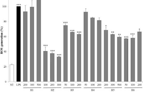
Antioxidant activities − Antioxidant activities of halophytes were measured using DPPH, hydroxyl radical scavenging assay and ROS generation (Table 2, Fig. 1 and 2). The DPPH radical scavenging activity was categorized into two groups: (1) the high activity group of L. tetragonum (0.020 mg/mL), S. brachyotus (0.056 mg/
mL), T. maritima (0.061 mg/mL), and S. glauca (0.095 mg/mL), and, (2) the low activity group of S. japonica (0.510 mg/mL), and S. europaea (0.570 mg/mL). In the hydroxyl radical scavenging activity test, S. brachyotus (43.08 and 71.38%) fared better than other species [L.
tetragonum (6.48 and 11.13%), S. glauca (16.87 and 18.26%), S. japonica (18.44 and 36.56%), T. maritima (10.71 and 4.51%), and S. europaea (13.08 and 47.46%)]
using concentrations of 0.625 and 1.250 mg/mL.
To investigate cytotoxicity before inhibition of intracellular ROS activity, we evaluated the effects of halophyte extracts on cell viability. The halophyte extracts did not affect cell viability in LPS-stimulated RAW 264.7 cells (Fig. 3). As shown in Fig. 2, treatment with the extract of halophyte tended to decrease ROS generation. S. brachyotus, at a concentration 100 µg/mL, lowered the level of ROS generation (40.50%) compared to the LPS-stimulated RAW 264.7 cells (control). In this study, L. tetragonum and S.
brachyotus showed the strongest antioxidant activity.
Anti-inflammatory activity − Anti-inflammatory activity was evaluated by measuring NO production and PGE
2released from cells after treatment with halophyte extract.
Table 1. Total polyphenol content of six halophyte extracts expressed as equivalent of gallic acid (mg of GA/g of extract)
Species Total polyphenol content (mg/100 g dry weight) Plumbaginaceae
H1 14.27± 0.26a
Asteraceae
H2 59.34± 0.32
Chenopodiaceae
H3 54.29± 0.15
H4 52.84± 0.46
H6 58.71± 0.29
Juncaginaceae
H5 53.33± 0.37
a Data are expressed as the mean± SD (n = 3).
Table 2. DPPH radical scavenging activity of six halophyte extracts expressed as half maximal inhibitory concentration (IC50)
Species IC50 (mg/ml)
Plumbaginaceae
H1 0.020± 0.001a
Asteraceae
H2 0.056± 0.001
Chenopodiaceae
H3 0.095± 0.003
H4 0.510± 0.003
H6 0.061± 0.001
Juncaginaceae
H5 0.570± 0.009
Control
Ascorbic acidb 0.019± 0.004
a Data are expressed as the mean± SD (n = 3).
b Ascorbic acid was used as a positive control.
Fig. 1. Hydroxyl radical scavenging activity of the extracts from six halophytes. Experiment was performed in triplicate and data are represented as mean± SD.
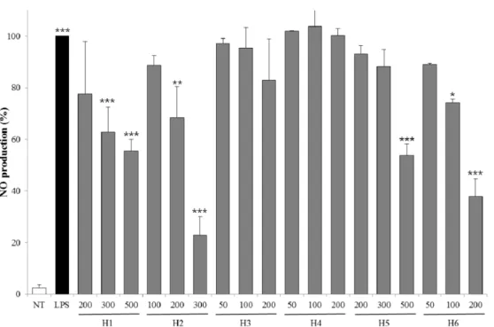
MTT assays were used to rule out the possibility that the concentrations of the six halophyte extracts used did not affect cell viability (Fig. 3). As shown in Fig. 4, the extracts of six halophytes suppressed NO production.
Especially, 300 µg/mL of S. brachyotus showed the best inhibition of NO production (22.90%) compared to LPS-
stimulated RAW 264.7 cells, and 200 µg/mL of S.
europaea showed good inhibition of NO production (37.71%). Except for the two aforementioned samples, the other halophyte extract-treated cells showed more than 50% NO production.
In the PGE
2tests (Fig. 5), all halophyte extract-treated
Fig. 2. ROS generation of extracts from six halophytes. Experiment was performed in triplicate and data are represented as mean ± SD. * p < 0.05; ** p < 0.01; *** p < 0.001.Fig. 3. Cell viability of extracts from six halophytes. Experiment was performed in triplicate and data are represented as mean ± SD.
cells showed less than 50% inhibition of PGE
2release compared to LPS-stimulated RAW 264.7 cells. L.
tetragonum inhibited PGE
2release by 32.04%, 11.53%, and 5.49% at 200, 300, and 500 µg/mL, respectively. S.
brachyotus inhibited PGE
2release by 51.84%, 40.04%, and 12.11% at 100, 200, and 300 µg/mL, respectively. In
particular, S. europaea showed the best inhibition of the PGE
2release by 22.27%, 14.03%, and 8.48% at low concentrations (50, 100, and 200 µg/mL). In our study, L.
tetragonum, S. brachyotus, and S. europaea showed good anti-inflammatory activity.
Fig. 4. Inhibitory effects of NO production for extracts from six halophytes in LPS-RAW 264.7 cells. Experiment was performed in triplicate and data are represented as mean± SD. * p < 0.05; ** p < 0.01; *** p < 0.001.
Fig. 5. Inhibitory effects of PGE2 release for extracts from six halophytes in LPS-RAW 264.7 cells. Experiment was performed in triplicate and data are represented as mean± SD. * p < 0.05; ** p < 0.01; *** p < 0.001.
Discussion
Polyphenol compounds, including flavonoids, tannins, and lignans, are widely distributed in nature. Polyphenols have been reported to have various biological activities that result in antioxidant, anti-hyperlipidemic, anti-cancer, and anti-inflammatory effects.
21,22In our antioxidant test, L. tetragonum and S. brachyotus, that have high total polyphenol content, showed the strongest antioxidant activity. In previous studies, phyto- chemicals were reported to contain many polyphenol compounds such as myricetin-3-O- β-
D-galactopyranoside, myricetin-3-O- α-
L-rhamnopyranoside, quercetin-3-O- β-
D- glucopyranoside, and quercetin-3-O- β-
D-galactopyrano- side from L. tetragonum, and chlorogenic acid, luteolin-7- O- β-
D-rutinoside, luteolin-7-O- β-
D-glucopyranoside, and luteolin from S. brachyotus.
12,20In addition, L. tetragonum and S. brachyotus have been shown to have antioxidant activity, which was evaluated by measuring ROS generated in HT-1080 cells and by measuring DPPH and ABTS scavenging and lipid peroxidation inhibition, respectively.
10,12The antioxidant activity of flavonoids such as myricetin-3-O- β-
D-galactopyranoside, myricetin- 3-O- α-
L-rhamnopyranoside, quercetin-3-O- β-
D-glucopy- ranoside, quercetin-3-O- β-
D-galactopyranoside, luteolin- 7-O- β-
D-rutinoside, luteolin-7-O- β-
D-glucopyranoside, and luteolin have been studied and documented.
14,15Therefore, the polyphenols of L. tetragonum and S. brachyotus were thought to contribute to their antioxidant activity.
L. tetragonum, S. brachyotus, and S. europaea, that have high polyphenol content, showed good anti- inflammatory activity. Except for L. tetragonum and S.
brachyotus mentioned above, S. europaea contains various polyphenol compounds including isorhamnetin-3-O- β-
D- glucopyranoside, quercetin-3-O- β-
D-glucopyranoside, 3- caffeoyl, 4-dihydrocaffeoyl quinic acid, quercetin, iso- rhamnetin, quercetin-3-O- β-
D-glucopyranoside, quercetin- 3’,4’-di-O- β-
D-glucopyranoside, and rutin.
16-18L. tetragonum, S. brachyotus, and S. europaea have been shown to have anti-inflammatory activity, which was evaluated by inhi- bition of NO production and suppression of iNOS expression in LPS-activated RAW 264.7 macrophages.
30,31Specifically, flavonoids are known to have anti-inflamma- tory activity.
32,33Therefore, the polyphenols present in L.
tetragonum, S. brachyotus, and S. europaea are inferred to be associated with anti-inflammatory activity.
In conclusion, polyphenols of L. tetragonum, S.
brachyotus, and S. europaea were inferred to be associated with their antioxidant and anti-inflammatory activity. The differences in the degree of activity among
the six halophytes tested are probably due to the differences in the specific polyphenolic content in each plant. Therefore, additional studies need to be performed to isolate, identify, and measure the anti-oxidant and anti- inflammatory activity of individual polyphenols from S.
brachyotus and L. tetragonum.
Acknowledgments
This work was supported by National Marine Biodiversity Institute of Korea Research Program 2017M01400.
References
(1) El Shaer, H. M. Potential of halophytes as animal fodder in Egypt.
in: Lieth, H.; Mochtchenko, M. Part II: Chemical Contents. Cash Crop Halophytes: Recent Studies; Kluwer Academic Publishers: London, 2003, pp 111-119.
(2) Kim, N. -H.; Heo, J. -D.; Kim, T. B.; Rho, J. -R.; Yang, M. H.;
Jeong, E. J. Biol. Pharm. Bull. 2016, 39, 1022-1028.
(3) An, R. -B.; Sohn, D. -H.; Jeong, G. -S.; Kim, Y. -C. Arch. Pharm.
Res. 2008, 31, 594-597.
(4) Lee, S. -G.; Kim, J. -B.; Kang, H. Trop. J. Pharm. Res. 2016, 15, 1175-1181.
(5) Choi, J. -I.; Kim, Y. -J.; Kim, J. -H.; Song, B. -S.; Yoon, Y.; Byun, M. -W.; Kwon, J. -H.; Chun, S. -S.; Lee, J. -W. J. Korean Soc. Food Sci.
Nutr. 2009, 38, 131-135.
(6) Kang, H.; Koppula, S.; Kim, H. -K.; Park, T. -K. Trop. J. Pharm.
Res. 2013, 12, 351-356.
(7) Lellau, T. F.; Liebezeit, G. Pharm. Biol. 2003, 41, 293-300.
(8) Lellau, T. F.; Liebezeit, G. Mar. Biodivers. 2003, 32, 177-181.
(9) Nugroho, A.; Kim, M. -H.; Lee, C. M.; Choi, J. S.; Lee, S.; Park, H.
-J. Nat. Prod. Sci. 2012, 18, 39-46.
(10) Xia, D. -Z.; Yu, X. -F.; Zhu, Z. -Y.; Zou, Z. -D. Nat. Prod. Res.
2011, 25, 1893-1901.
(11) Cho, J. -Y.; Yang, X.; Park, K. -H.; Park, H. J.; Park, S. -Y.; Moon, J. -H.; Ham, K. -S. Food Sci. Biotechnol. 2013, 22, 1547-1557.
(12) Lee, J. I.; Kong, C. -S.; Jung, M. E.; Hong, J. W.; Lim, S. Y.; Seo, Y. Biotechnol. Bioprocess Eng. 2011, 16, 992-999.
(13) Qiu, P.; Wang, Q. -Z.; Yin, M.; Wang, M.; Zhao, Y. -Y.; Shan, Y.;
Feng, X. Zhong Yao Cai 2015, 38, 751-753.
(14) Agati, G.; Azzarello, E.; Pollastri, S.; Tattini, M. Plant Sci. 2012, 196, 67-76.
(15) Sun, L.; Zhang, J.; Lu, X.; Zhang, L.; Zhang, Y. Food Chem.
Toxicol. 2011, 49, 2689-2696.
(16) Hwang, Y. P.; Kim, H. G.; Choi, J. H.; Truong Do, M.; Tran, T. P.;
Chun, H. K.; Chung, Y. C.; Jeong, T. C.; Jeong, H. G. Mol. Nutr. Food Res. 2013, 57, 471-482.
(17) Kim, H. S.; Yoon, Y. S.; Cho, J. W. Korean J. Med. Crop Sci. 2008, 16, 231-237.
(18) Kong, C. -S.; Lee, J. I.; Kim, Y. A.; Kim, J. -A.; Bak, S. S.; Hong, J. W.; Park, H. Y.; Yea, S. S.; Seo, Y. Process Biochem. 2012, 47, 1073- 1078.
(19) Eyjólfsson, R. Phytochemistry 1970, 9, 845-851.
(20) Lee, K. S.; Kim, A. J.; Lee, K. Y. J. East Asian Soc. Diet. Life 2012, 22, 521-526.
(21) Kim, E. J.; Choi, J. Y.; Yu, M.; Kim, M. Y.; Lee, S.; Lee, B. -H.
Korean J. Food Sci. Technol. 2012, 44, 337-342.
(22) Kim, S. J.; Lee, G.; Moh, S. H.; Park, J.; Auh, C. -K.; Chung, Y.;
Ryu, T. K.; Lee, T. -K. J. Korea Acad. Industr. Coop. Soc. 2013, 14,
3081-3088.
(23) Singleton, V. L.; Rossi, J. A. Am. J. Enol. Viticult. 1965, 16, 144- 158.
(24) Blois, M. S. Nature 1958, 181, 1199-1200.
(25) Rosen, G. M.; Rauckman, E. J. Mol. Pharmacol. 1980, 17, 233- 238.
(26) Hansen, M. B.; Nielsen, S. E.; Berg, K. J. Immunol. Methods 1989, 119, 203-210.
(27) Engelmann, J.; Volk, J.; Leyhausen, G.; Geurtsen, W. J. Biomed.
Mater. Res. B 2005, 75B, 272-276.
(28) Kim, Y. -M.; de Vera, M. E.; Watkins, S. C.; Billiar, T. R. J. Biol.
Chem. 1997, 272, 1402-1411.
(29) Moon, J. H.; Kim, S. Y.; Lee, H. G.; Kim, S. U.; Lee, Y. B. Exp.
Mol. Med. 2008, 40, 11-18.
(30) Rhee, M. H.; Park, H. -J.; Cho, J. Y. J. Med. Plants Res. 2009, 3, 548-555.
(31) Yang, E. -J.; Yim, E. -Y.; Song, G.; Kim, G. -O.; Hyun, C. -G.
Interdiscip. Toxicol. 2009, 2, 245-249.
(32) García-Lafuente, A.; Guillamón, E.; Villares, A.; Rostagno, M. A.;
Martínez, J. A. Inflamm. Res. 2009, 58, 537-552.
(33) Read, M. A. Am. J. Pathol. 1995, 147, 235-237.
Received September 1, 2017 Revised October 26, 2017 Accepted October 27, 2017