Introduction
Idiopathic pulmonary fibrosis (IPF) is the irreversible form of the idiopathic interstitial pneumonia that has symptoms of chronic and progressive dyspnea by unknown causes
1,2. IPF is etiologically characterized with alveolar epithelial cell injury, fibroblast/myofibroblast proliferation, and excessive extracel- lular matrix (ECM) deposition leading to the destruction of the alveolar structure. Recently, the incidence and prevalence of IPF have been increased with emerging medical attention in public. In addition, environmental factors including stone, sand, silica, cigarette smoke and wood/metal/micro dust are seriously considered to IPF associated risk factors
3-5. Those
Bleomycin Inhibits Proliferation via Schlafen-Mediated Cell Cycle Arrest in Mouse Alveolar Epithelial Cells
Soojin Jang, M.S. 1 , Se Min Ryu, M.D., Ph.D. 1 , Jooyeon Lee, M.S. 1 , Hanbyeol Lee, M.S. 1 , Seok- Ho Hong, Ph.D. 2 , Kwon-Soo Ha, Ph.D. 3 , Won Sun Park, Ph.D. 4 , Eun-Taek Han, Ph.D. 5 and Se-Ran Yang, D.V.M., Ph.D. 1
Departments of
1Thoracic and Cardiovascular Surgery,
2Internal Medicine,
3Molecular and Cellular Biochemistry,
4Physiology, and
5
Medical Environmental Biology and Tropical Medicine, Kangwon National University School of Medicine, Chuncheon, Korea
Background: Idiopathic pulmonary fibrosis involves irreversible alveolar destruction. Although alveolar epithelial type II cells are key functional participants within the lung parenchyma, how epithelial cells are affected upon bleomycin (BLM) exposure remains unknown. In this study, we determined whether BLM could induce cell cycle arrest via regulation of Schlafen (SLFN) family genes, a group of cell cycle regulators known to mediate growth-inhibitory responses and apoptosis in alveolar epithelial type II cells.
Methods: Mouse AE II cell line MLE-12 were exposed to 1–10 μg/mL BLM and 0.01–100 μM baicalein (Bai), a G1/G2 cell cycle inhibitor, for 24 hours. Cell viability and levels of pro-inflammatory cytokines were analyzed by MTT and enzyme- linked immunosorbent assay, respectively. Apoptosis-related gene expression was evaluated by quantita tive real-time reverse transcription‒polymerase chain reaction (qRT-PCR). Cellular morphology was determined after DAPI and Hoechst 33258 staining. To verify cell cycle arrest, propidium iodide (PI) staining was performed for MLE-12 after exposure to BLM.
Results: BLM decreased the proliferation of MLE-12 cells. However, it significantly increased expression levels of interleukin 6, tumor necrosis factor α, and transforming growth factor β1. Based on Hoechst 33258 staining, BLM induced condensation of nuclear and fragmentation. Based on DAPI and PI staining, BLM significantly increased the size of nuclei and induced G2/M phase cell cycle arrest. Results of qRT-PCR analysis revealed that BLM increased mRNA levels of BAX but decreased those of Bcl2. In addition, BLM/Bai increased mRNA levels of p53, p21, SLFN1, 2, 4 of Schlafen family.
Conclusion: BLM exposure affects pulmonary epithelial type II cells, resulting in decreased proliferation possibly through apoptotic and cell cycle arrest associated signaling.
Keywords: Idiopathic Pulmonary Fibrosis; Alveolar Epithelial Cells; Cell Cycle Arrest; Schlafen; Bleomycin
Address for correspondence: Se-Ran Yang, D.V.M., Ph.D.
Department of Thoracic and Cardiovascular Surgery, Kangwon National University School of Medicine, 1 Gangwondaehak-gil, Chuncheon 24341, Korea Phone: 82-33-250-7883, Fax: 82-33-255-8809
E-mail: [email protected] Received: Dec. 12, 2017 Revised: Mar. 20, 2018 Accepted: Apr. 30, 2018 Published online: Jun. 19, 2018
cc
It is identical to the Creative Commons Attribution Non-Commercial License (http://creativecommons.org/licenses/by-nc/4.0/).
Copyright © 2019
The Korean Academy of Tuberculosis and Respiratory Diseases.
risk factors cause the release of pro-inflammatory cytokines, ECM deposition leading to irreversible alveolar destruction
6. Representatively, pirfenidone and nintedanib have been eval- uated in clinical trials of patient with IPF due to anti-fibrotic ef- fect, however, these therapeutics exhibit the context of certain limitations regarding safety and specific efficacy
7,8. Although the pathogenesis of IPF which is associated with progressive fibro-proliferative as well as destructive pathway of alveolar epithelial cell, the underlying mechanism is still unknown.
Bleomycin (BLM) is a glycopeptide antibiotic with potent tumor therapeutic in various squamous carcinoma of testicu- lar and lymph nodes. However, its adverse effect has been associated with lung injury. BLM-induced pulmonary toxicity is chemotoxic related to pulmonary fibrosis with symptoms of irregular respiration and cough
9,10. In this aspect, the animal and cellular models of BLM induced lung fibrosis injury have been widely used due to the cytotoxic effects. In pulmonary parenchyma of IPF, alveolar epithelial cells have been con- sidered as primary damaged cells of surrounding inflamma- tory
11. Sisson et al.
12has been reported that alveolar epithelial type II cells (AE II) in diphtheria toxin-exposed mouse lung functionally inhibited fibroblast proliferation and collagen synthesis. Moreover, BLM has promoted the release of pro- inflammatory cytokines such as tumor necrosis factor α (TNF-α), interleukin-6 (IL-6), and transforming growth factor β1 (TGF-β1)
13. It has been generally accepted that fibrotic dis- eases are promoted by TGF-β1 and appear AE II cells apopto- sis, accumulation of collagen and ECM, and fibroblast prolif- eration
10,14. Emodin, an antibacterial anthraquinone exhibited the down-regulation of anti-apoptotic protein Bcl2 and up- regulation of pro-apoptotic protein BAX promoted apoptosis in TGF-β1–stimulated human fibroblasts
15. In addition, BLM- induced DNA double-strand breaks increased expression of the p21/p53 pathway that involved cell cycle arrest and cell apoptosis
16. Recently, Schlafen (SLFN) family are proteins that have biological functions the effect that regulation of lympho- cyte differentiation, growth and T-cell development/activa- tion
17. It has been reported that SLFN family have regulation of cell cycle progression and growth arrest
18,19. Baicalein (Bai) induced G1 cell cycle arrest and apoptosis in osteosarcoma cell
20. Moreover, Bai induced G1/G2 cell cycle arrest in rat heart endothelial cells
21. We used Bai in order to inhibit G1/
G2 cell cycle of MLE-12 cells. Therefore, in this study, we de- termined whether SLFN family is involved in the regulation of cell proliferation and apoptosis in BLM or Bai exposed mouse AE II (MLE-12 cells).
Materials and Methods
1. Chemicals
BLM was purchased from Tokyo Chemical Industry
(#B3972; Tokyo, Japan). Methylthiazolyldiphenyl-tetrazolium bromide (MTT) were bought from Sigma-Aldrich (#M5655;
St. Louis, MO, USA). Hoechst 33258 were bought from Sigma- Aldrich (#94403). Bai was purchased from Sigma-Aldrich (#465119).
2. Cell culture and treatment
MLE-12 cells (#CRL-2110; American Type Culture Col- lection, Manassas, VA, USA) maintained in DMEM/F-12 (#SH30023.1; HyClone, South Logan, UT, USA) with 2% fetal bovine serum (FBS; #FBSUS500-S; AusGeneX, CA, Santa Clara, USA), 1% penicillin/streptomycin, 0.005 mg/mL insu- lin, 30 nM Sodium selenite, 10 nM Hydrocortisone, and 10 nM β-estradiol and incubated in incubator (37°C, 95% air/5%
CO
2). MLE-12 cells maintained FBS-free media at 12 hours before BLM treatment. After starvations, BLM treated dose of 1–10 μg/mL for 24 hours in MLE-12 cells. Bai dissolved in dimethyl sulfoxide (DMSO) and treated dose of 0.01–100 μM for 24 hours in MLE-12 cells.
3. MTT assay
MLE-12 cells were grown in a 96-well plate and it was treated BLM or Bai for 24 hours. After treatment, 20 μL/mL of MTT reagent were added 100 μL per well for 2 hours at 37°C.
The reagent was removed and each well was added 100 μL of DMSO and dissolved purple formazan crystals. The measure- ment was wavelength 570 nm of absorbency using microplate reader (BioTek, Winooski, VT, USA). Calculation of cell viabil- ity was divided BLM treatment group into non-treatment.
4. Quantitative real-time reverse transcription‒poly- merase chain reaction
Total RNA from MLE-12 cells was isolated using Trizol Iso- lation Reagent (#79306; Qiagen, NRW, Düsseldorf, Germany).
For reverse transcription–polymerase chain reaction, 1 μg of total RNA was reverse transcribed for cDNA synthesis in each tube using Revers Transcription-premix (#EBT-1514; Elpis Biotech, Daejeon, Korea). Quantitative real-time reverse tran- scription–polymerase chain reaction (qRT-PCR) was used with SYBR Green (#RT501; Enzynomics, Daejeon, Korea) for gene expression of mRNA levels. Two pairs of primers are shown in Table 1. Relative quantification of gene expression was normalized Ct values of β-actin each sample and calcu- lated by 2
-ΔΔCtmethods.
5. Enzyme-linked immunosorbent assay
After BLM treatment in MLE-12 cells for 24 hours were
measured TNF-α (#DY410), IL-6 (#DY406), and TGF-β1
(#DY1679) cytokine levels. Measurement of cytokine level
was used DuoSet enzyme-linked immunosorbent assay kit (R&D Systems, Minneapolis, MN, USA) in MLE-12 cell culture supernatant. All data were obtained duplicated experiments repeated at least three.
6. Hoechst 33258 staining
MLE-12 cells were grown at on sterile cover glass and treated BLM for 24 hours. The culture medium was removed through suction and the cells were washed that used with phosphate buffered saline. The cells were fixed with 4% para- formaldehyde for 10 minutes at room temperature (RT) and permeabilized with 0.2% Tripton X-100 for 10 minutes at RT.
The cells were incubated with 1 μg/mL Hoechst 33258 for 10 minutes at 37°C and were mounted on the slide. Photographs were observed using a fluorescence microscope (Olympus, Tokyo, Japan). The abnormal index was measured by and cal- culated percentages (%).
7. Immunocytochemistry
MLE-12 cells were grown at on sterile cover glass and treat- ed BLM for 24 hours. The cells were fixed with 4% parafor-
maldehyde for 10 minutes, permeabilized with 0.2% Tripton X-100 for 10 minutes. The cells were stained nuclei with DAPI (#F6057; Sigma-Aldrich). Photographs were studied using a confocal laser-scanning microscope (#LSM 510; Carl Zeiss, Stuttgart, Germany). The nuclear size was obtained by nuclear area measured using ImageJ.
8. Propidium iodide staining
The cells were grown at 6-well plate and treated BLM for 24 hours. The cells harvested and fixed in 70% ethanol for 15 minutes at –20°C. DNA staining was using propidium iodide (PI) staining reagent (#550825; BD Sciences, San Jose, CA, USA) for 15 minutes at RT. Results were analyzed using Flow Cytometry (Accuri C6; BD Biosciences).
9. Statistical analysis
All results were shown as the mean±standard deviation.
One-way analysis of variance (ANOVA) was calculated for comparisons of multiple groups and Dunnett’s test was used by post hoc test. Statistically significant was considered the value of p<0.05, p<0.01, and p<0.001. All results were analyzed Table 1. Sequences of the primers in the qRT-PCR analysis
Gene Sequence (5′→3′) Size (bp) NM_number
Surfactant protein C (SPC) F: GGA GCA CCG GAA ACT CAG AA 80 NM_011359
R: CCA GTG GAG CCG ATG GAA
Bcl-2–like protein 4 (BAX) F: AGG ATG CGT CCA CCA AGA AG 80 NM_007527
R: CCT CTG CAG CTC CAT ATT GCT
B-cell lymphoma 2 (Bcl2) F: GAA GGG CTT CAC ACC CAA ATC 80 NM_009741
R: CTT CTA CGT CTG CTT GGC TTT GA
Cyclin-dependent kinase inhibitor 1 (p21) F: GAG GCA GAC CAG CCT GAC A 80 NM_007669 R: CGT GGG CAC TTC AGG GTT T
Tumor protein p53 (p53) F: CAC CTC ACT GCA TGG ACG AT 80 NM_011640
R: CAC TCG GAG GGC TTC ACT TG
Proliferating cell nuclear antigen (PCNA) F: GCA ACT TGG AAT CCC AGA ACA 80 NM_011045 R: GGT CTC GGC ATA TAC GTG CAA
Schlafen 1 (SLFN1) F : CCT ACC CCT TCC CCA TGC T 80 NM_011407
R: ACC ATC AGG GCG CGT ATT AT
Schlafen 2 (SLFN2) F : AAT CTT TGG GCT GCC TAT TGG 80 NM_011408
R: CGG CGC TCA CTT GTA CAG AA
Schlafen 4 (SLFN4) F: CAA ACG CTG CCT GTC ACT CAC T 80 NM_011410
R: GAT TTG TGC ACT TCG ATG AAT TTG
β-Actin F: AGG CCA ACC GTG AAA AGA TG 80 NM_007393
R: CAC AGC CTG GAT GGC TAC GT
qRT-PCR: quantitative real-time reverse transcription–polymerase chain reaction.
using Prism 5 and repeated experiments at least three.
Results
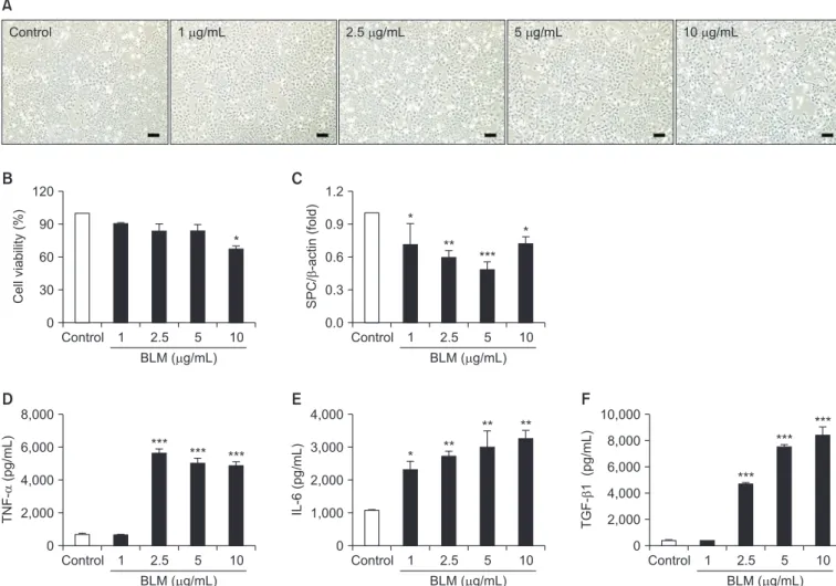
1. BLM increased pro-inflammatory cytokines with loss of cell viability
In order to determine whether BLM affects cellular prolif- eration, MLE-12 cells were treated with BLM (1–10 μg/mL) for 24 hours. The morphological change occurred flatten and enlargement of cell body in MLE-12 cells exposed to BLM under the microscope (Figure 1A). In MTT assay, cell viability was significantly decreased in treatment up to 10 μg/mL BLM (Figure 1B). In qRT-PCR analysis, mRNA level of surfactant
protein C (SPC) which is a specific marker of lung AE II cell was decreased (Figure 1C). Moreover, pro-inflammatory cy- tokines including TNF-α, IL-6, and TGF-β1 were significantly released in response to BLM treatment in MLE-12 cells (Figure 1D–F).
2. BLM-induced apoptosis and cell cycle arrest with nuclear enlargement
We determine whether BLM induces apoptosis and cell cycle arrest in MLE-12 cells. In Hoechst 33258 staining, the nucleus of BLM treated MLE-12 cells were occurred con- densation and fragmentation in a dose-dependent manner under the microscope (Figure 2A). Moreover, in qRT-PCR analysis, BLM treatment increased mRNA expression of a
Figure 1. In the MLE-12 cells was evaluated toxicity effects of by bleomycin (BLM). MLE-12 cells were treated BLM (1–10 μg/mL) for 24
hours. (A) MLE-12 cells were induced morphological change by BLM. Scale bars=100 μm. (B) MLE-12 cells were treated with BLM and deter-
mined cell viability using MTT assay. (C) Expression of alveolar epithelial cell marker, surfactant protein C (SPC) was measured by quantita-
tive real-time reverse transcription–polymerase chain reaction. The graph was measured inflammatory cytokines of tumor necrosis factor α
(TNF-α) (D), interleukin-6 (IL-6) (E), and transforming growth factor β1 (TGF-β1) (F) in cell culture supernatant in enzyme-linked immuno-
sorbent assay. *p<0.05, **p<0.01, ***p<0.001.
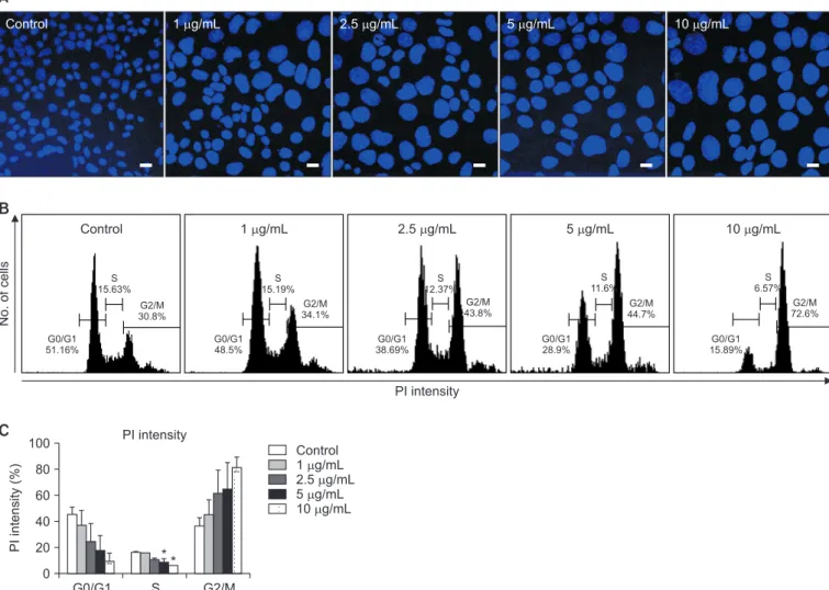
pro-apoptotic factor (BAX) while decreased expression of anti-apoptotic factor (Bcl2) (Figure 2B). Cell cycle arrest was analyzed using DAPI and PI staining. In DAPI staining, cel- lular nucleus enlargement was morphologically found with loss of cell number (Figures 2A, 3A, B). It has been shown that increased size of nucleus is associated with cell cycle arrest leading to senescence
22. Therefore, we measured nuclear size and the abnormal index. As a result, nucleus size and abnor- mal index were significantly increased in BLM exposed in MLE-12 cells (Table 2). In addition, in flow cytometry analysis, BLM decreased G0/G1 whereas increased S and G2/M phase in MLE-12 cells (Figure 3B). These findings suggest that BLM increased nuclear enlargement and it is associated with apop- tosis and cell cycle arrest.
3. BLM-mediated the alteration of cell cycle regulatory gene expression in MLE-12 cells
We next determined the alteration of cell cycle arrest- related gene by BLM treatment for 24 hours in MLE-12 cells.
In qRT-PCR analysis, the mRNA levels of p21 and p53 were significantly increased whereas the mRNA level of Pcna was deceased in MLE-12 cells treated with BLM for 24 hours (Figure 4A, B). SLFN family has been implicated the cell cycle regulation and cell growth arrest
19. Therefore, we conducted qRT-PCR analysis in BLM-exposed MLE-12 cells, and mRNA levels of SLFN2 and SLFN4 were significantly increased rather than the mRNA expression of Slfn1 (Figure 4D–F). These data
suggest that cell cycle arrest–related genes including p21, p53, and SLFN family are increased by BLM treatment leading to apoptosis in MLE-12 cells.
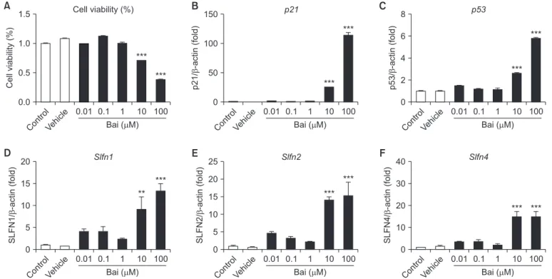
4. Bai-mediated the alteration of SLFN family gene expression in MLE-12 cells
To determine whether cell cycle arrest-related gene and SLFN family gene expression level were changed by Bai that G1/G2 phase cell cycle arrest inhibitor. The cell viability was significantly decreased by Bai in dose-dependent manner (Figure 5A). The mRNA level of cell cycle arrest-related gene such as p21, p53, Slfn1, Slfn2, and Slfn4 were significantly in- creased by Bai (Figure 5B–F). These data suggest that SLFN1, SLFN2, and SLFN4 associated G1/G2 cell cycle arrest in MLE- 12 cells.
Discussion
Injury of AE II cells has been implicated the pathological feature to trigger the loss of type I alveolar epithelial cells leading to induce the migration of activated fibroblasts in interstitial compartment of distal alveoli in the pathogenesis of pulmonary fibrosis
23. Injury of AE II cells includes cellu- lar functional deficiency such as decreased SPC expression which functions to decrease collagen accumulation and inflammation
24, increased secretion of pro-inflammatory cy-
***
***
***
** *
*** ** **
***
Control 1 g/mL 2.5 g/mL 5 g/mL 10 g/mL
A
C B
Control 2.0 1.5 1.0 0.5 0.0
1 2.5 5 10
BLM ( g/mL) BAX
BAX/ -actin (fold)
Control 1.2
0.8
0.4
0.0
1 2.5 5 10
BLM ( g/mL) Bcl2
Bcl2/ -actin (fold) BAX/Bcl2 (fold)
D
Control 3 2 1 4
1 2.5 5 10
BLM ( g/mL) BAX/Bcl2
0
Figure 2. The MLE-12 cells induced apoptosis by bleomycin (BLM). (A) MLE-12 cells were stained with Hoechst 33258 staining after BLM treatment for 24 hours. Scale bars=100 μm. Expression of cell apoptosis marker, BAX (B) and Bcl2 (C) was measured by quantitative real- time reverse transcription‒polymerase chain reaction (qRT-PCR). BAX/Bcl2 ratio (D) was divided (B, C) that measured by qRT-PCR. *p<0.05,
**p<0.01, ***p<0.001.
tokines
25, and accelerated the cellular senescence of alveolar epithelium
26. In human A549 cell line and rat primary AE II cells treated with BLM, senescence-associated β-gal activity
and flat/enlarged morphology has been exhibited
27. In this perspective, the nuclear enlargement and decreased viability of AE II cells in our findings agree with BLM-induced cellular senescence. Moreover, cellular senescence accelerates to un- dergo cell cycle arrest with distinct morphological feature. Al- though the cellular senescence in tissue fibrosis is unclear, it is generally accepted that telomeres are shortened in AE II cells of IPF patients
28. Recently, Naikawadi et al.
29have been shown that deletion of telomere repeat binding factor 1 (TRF1) which is a component protein in shelterin complex of telomeres of AE II cells causes pulmonary fibrosis in SPC-CreTrf1
fl/flmice.
When SPC-CreTrf1
fl/flmice are induced fibrosis, the prolonged telomere dysfunction caused AE II cell hyperplasia during lung remodeling. In contrast, Povedano et al.
30demonstrated that Trf1 deletion develops 90% of AE II cells apoptosis and pulmonary fibrosis through induction of telomere damage using lungs of Trf1
∆/∆mouse. In addition, Cui et al.
31have been shown that up-regulated miR-34a alveolar epithelial dysfunc-
Control 1 g/mL 2.5 g/mL 5 g/mL 10 g/mL
A
No. of cells
Control 1 g/mL 2.5 g/mL 5 g/mL 10 g/mL
G0/G1 51.16%
S 15.63%
G2/M 30.8%
G0/G1 48.5%
S 15.19%
G2/M 34.1%
G0/G1 38.69%
S 12.37%
G2/M 43.8%
G0/G1 28.9%
S 11.6%
G2/M 44.7%
G0/G1 15.89%
S 6.57%
G2/M 72.6%
B
PI intensity
PI intensity (%) *
G0/G1 100
80 60 40 20 0
C PI intensity
Control 1 g/mL 2.5 g/mL 5 g/mL 10 g/mL
S G2/M
*
Figure 3. Evaluation of nuclear enlargement and cell cycle arrest induced in MLE-12 cells by bleomycin (BLM). (A) The image was obtained using DAPI staining in MLE-12 cells after BLM treatment. Scale bars=200 μm. (B) Propidium iodide (PI) staining was performed for cell cycle arrest in MLE-12 cells after BLM treatment. (C) Statistical analysis was each percentage of the field in the samples. *p<0.05.
Table 2. Nuclear size and apoptotic index by bleomycin treatment in MLE-12 cells
Group Nucleus size (μm) Abnormal index (%)
†Control 72.6 32.1
Bleomycin 1 μg/mL 136.5*** 62.6**
Bleomycin 2.5 μg/mL 129.9*** 90.0***
Bleomycin 5 μg/mL 128.3*** 95.0***
Bleomycin 10 μg/mL 167.5*** 88.6***
**p<0.01, ***p<0.001.
†