Vascular Specialist International
Vol. 36, No. 2, June2020
pISSN 2288-7970 • eISSN 2288-7989
INTRODUCTION
Endovascular treatment has the advantage of low peri- procedural risks, short hospital stays, and repeatable in-
terventions. The efficacy of endovascular treatment has been increasing steadily and is now becoming mainstream for femoropopliteal occlusive disease [1,2]. However, as the number of patients with diabetes and renal insufficiency
The Effect of Severe Femoropopliteal Arterial Calcification on the Treatment Outcome of Femoropopliteal Intervention in Patients with Ischemic Tissue Loss
Hyun Yong Lee
1, Ui Jun Park
2, Hyoung Tae Kim
2, and Young-Nam Roh
21Department of Surgery, Soonchunhyang University Cheonan Hospital, Cheonan, 2Division of Transplantation and Vascular Surgery, Department of Surgery, Keimyung University Dongsan Medical Center, Daegu, Korea
Purpose: We investigated the effect of severe calcification of the femoropopliteal artery on intervention outcomes in patients with ischemic tissue loss.
Materials and Methods: A retrospective review of the first endovascular treatment of the femoropopliteal artery for ischemic tissue loss between May 2010 and Feb- ruary 2018 was performed. The calcification of femoropopliteal lesions was esti- mated by the Compliance 360° score, and lesions with a score of 4 were defined as severe calcification lesions.
Results: Overall, 135 first femoropopliteal endovascular procedures on 135 limbs from 112 patients were included in this study. Among the 135 limbs that received treatement of the femoropopliteal arteries, 74 limbs had Trans-Atlantic Inter Soci- ety Consensus (TASC) A or B lesions and 61 limbs had TASC C or D lesions. Among 61 cases of TASC C or D lesions, 21 limbs (34.4%) had severe calcification; there was no statistically significant difference in limb salvage (P=0.75), and amputation- free survival (P=0.11) based on the degree of calcification. However, the survival rate in TASC C or D lesions was significantly different between the two groups (non-severe calcification group vs severe calcification group at 1-year, 2-years, and 3-years: 88.6%, 79.7%, and 61.0% vs 70.0%, 56.0%, and 28.0%, respectively, P=0.01). In multivariate analysis of influencing factors for poor survival in TASC C or D using the Cox proportional hazards model, severe calcification (hazard ratio, 2.362; 95% confidence interval, 1.035-5.391; P=0.041) was a statistically signifi- cant risk factor.
Conclusion: Severe femoropopliteal artery calcification was associated with poor survival, especially in TASC C or D lesions.
Key Words: Ischemic tissue loss, Arterial calcification, Endovascular treatment
Received January 29, 2020 Revised April 20, 2020 Accepted May 18, 2020
Corresponding author: Young-Nam Roh Division of Transplantation and Vascular Surgery, Department of Surgery, Dongsan Medical Center, Keimyung University School of Medicine, 1035 Dalgubeol- daero, Dalseo-gu, Daegu 42601, Korea Tel: 82-53-258-4708
Fax: 82-53-258-4710
E-mail: [email protected] https://orcid.org/0000-0002-0799-1578
Copyright © 2020, The Korean Society for Vascular Surgery
This is an Open Access article distributed under the terms of the Creative Commons Attribution Non-Commercial License (http://creativecommons.org/licenses/by-nc/4.0) which permits unrestricted non-commercial use, distribution, and reproduction in any medium, provided the original work is properly cited.
Vasc Specialist Int 2020;36(2):96-104 • https://doi.org/10.5758/vsi.200005
increases, vascular calcification, one of the important bar- riers in peripheral vascular intervention, is also increasing.
Vascular calcification is known to affect the outcome and the technical success rate of the endovascular procedure [3].
Currently, most of the United States Investigational Device Exemption protocols for endovascular devices are not ap- plied for patients with severe calcification [4], as it relates to the occurrence of adverse events, such as dissection, vessel perforation, and atheroembolism.
Many studies reported the risk factors associated with the prognosis of endovascular treatment of femoropopliteal lesions [5-8]. Critical limb-threatening ischemia (CLTI) is known to be associated with a high risk of limb amputa- tion and mortality [9]. CLTI represents the terminal stage of peripheral arterial disease. It occurs when arterial vessels are improperly perfused and unable to maintain tissue vi- ability [10]. However, there are few data on the intermediate or long-term effects of vascular calcification on the endo- vascular treatment outcome for femoropopliteal lesions in patients with CLTI.
The aim of this study was to investigate the outcomes after endovascular treatment according to the degree of femoropopliteal arterial calcification in patients with isch- emic tissue loss.
MATERIALS AND METHODS
A database of patients who underwent endovascular treatment of the femoropopliteal artery for ischemic tissue loss between May 2010 and February 2018 was evaluated.
Procedures for chronic femoropopliteal atherosclerotic steno-occlusive disease were included in the study. In- terventions in patients with acute limb ischemia, in-stent restenosis, trauma, popliteal aneurysm, re-intervention, and procedures for bypass graft were excluded. The defini- tion of technical success was no residual stenosis (≤50% in angioplasty alone; ≤30% in stent use) and the absence of flow-limiting dissection in treated femoropopliteal artery confirmed by digital subtraction angiography [11]. Major amputation was defined as any procedure that results in an amputation at the level of the ankle or above [11]. A major amputation indication was major tissue loss extend- ing above the metatarsal level (Rutherford category 6). Of 252 limbs that underwent procedures for de novo chronic femoropopliteal lesions in patients with ischemic tissue loss, we excluded interventions for patients with vasculitis (n=2), technical failure (n=7), and major tissue loss, which inevita- bly necessitates major amputation at presentation, such as involving the metatarsal area (n=8). Additionally the cases without sufficient data or wound follow-up (n=100) were excluded. Finally, 135 first femoropopliteal endovascular
procedures on 135 limbs from 112 patients were included in this study.
The number of technical failures was small (n=7) be- cause it included only cases where guidewire passage was attempted, except for cases without intervention or only diagnostic angiography. For wound healing, it is ideal to make direct in-line flow up to ankle level. However, in this study, technical success focused on the treatment of the femoropopliteal artery. Therefore, if femoropopliteal inter- vention was successful, even if direct in-line tibial flow up to ankle level was not obtained, it was considered a techni- cal success.
We reviewed demographic data, comorbidities, details of endovascular procedures, and clinical outcomes. Comorbidi- ties included hypertension, diabetes mellitus, ischemic heart disease (history of medical treatment, coronary bypass, or percutaneous coronary interventions), renal insufficiency, renal replacement therapy, and cerebrovascular disease (stroke or transient ischemic attack).
Computed tomography angiography (CTA) has been used limitedly in patients with renal insufficiency. However, CTA was performed without restriction in patients already un- dergoing renal replacement. Since the degree of calcifica- tion was evaluated based on CTA in our study, 11 patients who did not undergo CTA due to renal insufficiency were excluded, and these patients were included in the 100 cases treated with insufficient data. The degree of lesion calcifi- cation was analyzed using the Com pliance 360° score [11,12].
Com pliance 360° score is divided into 5 grades: grade 0, the calcium deposit is absent from the circumference and length; grade 1, the calcium deposit has a circumference less than 180° and less than half of lesion length; grade 2, the calcium deposit has a circumference less than 180° and greater than half of lesion length; grade 3, the calcium de- posit has a circumference greater than 180° and less than half of lesion length; and grade 4, the calcium deposit has a circumference greater than 180° and greater than half of lesion length. We divided grade 0 through 3 into the non- severe calcification group and grade 4 into the severe cal- cification group. Classification of femoropopliteal lesions according to degree of calcification is shown in Fig. 1.
The patients who had a successful endoluminal inter- vention received dual anti-platelets. Patients underwent routine clinical ankle–brachial index (ABI) measurements 1, 3, and 6 months after the procedure and every 6 months thereafter. During follow-up, a duplex scan or CTA was per- formed if patients were symptomatic or if noninvasive tests suggested the presence of restenosis or an occlusion.
Hypertension was defined as using antihypertensive
medication for high blood pressure or having a systolic
blood pressure ≥140 mm Hg and/or diastolic blood pres-
sure ≥90 mm Hg. Diabetes mellitus was defined as using blood glucose-lowering medication and/or having a fasting glucose level of ≥126 mg/dL. Dyslipidemia was defined as using lipid-lowering medication and/or having a cholesterol level >200 mg/dL and/or low-density lipoprotein cholesterol level >130 mg/dL. Renal insufficiency was defined as hav- ing a serum creatinine level >1.5 mg/dL or being on renal replacement therapy.
Clinical outcome variables included limb salvage, patient survival, and amputation-free survival. Limb salvage was defined as not receiving any major amputations. Amputa- tion-free survival was defined as not receiving above-ankle amputation of the index limb or dying. Primary patency was defined as treated vessel without a significant steno- sis (a peak systolic velocity ratio >2.4 on duplex scan and
>50% stenosis on angiography). Secondary patency was defined patency of the target lesion after treatment of a re- occlusion of the index lesion.
All statistical analyses were performed using IBM SPSS Statistics for Windows, version 21.0 (IBM Corp, Armonk, NY, USA). The Kaplan–Meier method with the log-rank test was used to calculate primary patency, secondary patency, limb salvage, patient survival, and amputation-free surviv- al. Chi-squared and Fisher exact tests were used to compare the degree of calcification and various parient characteris- tics between the groups. Multivariate analysis of prognos- tic relevance was evaluated by multivariate Cox regression analysis. A value of P<0.05 was considered statistically sig- nificant in all analyses.
The study protocol was developed in accordance with
AB
C
Fig. 1. Computed tomogra- phy angiography (CTA) images showed different degree of calcification in femoropopli- teal artery. (A) Circumference of femoal artery calcification in cross-sectional image; left,
<180°; right, ≥180°. (B) CTA
images of non-severe calcifica-
tion. (C) CTA images of severe
calcification.
the Declaration of Helsinki, and approved by the Dongsan Medical Center Institutional Review Board (DSMC no. 2019- 07-008). The requirement for informed consent was waived due to the retrospective nature of study.
RESULTS
1) Patient population
A comparison of demographics and cardiovascular co- morbidities are shown in Table 1. The distribution rates of non-severe (n=100) and severe (n=35) calcification groups
were 74.1% (77.0% male; age 71.5±8.3 years) and 25.9%
(62.9% male; age 68.6±9.7 years), respectively. There was no difference in the prevalence of diabetes, hypertension, and smoking. However, the prevalence of ischemic heart disease (P<0.001), renal insufficiency (P<0.001), and renal replacement therapy (P<0.001) were significantly higher in the severe calcification group.
2) Lesion characteristics and procedural details
The lesion characteristics and procedural details are shown in Table 1. In the comparison of the Trans-Atlantic
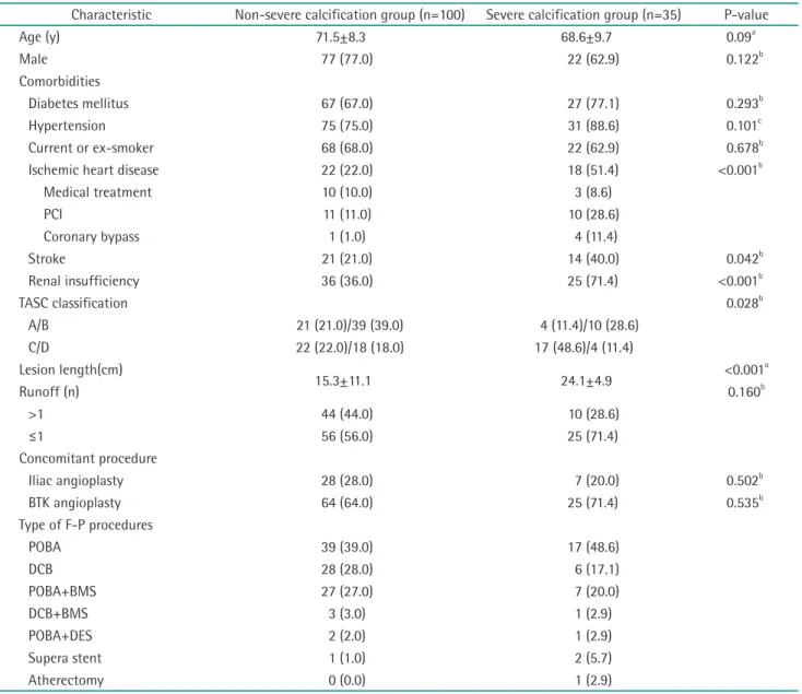
Table 1. Patient, lesion and procedure characteristics
Characteristic Non-severe calcification group (n=100) Severe calcification group (n=35) P-value
Age (y) 71.5±8.3 68.6±9.7 0.09a
Male 77 (77.0) 22 (62.9) 0.122b
Comorbidities
Diabetes mellitus 67 (67.0) 27 (77.1) 0.293b
Hypertension 75 (75.0) 31 (88.6) 0.101c
Current or ex-smoker 68 (68.0) 22 (62.9) 0.678b
Ischemic heart disease 22 (22.0) 18 (51.4) <0.001b
Medical treatment 10 (10.0) 3 (8.6)
PCI 11 (11.0) 10 (28.6)
Coronary bypass 1 (1.0) 4 (11.4)
Stroke 21 (21.0) 14 (40.0) 0.042b
Renal insufficiency 36 (36.0) 25 (71.4) <0.001b
TASC classification 0.028b
A/B 21 (21.0)/39 (39.0) 4 (11.4)/10 (28.6)
C/D 22 (22.0)/18 (18.0) 17 (48.6)/4 (11.4)
Lesion length(cm)
15.3±11.1 24.1±4.9 <0.001a
Runoff (n) 0.160b
>1 44 (44.0) 10 (28.6)
≤1 56 (56.0) 25 (71.4)
Concomitant procedure
Iliac angioplasty 28 (28.0) 7 (20.0) 0.502b
BTK angioplasty 64 (64.0) 25 (71.4) 0.535b
Type of F-P procedures
POBA 39 (39.0) 17 (48.6)
DCB 28 (28.0) 6 (17.1)
POBA+BMS 27 (27.0) 7 (20.0)
DCB+BMS 3 (3.0) 1 (2.9)
POBA+DES 2 (2.0) 1 (2.9)
Supera stent 1 (1.0) 2 (5.7)
Atherectomy 0 (0.0) 1 (2.9)
Values are presented as mean±standard deviation or number (%).
PCI, percutaneous coronary intervention; TASC, Trans-Atlantic Inter Society Consensus; BTK, below-the-knee; POBA, plain old balloon angioplasty; DCB, drug-coated balloon; BMS, bare metal stent; DES, drug-eluting stent.
aStudent t-test. bChi-squre test. cFisher’s exact test.
Inter Society Consensus (TASC) classification of the femoro- popliteal artery, there was a significant difference between the non-severe and severe calcification group (P=0.028).
However, there was no difference in the patent tibial runoff evaluated by intraoperative angiography (P=0.160). In the non-severe calcification group, angioplasties of the iliac and below-the-knee (BTK) arteries were performed simul- taneously in 28.0% and 64.0% of limbs, respectively. With regards to the balloon angioplasty, a plain balloon was used in 68.0% of limbs, and a drug-coated balloon (DCB) was used in 32.0%. A bare-metal stent (BMS) after plain bal- loon angioplasty was used in 27.0% of limbs for residual stenosis or flow-limiting dissection. In the severe calcifica- tion group, angioplasties of the iliac and BTK arteries were performed simultaneously in 20.0% and 71.4% of limbs, respectively. For the balloon angioplasty, plain balloons and DCBs were used in 71.5% and 28.5% of limbs, respectively.
A BMS after plain balloon angioplasty was used in 20.0%
of limbs for residual stenosis or flow-limiting dissection.
A BMS after angioplasty using a DCB was used in 2.9% of limbs.
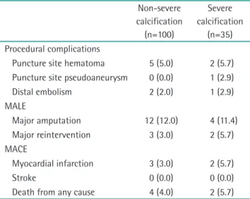
3) Early clinical outcome
Early clinical outcomes within 30 days after the proce- dure are shown in Table 2. Puncture site hematoma oc-
curred immediately after procedures in 5.0% (n=5) of cases in the non-severe calcification group and 5.7% (n=2) in the severe calcification group. Distal embolization occurred in 2.0% (n=2) of cases in the non-severe calcification group and 2.9% (n=1) in the severe calcification group. Major amputation immediately after the procedure occurred in
Table 2. Immediate safety outcome (<30 days)
Non-severe calcification(n=100)
Severe calcification
(n=35) Procedural complications
Puncture site hematoma 5 (5.0) 2 (5.7) Puncture site pseudoaneurysm 0 (0.0) 1 (2.9)
Distal embolism 2 (2.0) 1 (2.9)
MALE
Major amputation 12 (12.0) 4 (11.4) Major reintervention 3 (3.0) 2 (5.7) MACE
Myocardial infarction 3 (3.0) 2 (5.7)
Stroke 0 (0.0) 0 (0.0)
Death from any cause 4 (4.0) 2 (5.7) Values are presented as number (%).
MALE, major adverse limb events; MACE, major adverse cardiovas- cular events.
Fig. 2. Kaplan–Meier (KM) curves of primary and secondary patency rates comparing non-severe and severe calcification groups. Patencies were significantly worse in severe calcification group.
0 100
90 80 70 60 50 40 30 20 10
36
%
Month 0
Non-severe calcification (n=100)
1 yr
24 12
Severe calcification (n=35)
KM estimate
# at risk KM estimate
# at risk
2 yr 3 yr
88.7%
80 64.1%
18
77.2%
54 49.3%
11
60.6%
5 44.4%
3
0 100
90 80 70 60 50 40 30 20 10
36
%
Month 0
Non-severe calcification (n=100)
1 yr
24 12
Severe calcification (n=35)
KM estimate
# at risk KM estimate
# at risk
2 yr 3 yr
93.8%
84 79.1%
21
92.6%
64 75.3%
15
76.8%
6 75.3%
3 Non-severe calcification
Severe calcification
Non-severe calcification Severe calcification
Log rank P=0.009 Log rank P=0.034
Primary patency Secondary patency
12.0% (n=12) of cases in the non-severe calcification group and 11.4% (n=4) in the severe calcification group. Reinter- vention in each group was 3.0% (n=3) and 5.7% (n=2), re- spectively. Death immediately after the procedure occurred in 4.0% (n=4) of cases in the non-severe calcification group and 5.7% (n=2) in the severe calcification group.
4) Midterm outcome
Fig. 2 shows the primary patency and secondary patency according to the degree of calcification. The primary pa- tency was significantly different between the two groups (non-severe calcification group vs severe calcification group at 1-year, 2-years, and 3-years: 88.7%, 77.2%, and 60.6% vs. 64.1%, 49.3%, and 44.4%, respectively, P=0.009).
0 100
90 80 70 60 50 40 30 20 10
36
%
Month 0
Non-severe calcification (n=40)
1 yr
24 12
Severe calcification (n=21)
KM estimate
# at risk KM estimate
# at risk
2 yr 3 yr
81.5%
30 84.0%
15
81.5%
22 84.0%
11
81.5%
5 84.0%
2 Non-severe calcification Severe calcification
Log rank P=0.75
A
0 100
90 80 70 60 50 40 30 20 10
36
%
Month 0
Non-severe calcification (n=40)
1 yr
24 12
Severe calcification (n=21)
KM estimate
# at risk KM estimate
# at risk
2 yr 3 yr
73.6%
28 60.0%
15
64.4%
21 52.5%
12
54.5%
5 31.5%
2 Non-severe calcification Severe calcification
Log rank P=0.11
C
0 100
90 80 70 60 50 40 30 20 10
36
%
Month 0
Non-severe calcification (n=40)
1 yr
24 12
Severe calcification (n=21)
KM estimate
# at risk KM estimate
# at risk
2 yr 3 yr
88.6%
29 70.0%
17
79.7%
25 56.0%
13
61.0%
6 28.0%
3 Non-severe calcification Severe calcification
Log rank P=0.01
B
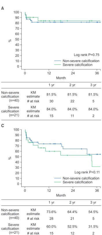
Fig. 3. Kaplan–Meier (KM) curves of limb salvage rate (A),
overall survival rate (B), and amputation-free survival rate
(C) in patients with Trans-Atlantic Inter Society Consensus
(TASC) C/D lesions. Overall survival rate was significantly
worse in severe calcification group.
Also, the secondary patency was significantly different be- tween the two groups (non-severe calcification group vs.
severe calcification group at 1-year, 2-years, and 3-years:
93.8%, 92.6%, and 76.8% vs. 79.1%, 75.3%, and 75.3%, respectively, P=0.034).
Among 74 limbs with TASC A or B lesions, there were 60 limbs (81.1%) with non-severe calcification lesions and 14 limbs (18.9%) with severe calcification lesions. For TASC A or B groups, there were no statistically significant dif- ferences in limb salvage (P=0.51), survival (P=0.13), and amputation-free survival (P=0.22), according to the degree of calcification. Among 61 limbs with TASC C or D lesions, there were 40 limbs (65.6%) with non-severe calcification lesions and 21 limbs (34.4%) with severe calcification le- sions. There was no statically significant difference in limb salvage between the non-severe calcification and severe calcification groups in patients with TASC C or D lesions (P=0.75). However, the survival rate was significantly dif- ferent between the two groups (non-severe calcification group vs severe calcification group at 1-year, 2-years, and 3-years: 88.6%, 79.7%, and 61.0% vs. 70.0%, 56.0%, and 28.0%, respectively, P=0.01). In comparing amputation- free survival rates, there was not a significant difference between the two groups in patients with TASC C or D le- sions (P=0.11) (Fig. 3). In multivariate analysis of influenc- ing factors for poor survival in TASC A or B using the Cox proportional hazards model, ischemic heart disease (hazard ratio [HR], 1.064; 95% confidence interval [CI], 0.390-2.904;
P=0.904), renal insufficiency (HR, 1.627; 95% CI, 0.642- 4.127; P=0.305), severe calcification (HR, 1.590; 95% CI, 0.569-4.442; P=0.376) showed no statistical significance.
In multivariate analysis of influencing factors for poor sur- vival in TASC C or D using the Cox proportional hazards model, ischemic heart disease (HR, 1.177; 95% CI, 0.470- 2.947; P=0.727), renal insufficiency (HR, 1.276; 95% CI,
0.566-2.878; P=0.557) showed no statistical significance.
However, severe calcification (HR, 2.362; 95% CI, 1.035- 5.391; P=0.041) was statistically significant risk factor (Table 3, Supplementary Table 1).
DISCUSSION
Endovascular therapy has been extensively used in the revascularization of peripheral arterial disease with low mortality and less invasive procedures compared to bypass surgery [13,14]. In previous treatment guidelines, endo- vascular therapy was recommended only for single, short, and focal lesions, but recommendations have changed to advocate endovascular therapy, rather than open surgery, for most lesions due to advances in technologies and tech- niques [15]. However, the presence of vascular calcification, particularly in the infrainguinal vasculature, represents a significant challenge to current endovascular device strategies [4]. Severe calcification may be associated with increased dissection, perforation, and atheroembolism. It is also known that balloon angioplasty for severely calci- fied lesions is limited by early elastic recoil and poor early and long-term outcomes [5]. Although several studies have investigated the effect of significant calcification on reste- nosis after endovascular treatment, we focused on clinical characteristics, such as limb salvage and survival according to degree of vascular calcification, because clinical out- comes, such as wound healing, limb salvage, and survival, are more ultimate endpoints of treatment than technical or anatomical endpoints [16].
Determining the relevant score components by selecting the appropriate calcification grading system is an impor- tant factor in correlating the outcome in this analysis [17].
The existing scoring system, the peripheral arterial calcium scoring system (PACSS), highlights the pathologic location of calcification (intima, media, or combined) along with the length of the segment affected [4]. However, the lack of a validated, quantitative calcium scoring system makes it dif- ficult to evaluate the outcomes of endovascular therapies in patients with calcified peripheral arteries. A calcifica- tion scoring system based on angiography is a useful tool for selecting treatment modality and device during the procedure [17]. However, objective scoring of vascular cal- cification by conventional angiography was impossible in this retrospective study. According to the protocol of our institution, pre-procedural CTA were obtained in all cases;
therefore, we evaluated the degree of vascular calcification based on CTA according to the score using the 360° trial [11,12].
Severe calcification is known to reduce the durability of endovascular therapy of femoropopliteal lesions. In our Table 3. Risk factors for poor survival in TASC A/B and TASC
C/D
Variable P HR (95% CI)
TASC A/B
Ischemic heart disease 0.904 1.064 (0.390-2.904) Renal insufficiency 0.305 1.627 (0.642-4.127) Severe calcification 0.376 1.590 (0.569-4.442) TASC C/D
Ischemic heart disease 0.727 1.177 (0.470-2.947) Renal insufficiency 0.557 1.276 (0.566-2.878) Severe calcification 0.041 2.362 (1.035-5.391) By Cox’s proportional hazard model.
TASC, Trans-Atlantic Inter Society Consensus; HR, hazard ratio; CI, confidence interval.
study, there was also statistically significant differences in primary patency and secondary patency according to the degree of calcification. de Athayde Soares et al. [15] report- ed that severe calcification were related to loss of primary patency. In their study, one tibial vessel or isolated popliteal artery runoff, high calcification grade, small vessel diam- eter (<4 mm), or primary angioplasty without stenting were associated with loss of primary patency. Okuno et al. [18]
reported that PACSS grade 4 was significantly associated with both major adverse life events and mortality.
In the present study, in patients with TASC C or D le- sions, limb salvage and amputation-free survivals were not affected by the degree of calcification, but the survival rate was. Clinical implications of the presence of calcification in the coronary artery leading to an increased cardiovas- cular risk have been well demonstrated [19]. However, little is known about the association between lower extremity arterial calcification and clinical outcomes [20]. Only a few studies reported that the calcium score in the iliac or tibial artery was associated with amputation and mortality in patients who had symptomatic peripheral arterial diase- ase [20,21]. In the present study, there were significantly more patients with comorbidities of ischemic heart disease, stroke, renal insufficiency, and renal replacement in the se- vere calcification group than in the non-severe calcification group. However, in the multivariate analysis of the TASC C/
D lesions, severe calcification was found to be a significant factor related with poor survival, independent of ischemic heart disease and renal insufficiency.
This study has some limitations. First, initial wound data and follow-up data were not available in many cases due to the retrospective design, and, thus, these cases were excluded. Also, because it was a retrospective study, WIFI classification was difficult to confirm, and all cases were classified as Rutherford category 5 as minor tissue loss.
Secondly, we did not distinguish whether the calcification was intimal or medial. Since this study did not distinguish between intimal and medical calcification, many of the cas- es classified as severe calcification were medial calcification cases. In the case of medial calcification, lesions seen in DSA are often less severe than those seen in CT. In addition, since with medial calcification lesions it is easy to guide wire passage and dissection does not occur easily, it seems that the severe calcification group required less bailout stenting than expectation. In this study, bailout stenting in the non-severe calcification group was 42.6% in plain old balloon angioplasty (POBA) and 9.7% in DCB, and in the severe calcification goup, was 32.0% in POBA and 14.3% in DCB. Also, in our center’s strategy, preballooning was per- formed using a plain balloon with a 1 mm-smaller diameter before DCB, which resulted in a protective effect against
dissection. For this reason, less bailout stenting were used when using DCB.
CONCLUSION
In conclusion, severe calcification of the femoropopliteal artery with TASC A or B lesions did not affect the clinical outcomes in patients with ischemic tissue loss. In TASC C or D lesions, severe calcification of femoropopliteal artery did not affect limb salvage in patients with ischemic tissue loss.
However, femoropopliteal arterial calcification was signifi- cantly associated with poor survival in TASC C or D lesions.
FUNDING
This study was supported by the Keimyung University Research Grant 2018.
SUPPLEMENTARY MATERIALS
Supplementary data can be found via https://doi.
org/10.5758/vsi.200005.
CONFLICTS OF INTEREST
The authors have nothing to disclose.
ORCID
Hyun Yong Lee
https://orcid.org/0000-0002-9814-1412 Ui Jun Park
https://orcid.org/0000-0002-5203-4216 Hyoung Tae Kim
https://orcid.org/0000-0002-9714-8778 Young-Nam Roh
https://orcid.org/0000-0002-0799-1578
AUTHOR CONTRIBUTIONS
Concept and design: YNR. Analysis and interpretation:
HYL, YNR. Data collection: HYL, UJP, HTK, YNR. Writing the article: HYL, YNR. Critical revision of the article: HYL, YNR. Final approval of the article: YNR. Statistical analysis:
YNR. Obtained funding: YNR. Overall responsibility: YNR.
REFERENCES
1) European Stroke Organisation, Ten- dera M, Aboyans V, Bartelink ML, Baumgartner I, Clément D, et al. ESC Guidelines on the diagnosis and treat- ment of peripheral artery diseases:
Document covering atherosclerotic disease of extracranial carotid and vertebral, mesenteric, renal, upper and lower extremity arteries: the Task Force on the diagnosis and treatment of peripheral artery diseases of the European Society of Cardiology (ESC).
Eur Heart J 2011;32:2851-2906.
2) Jaff MR, White CJ, Hiatt WR, Fowkes GR, Dormandy J, Razavi M, et al. An update on methods for revasculariza- tion and expansion of the TASC lesion classification to include below-the- knee arteries: a supplement to the inter-society consensus for the man- agement of peripheral arterial disease (TASC II). Vasc Med 2015;20:465-478.
3) Shin SH, Baril D, Chaer R, Rhee R, Makaroun M, Marone L. Limitations of the Outback LTD re-entry device in femoropopliteal chronic total occlu- sions. J Vasc Surg 2011;53:1260-1264.
4) Rocha-Singh KJ, Zeller T, Jaff MR.
Peripheral arterial calcification: preva- lence, mechanism, detection, and clinical implications. Catheter Cardio- vasc Interv 2014;83:E212-E220.
5) Capek P, McLean GK, Berkowitz HD.
Femoropopliteal angioplasty. Factors influencing long-term success. Circu- lation 1991;83(2 Suppl):I70-I80.
6) Clark TW, Groffsky JL, Soulen MC.
Predictors of long-term patency after femoropopliteal angioplasty: results from the STAR registry. J Vasc Interv Radiol 2001;12:923-933.
7) Hunink MG, Donaldson MC, Meyero- vitz MF, Polak JF, Whittemore AD, Kandarpa K, et al. Risks and benefits
of femoropopliteal percutaneous balloon angioplast y. J Vasc Surg 1993;17:183-192; discussion 192-194.
8) Strecker EP, Boos IB, Göttmann D.
Femoropopliteal artery stent place- ment: evaluation of long-term suc- cess. Radiology 1997;205:375-383.
9) Rollins KE, Jackson D, Coughlin PA.
Meta-analysis of contemporary short- and long-term mortality rates in patients diagnosed with critical leg ischaemia. Br J Surg 2013;100:1002- 1008.
10) Mustapha JA, Diaz-Sandoval LJ, Saab F. Infrapopliteal calcification patterns in critical limb ischemia: diagnostic, pathologic and therapeutic implica- tions in the search for the endovas- cular holy grail. J Cardiovasc Surg (Torino) 2017;58:383-401.
11) Patel MR, Conte MS, Cutlip DE, Dib N, Geraghty P, Gray W, et al. Evaluation and treatment of patients with lower extremity peripheral artery disease:
consensus definitions from Periph- eral Academic Research Consortium (PARC). J Am Coll Cardiol 2015;65:931- 941.
12) Dattilo R, Himmelstein SI, Cuff RF.
The COMPLIANCE 360° Trial: a ran- domized, prospective, multicenter, pilot study comparing acute and long- term results of orbital atherectomy to balloon angioplasty for calcified femoropopliteal disease. J Invasive Cardiol 2014;26:355-360.
13) Vartanian SM, Conte MS. Surgical intervention for peripheral arterial disease. Circ Res 2015;116:1614-1628.
14) Thukkani AK, Kinlay S. Endovascular intervention for peripheral artery dis- ease. Circ Res 2015;116:1599-1613.
15) de Athayde Soares R, Matielo MF, Bro- chado Neto FC, Pires APM, de Almeida
RD, de Jesus Martins M, et al. Impact of calcification and infrapopliteal out- flow on the outcome of endovascular treatment of femoropopliteal oc- clusive disease. JRSM Cardiovasc Dis 2019;8:2048004019828941.
16) Okazaki J, Matsuda D, Tanaka K, Ishida M, Kuma S, Morisaki K, et al.
Analysis of wound healing time and wound-free period as outcomes after surgical and endovascular revascular- ization for critical lower limb ischemia.
J Vasc Surg 2018;67:817-825.
17) Tepe G, Beschorner U, Ruether C, Fischer I, Pfaffinger P, Noory E, et al. Drug-eluting balloon therapy for femoropopliteal occlusive disease:
predictors of outcome with a special emphasis on calcium. J Endovasc Ther 2015;22:727-733.
18) Okuno S, Iida O, Shiraki T, Fujita M, Masuda M, Okamoto S, et al. Impact of calcification on clinical outcomes after endovascular therapy for superfi- cial femoral artery disease: assessment using the peripheral artery calcifica- tion scoring system. J Endovasc Ther 2016;23:731-737.
19) Chen NX, Moe SM. Vascular calcifica- tion: pathophysiology and risk factors.
Curr Hypertens Rep 2012;14:228-237.
20) Huang CL, Wu IH, Wu YW, Hwang JJ, Wang SS, Chen WJ, et al. Association of lower extremity arterial calcifica- tion with amputation and mortal- ity in patients with symptomatic peripheral artery disease. PLoS One 2014;9:e90201.
21) Allison MA, Hsi S, Wassel CL, Morgan C, Ix JH, Wright CM, et al. Calcified atherosclerosis in different vascular beds and the risk of mortality. Arterio- scler Thromb Vasc Biol 2012;32:140- 146.