Introduction
Stress is a risk factor for depression and insomnia (sleep problems), with insomnia being one of the key symptoms of common depressive disorder
1). Depressive disorders are characterized by changes in the mental status such as a marked loss of interest, a low mood, fatigue, and worthlessness
2). Several types of sedative antidepressant drugs are often used in the treatment of insomnia
3). Chronic restraint stress (CRS) is commonly induced in animal studies to mimic the development of clinical depression. CRS exerts common depressive-like symptoms, such as changes to corticosterone (CORT) and monoamine neurotransmitter (e.g. serotonin; 5-HT, dopamine; DA, and norepinephrine; NE) levels, and locomotor activity deficit
4,5). Serotonin and serotonin receptors (5-HT receptors) play a key role in depression. Activation of the 5-HT
2Areceptor, a Gq-coupled receptor, leads to an accumulation of IP
3, diacylglycerol (DAG), and activation of protein kinase C (PKC), which causes the release of Ca
2+, thereby activating the extracellular signal-regulated kinases 1/2 (ERK1/2) pathway. 5-HT
2Areceptors are expressed in a majority of
neocortical cells (mainly prefrontal cortex; PFC), and are involved in the regulation of sleep and mood
6).
Stauntonia hexaphylla (SH) belongs to the genus Lardizabalaceae, and is widely distributed in Korea, Japan, and China. It has been commonly used as a herbal medicine in China, especially for its analgesic, sedative, and diuretic properties
7-9). Previous studies have demonstrated that the SH leaf exerts pharmacological effects, including antidiabetic
10)and anti-osteoporosis
11)effects. We have previously reported that the SH fruit extract has anti-inflammatory effects in lipopolysaccharide (LPS)-activated RAW 264.7 cells and in carrageenan-induced paw edema rats
12). We also reported that the SH fruit extract has antioxidant and hepatoprotective effects on hydrogen peroxide-induced cytotoxicity in HepG2 cells
13). In addition, previous studies have shown that the constituents of SH extract includes triterpenoids, glucosides, flavonoids, and phenylpropanoids
14). Vaccinium bracteatum (VB) belongs to the genus Ericaceae, and its fruits are commonly known as the “oriental blueberry” in Korea. Previous studies have demonstrated that VB exert pharmacological effects, including anti-fatigue
15), antimicrobial
16), anti-diabetes
17,18),
Antidepressant-like and Hypnotic Effects of the Herbal Extract Combination of Stauntonia hexaphylla and Vaccinium bracteatum Fruit in Mice
Dool-Ri Oh, Yujin Kim, Ara Jo, Sojeong Im, Cho Een Kim, Myung-A Jung, Jawon Shin, Huwon Kang, Eun Jin Choi, Jaeyong Kim, Chulyung Choi*
Jeonnam Bioindustry Foundation, Jeonnam Institute of Natural Resources Research (JINR)
Stauntonia hexaphylla (SH) and Vaccinium bracteatum (VB) are herbal extracts widely used in food and traditional herbal medicine, and have the ability to perform a wide range of biological activities. We aimed to investigate the effects of the SH and VB combination (SHVB) on mice models of chronic restraint stress (CRS) and pentobarbital-induced sleeping behaviors to elucidate its possible mechanisms of action. CRS-exposed mice treated with SHVB showed significantly decreased immobility time, increased swimming and climbing times in the forced swim test (FST), and increased locomotor activity in the open field test (OFT). SHVB decreased serum CORT levels, but enhanced brain monoamine neurotransmitters. SHVB significantly decreased the sleep latency and increased total sleep duration in pentobarbital-induced sleeping behavior in mice. SHVB showed inhibitory effect on 5-HT
2Areceptor-mediated ERK1/2 phosphorylation. These results suggest that SHVB has antidepressant and hypnotic effects by regulating the 5-HT
2Areceptor.
keywords : Stauntonia hexaphylla , Vaccinium bracteatum , Chronic restraint stress, Antidepressants, Serotonin 2A receptor
* Corresponding author
Chulyung Choi, Jeonnam Bioindustry Foundation, Jeonnam Institute of Natural Resources Research (JINR), 288 Woodland-gil, Anyang-myeon, Jangheung-gun, Jeollanamdo 59338, Republic of Korea.
·E-mail : [email protected] ·Tel : +82-61-860-2620
·Received : 2020/01/21 ·Revised : 2020/02/27 ·Accepted : 2020/03/17
ⓒ The Society of Pathology in Korean Medicine, The Physiological Society of Korean Medicine pISSN 1738-7698 eISSN 2288-2529 http://dx.doi.org/10.15188/kjopp.2020.04.34.2.88
Available online at https://kmpath.jams.or.kr
anti-oxidant
16,19), retinal protection
20), anti-proliferative and anti-inflammatory effects
21,22). We have previously reported that the VB fruit has antidepressant properties, protective effects on oxidative stress-induced apoptosis, sedative, and hypnotic effects
23-25). However, no studies have reported the antidepressant-like effects of the combination of SH and VB extracts (SHVB), especially in models of CRS and related depression disorders.
In the present study, we investigated the effects of the SHVB on pentobarbital-induced sleeping behavior in CRS-exposed mice, through a forced swimming test (FST) and open field test (OFT). We also evaluated the levels of CORT and monoamine neurotransmitters (5-HT, DA, and NE) in the CRS-exposed mice. In addition, the effects of SHVB on sleep latency and total sleep duration associated with sleep behavior in pentobarbital-induced mice were studied. Moreover, we investigated the inhibitory effects of SHVB on 5-HT
2A-associated ERK1/2 phosphorylation in Chinese hamster ovary (CHO)-K1 cells transfected with the human 5-HT
2Areceptor.
Materials and Methods
1. Preparation of plant extract
The Stauntonia hexaphylla (Thunb.) Decne. fruits were collected from the Jangheung-gun County (Jeollanamdo, Republic of Korea). The Vaccinium bracteatum Thunb. fruits used in this study were collected from the Goheung County (Jeollanamdo, Republic of Korea). The herbs were extracted with water at 100 ℃ for 4 h. The extraction yield of Stauntonia hexaphylla (Thunb.) Decne. fruits and Vaccinium bracteatum Thunb. fruits was about 8.0 % and 12.5 %, respectively. The extracts were stored at 4 ℃ for further use. The SHVB (NET-1601, SH: VB = 1:1, w/w) used in the present study was the same sample used in the clinical trial, which was approved by Institutional Review Board (IRB) at Konkuk University Medical Center (clinical trials registration number KUMC 2019-07-033-001).
2. Animals
Male ICR mice (five-week-old, weight 24–27 g) were purchased from Samtako Bio Korea (Osan, Republic of Korea). The animals were maintained at a constant room temperature of 22 ± 3 ℃, with a humidity of 50 ± 15 %, and were kept at a 12/12 h light/dark cycle. Mice were given free access to water and food. The all mice were acclimatized for 7 days prior to the experiments. All animal experiments were approved by the Institutional Animal Care
and Use Committee (IACUC) at Jeollanamdo Institute of Natural Resources Research (approval no. JINR-1806-2018 and JINR-1808-2018).
3. Drug administration and chronic restraint stress (CRS) procedure
The SHVB (100 and 200 mg/kg/day), escitalopram oxalate (EO, 10 mg/kg/day) and vehicles (saline) were administered orally for 3 consecutive weeks. Thirty minutes after drug administration, CRS was induced for a period of 6 h by clear plastic tubes, under a 60 W light for 3 consecutive weeks (11:00 and 5:00 p.m.) in accordance with a previously described method
24). Mice were evaluated OFT and FST behavioral tests, consecutively.
4. Open field test (OFT)
General locomotor activity was evaluated using the OFT. Thirty minutes after the final drug administration, the mice were placed into a 60 cm × 60 cm wooden box with 20 cm boundary walls, divided into 25 equal squares. Each mouse was gently placed in a corner of the apparatus and counted in a 5 min session, accordance to the previously described method
23). After each trial, the wooden box apparatus was cleaned with ethanol solution (70 % v/v).
5. Forced swim test (FST)
The FST was conducted in mice similarly to previous reports, with slight modifications
26). The mice were individually placed in a plexiglass cylinder (diameter 15 cm) filled with 20 cm of water at 23 ± 2 ℃. Mice was exposed to 15 min of FST (pre-test session). After 24 h, the animals were forced to swim for a 6 min post-test session. The time of immobility, swimming, and climbing behavior during the last 5 min of the test were recorded, in accordance to the previously described method
23).
6. Serum and brain sampling
The mice were sacrificed immediately after the FST evaluation. The blood samples were collected during decapitation. The serum was separated by centrifuging the blood at 3,000 rpm for 20 min, and stored at –80 ℃ until further analysis. Their brains were quickly removed, and the PFC were rapidly isolated, immediately frozen in liquid nitrogen, and stored at –80 ℃ until further analysis.
7. Measurement of serum CORT levels
The serum CORT were determined by ELISA in
accordance with the manufacturer’s instructions (Abnova).
Briefly, the serum was diluted to 1:100 with the diluted assay buffer that was provided. The 50 µL sample or standard were added to the pre-coated antibody plate that was provided with the kit and then 25 µL of CORT conjugate and 25 µL of CORT antibody were immediately added to each well and incubated at room temperature for 1 h. After incubation, the plates were washed four times and 100 µL of 3,3',5,5'-tetramethylbenzidine (TMB) substrate was added to each well for 30 min and stopped with stop solution (50 µL). The absorbance was read at 450 nm. The results were expressed in pg/mL.
8. Measurement of 5-HT, NE, and DA levels
For the quantification of monoamine levels, the PFC was lysed in 10 volumes per tissue weight of PRO-PREPTM protein extraction solution (iNtRON Biotechnology, Sungnam, Korea) on ice and incubated for 2 h at 4 ℃ with shaking.
The lysate were divided centrifugation at 13,000 rpm for 20 min at 4 ℃. Protein concentration were determined using the bicinchoninic acid assay (BCA) protein assay reagent (Thermo Scientific, Rockford, IL, USA) with bovine serum albumin (BSA) used as the standard. The levels of 5-HT, NE, and DA were determined in the PFC lysate as per the procedure described by the manufacturer (Abnova Corp., Taipei City, Taiwan).
9. Pentobarbital-induced sleeping test and drug administration The pentobarbital sodium-induced sleeping behavior of the mice were evaluated according to the method described previously
27). For the evaluation of the SHVB activity on the combined administration with pentobarbital sodium at a hypnotic dosage, the mice were divided into four groups (n
= 5 per group). Group I was treated with saline for 28 days (control group); Group II was treated with diazepam for 45 min before the experiment (DZP; 2 mg/kg) which served as a positive control; Group III and Group IV was treated with SHVB 100 and 200 mg/kg/day for 28 days (SHVB 100 and SHVB 200). After administration of pentobarbital sodium (45 mg/kg, i.p.; Sam Eung Ind. Co. Ltd, Seoul, Korea), the mice were observed for the sleep latency (loss of righting reflex), and duration of sleep (time between loss and regain of the righting reflex).
10. CHO-K1 cells cultures and human 5-HT
2Areceptor gene transfection
CHO-K1 cells (American Type Culture Collection; ATCC, Manassas, VA, USA) were maintained in Roswell Park Memorial Institute 1640 (RPMI 1640; Invitrogen Inc., Grand
Island, NY, USA) supplemented with 10 % fetal bovine serum (FBS; Invitrogen Inc.) at 37 ℃ in a humidified atmosphere containing 5 % CO
2. For western blot analysis, CHO-K1 cells (approximately 1 × 10
6cells/mL) were seeded in 6-well plates and incubated overnight. Cells were transiently transfected with the human 5-HT
2Areceptor plasmid DNA (NM 000621) using the transfection reagent Lipofectamine™ 2000 (Invitrogen Inc.) for 48 h following the manufacturer’s instructions.
11. Cell viability assay
Cell viability was assessed by the MTT assay. CHO-K1 cells were seeded at a density of 1.5 × 10
4cells/well in a 96-well plate for 24 h, and exposed to various concentrations of SHVB for 24 h. At the end of the treatment, MTT solution (5 mg/mL; 20 μL/well) was added to each well and incubated for 4 h. Subsequently, the supernatants were removed and the formazan crystals were solubilized in 150 μL dimethyl sulfoxide. The optical density (OD) was determined at 540 nm using a spectrophotometer.
12. Immunoblot analysis
For the evaluation of the 5-HT
2Areceptor-related activity, CHO-K1 cells transfected with the 5-HT
2Areceptor were incubated with serum-free RPMI1640, and treated with SHVB (50 or 100 µg/mL) or risperidone (10 µM, 5-HT
2Areceptor antagonist) alone for 30 min. The antagonistic effects were evaluated by exposing cells to SHVB or risperidone for 15 min followed by treatment with 5-HT (100 μM) for another 15 min. Cells were washed with cold phosphate buffered saline (PBS) and lysed with PRO-PREP
TMProtein Extraction Solution on ice for 20 min, and centrifuged separately at 13,000 rpm for 5 min at 4 ℃. The supernatants were collected and used immediately or stored at −80 ℃ until assayed.
Protein concentration was determined by the BCA protein assay reagent using BSA (Sigma Chemical Co., St.
Louis, MO, USA) as a standard. Protein lysates were separated on 10 % sodium dodecyl sulfate polyacrylamide gel electrophoresis using the Power Pac Basic electrophoresis apparatus (Bio-Rad, Hercules, CA, USA). The protein were transferred to a polyvinylidene difluoride membrane (0.45 mm pore size, Merck Millipore, Darmstadt, Germany). The membranes were blocked with 1 × TBS/0.2
% Tween-20 supplemented with 5 % skim milk for 1 h.
Immunoblotting was performed overnight at 4 ℃ with β
-actin, phospho and total ERK1/2 antibodies (Cell Signaling
Technology, Beverly, MA, USA). The membranes were
incubated with diluted horseradish peroxidase (HRP)-conjugated anti-rabbit IgG secondary antibodies (Cell Signaling Technology) for 1 h. The proteins were detected using a chemiluminescent substrates kit (Merck Millipore) in accordance with the instructions provided the manufacturer.
13. High-performance liquid chromatography (HPLC) Analysis
The analysis was performed using the SHIMADZU series Ultra-fast liquid chromatography system (LC-20AD, shimadzu, Kyoto, Japan) comprised of a diode array detector (SPD-M20A). The column was a Carotenoid-C30 (250 mm x 4.6 mm, 5 μm, YMC, Japan) and the detection wavelength was set at 340 nm for SHVB extract. The column temperature was set to 35 ℃. Mobile phase A was water and mobile phase B was acetonitrile with the elution profile as follows: 86 – 84 % A, 0 – 40 min, 84 – 100 % A;
40 – 42 min, 100 % A; 42 – 52 min, 100 – 86 % A; 52 – 53 min, 86 % A; 53 – 60 min. The flow rate was 1 mL/min, and the injection volume was 10 μL. These analysis were approved by Korea Health Supplement Institute (approval no. D2019072484).
14. Statistical analysis
The data are presented as the mean ± standard error of the mean (SEM). Data were statistically evaluated by a Student’s t-test or one-way analysis of variance (ANOVA) using the GraphPad Prism version 5.00 for Windows (GraphPad software, San Diego, CA, USA) software program.
The differences between the groups were assessed by using Duncan’s multiple range tests. A value of P < 0.05 was considered statistically significant.
Results
1. Effects of SHVB on behavioral immobility in FST in CRS-exposed mice
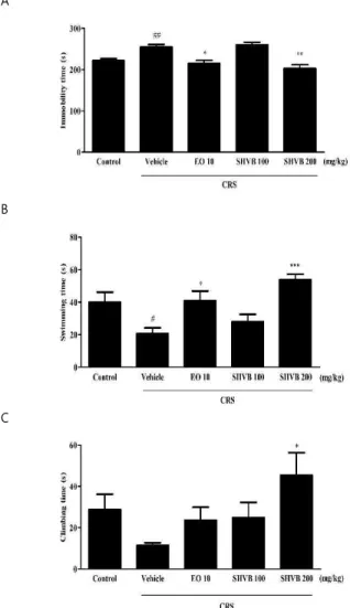
In order to investigate the effects of SHVB on the duration of immobility, swimming, and climbing behavior, we performed an FST. As shown in Fig. 1A, CRS-exposed mice showed a significant increase in the immobility time compared with the control group ( P < 0.01), while treatment with EO (10 mg/kg, positive control; P < 0.05) and SHVB (200 mg/kg, P < 0.01) significantly led to a decrease in the immobility time than the CRS group. In contrast, as shown in Fig. 1B and C, CRS-exposed mice showed a significant decrease in swimming ( P < 0.05) and climbing (not
significant) behavior compared with the control group.
However, SHVB treatment (200 mg/kg) significantly induced an increase in the climbing and swimming behavior ( P <
0.05 and P < 0.001, respectively) compared with the CRS group.
A
B
C
Fig. 1. Effects of SHVB treatment in the forced swim test (FST) in CRS-exposed mice. The effects of SHVB (100, and 200 mg/kg/day) on the duration of immobility (A), swimming (B), and climbing (C) behavior in the FST in CRS-induced mice. CRS, chronic restraint stress;
EO 10, escitalopram oxalate 10 mg/kg; SHVB 100, Stauntonia hexaphylla and Vaccinium bracteatum water extract 100 mg/kg; SHVB 200, Stauntonia hexaphylla and Vaccinium bracteatum water extract 200 mg/kg. The values are expressed as the mean ± standard error of the mean (n = 5).
#P < 0.05 and
##P < 0.01 compared with the control group;
*P < 0.05,
**P < 0.01, and
***P < 0.001 compared with the CRS group.
2. Effects of SHVB on the movement activity in CRS-exposed mice
The effects of SHVB (100 and 200 mg/kg/day) on the
movement activity are shown in Fig. 2. CRS-exposed mice
showed a significant decrease in the number of crossings
compared with the control group ( P < 0.01). However,
treatment with SHVB (100 and 200 mg/kg, P < 0.05, respectively) and EO (10 mg/kg, positive control, P < 0.01) showed higher locomotor activity compared with the CRS group.
Fig. 2. Effects of SHVB on locomotor activity in CRS-exposed mice.
CRS, chronic restraint stress; EO 10, escitalopram oxalate 10 mg/kg; SHVB 100, Stauntonia hexaphylla and Vaccinium bracteatum water extract 100 mg/kg; SHVB 200, Stauntonia hexaphylla and Vaccinium bracteatum water extract 200 mg/kg. The values are expressed as the mean ± standard error of the mean (n = 5).
##P < 0.01 compared with the control group;
*P <
0.05, and
**P < 0.01 compared with the CRS group.
3. Effects of SHVB on serum CORT levels in CRS-exposed mice
As shown in Fig. 3, the levels of serum CORT significantly increased in CRS-exposed mice ( P < 0.05) compared with the control group. However, SHVB (100 and 200 mg/kg) treatment decreased levels of CORT ( P < 0.05, respectively) levels than the CRS group.
Fig. 3. Effects of SHVB on serum CORT levels in CRS-exposed mice.
The levels of CORT were determined by ELISA kit (Abnova Corp.). CORT, corticosterone; CRS, chronic restraint stress; EO 10, escitalopram oxalate 10 mg/kg; SHVB 100, Stauntonia hexaphylla and Vaccinium bracteatum water extract 100 mg/kg; SHVB 200, Stauntonia hexaphylla and Vaccinium bracteatum water extract 200 mg/kg. The values are expressed as the mean ± standard error of the mean (n = 5).
#P < 0.05 compared with the control group;
*P < 0.05 compared with the CRS group.
4. Effects of SHVB on monoamine neurotransmitter levels in CRS-exposed mice
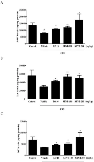
The levels of 5-HT, DA, and NE in the PFC when mice were exposed to CRS for 21 days are shown in Fig. 4. The
5-HT ( P < 0.05), DA (not significant), and NE (not significant) levels in the PFC were decreased compared with those in the control group. In contrast, daily administration of SHVB (100 and 200 mg/kg) significantly increased the 5-HT ( P < 0.01 and P < 0.05, respectively) (Fig. 4A), DA ( P <
0.01 and P < 0.05, respectively) (Fig. 4B), and NE ( P < 0.05, respectively) (Fig. 4C) levels in the PFC compared with those in the CRS group. Moreover, EO (10 mg/kg) also increased the 5-HT ( P < 0.05), DA ( P < 0.05), and NE ( P < 0.05) levels in the PFC compared with those in the CRS group.
A
B
C
Fig. 4. Effects of SHVB on monoamine neurotransmitters levels in CRS-exposed mice: (A) 5-HT, (B) DA, and (C) NE levels in the PFC.
CRS, chronic restraint stress; DA, dopamine; EO 10, escitalopram oxalate 10 mg/kg; 5-HT, serotonin; NE, norepinephrine; PFC, prefrontal cortex; SHVB 100, Stauntonia hexaphylla and Vaccinium bracteatum water extract 100 mg/kg; SHVB 200, Stauntonia hexaphylla and Vaccinium bracteatum water extract 200 mg/kg. The values are expressed as the mean ± standard error of the mean (n = 5).
#P < 0.05 compared with the control group;
*P <
0.05, and
**P < 0.01 compared with the CRS group.
5. Effects of SHVB on pentobarbital sodium-induced sleep in mice
After hypnotic pentobarbital (45 mg/kg, i.p.) injection,
the latency of sleep and total sleep time were observed, and the results are shown in Fig. 5. Treatment with SHVB (200 mg/kg/day, 163.14 ± 4.17 s, P < 0.05) led to significantly lower sleep latency compared with the control group (326.08
± 26.12 s) (Fig. 5A). In addition, treatment with SHVB (100 and 200 mg/kg/day, 6760.12 ± 299.90 s and 7890.08 ± 911.73 s, P < 0.05, respectively) led to a significantly higher total sleep duration compared with the control group (3188.51 ± 301.79 s) (Fig. 5B). Similarly, the DZP (diazepam, 2 mg/kg) treated mice also showed a decrease in the sleep latency and an increase in the total sleep duration compared with the control group (147.66 ± 3.13 s and 13770.33 ± 2897.43 s, P < 0.01 and P < 0.05, respectively).
A
B
Fig. 5. Effects of SHVB on the onset and duration of sleep in pentobarbital-treated mice. The sleep latency (A) and total sleeping time (B) were measured. DZP 2, diazepam 2 mg/kg; SHVB 100, Stauntonia hexaphylla and Vaccinium bracteatum water extract 100 mg/kg; SHVB 200, Stauntonia hexaphylla and Vaccinium bracteatum water extract 200 mg/kg.
The values are expressed as the mean ± standard error of the mean (n = 5).
*P < 0.05 and
**P < 0.01 compared with the control group.
6. Effects of SHVB on ERK1/2 pathways in 5-HT
2Areceptor expressed in CHO-K1 cells
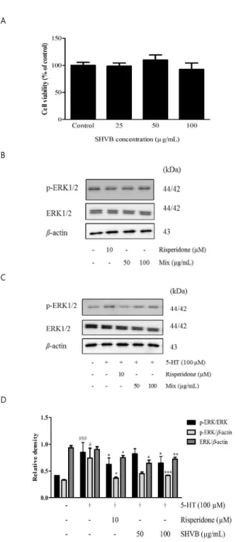
We next investigated whether the ERK1/2 phosphorylation pathway in CHO-K1 cells transfected with the human 5-HT
2Areceptor was involved in the antidepressant-like effects caused by SHVB. We measured the cytotoxicity of SHVB using the MTT assay. The results indicated that SHVB at concentrations of 25, 50, and 100 µg/mL did not alter the viability of CHO-K1 cells (Fig. 6A).
As shown in Fig. 6B, treatment with either the 5-HT
2Areceptor antagonist, risperidone, (10 µM) or SHVB (50 and 100 µg/mL) did not affect ERK1/2 phosphorylation at 30 min. As shown in Fig. 6C and D, treatment with 5-HT (100 µM) for 15 min increased ERK1/2 phosphorylation, while pre-treatment with SHVB (50 and 100 µg/mL) or risperidone (10 µM) decreased 5-HT-mediated ERK1/2 phosphorylation.
A
B
C
D
Fig. 6. Effects of SHVB on ERK1/2 phosphorylation in CHO-K1 cells
transfected with the human 5-HT
2Areceptor. (A) CHO-K1 cells were
treated with SHVB for 24 h and cell viability was determined by MTT
assay. (B) CHO-K1 cells transfected with the human 5-HT
2Areceptor were
treated with SHVB (50 or 100 µg/mL) or risperidone (5-HT
2Areceptor
antagonist, 10 µM) for 30 min. (C) For the evaluation of antagonistic
effects, CHO-K1 cells transfected with the human 5-HT
2Areceptor were
treated with 5-HT (100 µM) for 15 min after the pre-treatment with SHVB
(50 or 100 µg/mL) or risperidone (10 µM) for 15 min. (D) Quantitative analysis of relative ERK1/2 phosphorylation. 5-HT
2A, serotonin receptor subunit 2A; CHO-K1, Chinese Hamster Ovary K1 cells; ERK, extracellular signal-regulated kinases; SHVB, Stauntonia hexaphylla and Vaccinium bracteatum water extract. The values are expressed as the mean ± standard error of the mean (n = 3).
#P < 0.05 and
###P < 0.001 compared with the control group;
*P < 0.05,
**P < 0.01 and
***P < 0.001 compared with the 5-HT group.
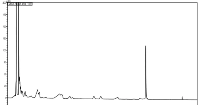
7. HPLC Analysis of the SHVB
We identified the SHVB by HPLC analysis. An illustration of the retention times is shown in Fig. 7.
0.0 5.0 10.0 15.0 20.0 25.0 30.0 35.0 40.0 45.0 50.0 55.0 min
0 25 50 75 100 125 150 175 200mAU
Extract-340nm,4nm (1.00)