울금으로부터 식품부패미생물에 대한 항균성 물질의 분리 및 동정
최 해 연
숙명여자대학교 식품영양학과
Isolation and Identification of Antimicrobial Compound from UlGeum (Curcuma longa L.)
Hae-Yeon Choi
Dept. of Food and Nutrition, Sookmyung Women's University, Seoul 140-742, Korea
Abstract
Antimicrobial activity of UlGeum (Curcuma longa L.) was investigated. Methanol extract of dried UlGeum was fractionated to hexane, chloroform, ethyl acetate, butanol and water fraction. The antimicrobial activity of five crude fractions were examined using impregnated paper disk agar diffusion. Ethyl acetate fraction showed the highest inhibitory effect on the microorganisms such as B. subtilis, S. aureus, E. coli, L. monocytogenes andV. parahaemolyticus at 1,000 μg/disc. Ethyl acetate fraction was further fractionated by silica gel column and thin layer chromatography (TLC). The antimicrobial compound was isolated from their fractions and its chemical structure was identified as a 2,3-dihydrobenzofuran by GC-MS and 1H-NMR.
Key words: Curcuma longa L., UlGeum, antimicrobial activity, isolation, identification
E-mail: [email protected]
Phone: 82-2-710-9471, Fax: 82-2-710-9479
서 론
천연물에 존재하는 항균성 물질에 대한 연구는 국내외에 서 오래 전부터 시도되고 있다. 특히 천연 보존료 개발의 일환으로 허브류와 향신료의 항균성에 관한 연구가 많이 이 루어지고 있는데 천연물의 항균성은 오래 전부터 알려져 왔 고, 이를 이용하여 식품이나 인체에서 발생하는 기생성 진균 류의 억제에 관한 연구가 활발히 이루어지고 있다(1). 이 중 천연색소들은 오랫동안 사용되어 안전하다고 간주된 것들 로 대부분 항산화, 항염증효과 등의 다양한 생리활성이 있는 것으로 알려져 있어 그 기능적 가치가 더욱 높아지고 있다.
식물색소의 자원이 되는 것은 감귤, 꼭두서니, 잇꽃, 지치, 치자, 황금, 단삼, 사프란, 울금 등이다(2). 우리나라의 천연 항균제 개발에 관한 연구로는 마늘, 양파, 백리향(3-5), 정향, 계피(6) 등의 향신료, 목단피(7), 단삼(8), 홍경천(9), 상백피 (10) 등의 약용식물, 미역(11), 지충이(12) 등의 해양자원 등 이 있으며, 이런 생리활성을 지닌 식품소재를 추출하거나 그대로 떡이나 국수 등 식품에 첨가하여 저장성을 연장하는 등 항균효과를 검색하는 연구도 진행되고 있다(13-17).
울금(Curcuma longaL.)은 생강과에 속하는 다년생 초본 으로 카레 등의 노란색을 나타내는 향신료의 성분이며 이들 성분들은 해독작용이 있어 전통적으로 염증치료의 약용식
물로 사용되어 왔고, 열대 및 아열대 지방에서 재배하며 남 아시아와 동남아시아에서 재배되어 왔다(18). 울금의 근경 을 수확하여 겉껍질을 벗기고 삶아서 말린 다음 가루로 만든 것이 turmeric이라고 하는 향신료로 카레분말의 주원료이다 (19). 울금에는 황색 색소로서 curcumin이 0.3% 들어있고 특유한 향이 있는 정유로써 turmerone과 dehydroturmerone 이 1~5% 들어있다. 울금의 주요성분은 curcumin 이외에 demethoxycurcumin, bisdemethoxycurcumin, cyclocur- cumin, calebin 등이 존재하며, 식물성 sterols 및 정유성분인 β-sitosterol, zingiberene, campesterol, stigmasterol, mono- 및 di-enoic acid, tumerone, zingiberone, borneol, eugenol, camphor, curdion, α-phellandrene, cineol 등이 전 체 4.2~4.5% 내외로 포함되어 있다(20-23). 울금의 생리활 성에 관한 연구를 보면 curcuminoids의 항산화작용(24), curcumin의 항산화․항암성․항돌연변이성․항염증(18,25) 및 항균성(26), tumerone 및ar-tumerone의 항뱀독성(27) 효과 등이 보고되었다.
본 연구에서는 천연식품 보존제 개발의 일환으로 천연색 소자원인 울금을 메탄올로 추출하여 여러 용매로 분획하여 식품부패미생물의 증식억제 효과를 검색하고, 항균성을 나 타내는 물질을 분리, 동정하였다.
재료 및 방법 실험재료
본 실험에 사용한 울금은 충남 공주산으로 건조된 상태로 구입하여 blender(FM-680w, HANIL Co., Wom Joo, Korea) 로 분쇄 후, 50 mesh 체에 내려 폴리에틸렌 백에 넣어 -40oC deep freezer에 보관하면서 사용하였다.
시험균주 및 배양
본 연구에 사용한 균주는 자연계에 널리 분포하여 식품을 변질시키는 유포자세균인 Bacillus subtilisKCTC 1021, 저 온에서도 생육하여 냉동, 냉장 식품에서 감염형 식중독의 원인이 되는 Listeria monocytogenesKCCM 40307, enter- otoxin을 생성하여 식중독의 원인이 되는 Staphylococcus aureusKCTC 1916, Gram 음성균으로 오염의 지표균이면 서 부패세균인 Escherichia coliKCTC 2441, 그리고 호염성 Gram 음성균으로 장염의 원인균이며 감염형 식중독을 일으 키는Vibrio parahaemolyticusKCTC 2471을 한국생명공학 연구원에서 분양을 받아 사용하였다.
배지는 tryptic soy broth(Difco, Detroit, USA)와 nutrient agar(Difco)를 사용하였고 V. parahaemolyticus는 위와 같 은 배지에 식염(NaCl)을 3%가 되도록 첨가하여 사용하였 다.B. subtilis는 TSB agar 배지로 30oC 16시간, 나머지 균 주는 Nutrient agar(Difco) 배지로 37oC에서 16시간동안 균 주의 생육특성에 맞추어 배양하였다.
울금 추출물의 항균성 검색
울금의 항균활성을 알아보기 위하여 분말화한 울금을 80oC에서 3시간 동안 메탄올로 3회 반복 추출하여 여과, 농 축 후 메탄올 추출물을 얻었다. 메탄올 추출물의 항균성 검 색은 액체배지 희석법(28)으로 하였는데 사면배지에 배양한 균주를 1백금이 취해 TSB(Tryptic soy broth, Difco)가 10 mL가 든 시험관에 접종하고 37oC에서 8시간 배양하여 UV/VIS Spectrophotometer(Jasco V-530, Tokyo, Japan) 를 이용하여 660 nm에서 O.D.값이 0.2가 되도록 희석하였 다. TSB에는 울금의 methanol 추출물을 500~2,000 μg/mL 가 되게 첨가한 후 균주를 접종하여 24시간 동안 37oC in- cubator(IB-05, Jeio Tech, Seoul, Korea)에서 배양시켜 660 nm에서 흡광도를 측정하여 실시하여 다음 식에 의해 억제 효과를 산출하였다.
% Inhibitory effect=
[{(control-control blank)-(treatment-treatment blank)}/
(control-control blank)]×100 울금의 용매 분획별 항균성 검색
각 계통분획물의 항균성 검색은 paper disc법(29)으로 하 였다. 시험용 평판배지는 nutrient agar를 멸균 후 직경 9 cm인 petri dish에 15 mL씩 분주하여 clean bench에서 하룻
밤 건조시키고 그 위에 각 균주의 배양액 100 μL를 도말하였 다. 각 용매 분획별 분획물의 농도는 500~2,000 μg/disc로 하였다. 이를 멸균된 disc(직경 8 mm, Toyo Seisakusho Co., Tokyo, Japan)에 흡수, 건조시켜 균주가 도말된 plate 표면에 올려놓은 후 37oC의 incubator에서 24시간 배양하여 disc 주 위에 생성된 clear zone의 직경으로 항균활성을 측정하였다.
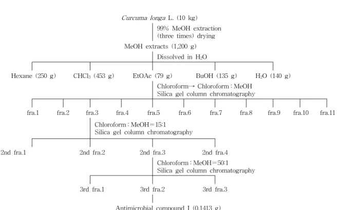
울금의 추출 및 분획
예비 실험에서 항균활성을 보인 분말화한 울금 10 kg을 중량대비로 10배의 메탄올로 80oC 수욕상에서 환류냉각하 면서 3시간씩 2회 반복추출하고, 감압장치(EYELA N-N, Rikakikai Co., Tokyo, Japan)를 이용하여 여과 및 농축한 후 메탄올 추출물 1,200 g을 얻었다. 메탄올 추출물을 증류 수에 1:1의 비율로 현탁한 후 Fig. 1과 같이n-hexane을 가 하여 분획한 후 여과, 감압 농축하여 분획물 250 g을 얻었다.
이와 같은 방법으로 chloroform, ethyl acetate, n-butanol 및 물로 극성이 낮은 용매에서 극성이 높은 용매로 순차적 으로 계통분획하여 chloroform 분획물 453 g, ethyl acetate 분획물 79 g,n-butanol 분획물 135 g 그리고 물 분획물 140 g을 얻었다.
Ethyl acetate 분획으로부터 항균성 물질의 분리
울금의 ethyl acetate fraction을 silica gel column chro- matography(10×120 cm)를 이용하여 chloroform→chloro- form : methanol 순으로 단계적으로 극성을 높여가면서 col- umn chromatography를 실시하였고 TLC로 monitoring 하 면서 11개의 소분획(sub-fraction)을 얻었다(Fig. 1). 11개의 fraction을 다시 상기 5종의 시험균주를 이용하여 항균활성 을 측정한 후 항균성이 높게 나타난 sub-fraction 3을 다시 silica gel chromatography와 TLC를 이용하여 chloroform : methanol=15:1 용매로 시작하여 극성을 높여가며 분리하 여 4개의 2차 소분획(2nd sub-fraction)을 얻었고, 이를 다시 항균활성 시험을 하여 항균성이 높게 나타난 2nd sub-frac- tion 3을 chloroform : methanol=50:1 용매로 시작하여 반복 분리하여 3차 소분획(3rd sub-fraction) 3개를 얻었으며, 이 중 3rd sub-fraction 2에서 항균성 물질을 얻었다. 추출과 silica gel column chromatography용 용매는 시약용 1급을 사용하였고 column chromatography용 silica gel을 Kieselgel 60(70-230 mesh, art. 7734, Merck, Darm-stadt, Germany) 을, TLC plate는 Kieselgel 6060 F254(art. 5715, Merck)를 사용하였다.
항균성 물질의 동정
항균성이 높게 나타난 물질은 GC-MS와1H-NMR을 통 하여 물질을 구명하였으며 동정된 물질을 paper disc 법으로 5균주에 대해 항균성을 실험하였다. Mass spectrum(MS)은 Hewlett-Packard 6890 gas chromatography와 Hewlett- Packard 5793 MSD를 사용하여 다음과 같은 조건으로 분석
Curcuma longa L. (10 kg) 99% MeOH extraction (three times) drying MeOH extracts (1,200 g)
Dissolved in H2O
Hexane (250 g) CHCl3(453 g) EtOAc (79 g) BuOH (135 g) H2O (140 g) Chloroform→ Chloroform : MeOH
Silica gel column chromatography
fra.1 fra.2 fra.3 fra.4 fra.5 fra.6 fra.7 fra.8 fra.9 fra.10 fra.11 Chloroform : MeOH=15:1
Silica gel column chromatography
2nd fra.1 2nd fra.2 2nd fra.3 2nd fra.4
Chloroform : MeOH=50:1
Silica gel column chromatography
3rd fra.1 3rd fra.2 3rd fra.3
Antimicrobial compound I (0.1413 g)
Fig. 1. Fractionation and isolation procedures of antimicrobial substance from Curcuma longa L.
Table 1. Antimicrobial activity of methanol extract from Curcuma longa L. against various microorganisms Conc.
(μg/mL) Antimicrobial activity (%)
B. subtilis S. aureus L. monocytogenes E. coli V. parahaemolyticus 1,000500
1,500 2,000
89.91 77.85 86.24 86.63
87.83 93.05 100100
50.96 35.78 37.86 46.17
94.95 100100 100
100100 100100 하였다. Column은 HP5-Ms(30 m×250 μm×0.25 μm), col-
umn 온도는 100oC에서 2분간 유지시킨 후 10oC/min으로 승 온하여 280oC, 10 min 조건으로 분석하였다. Injector 온도는 280oC, detector 온도는 280oC, carrier gas는 He(1.0 mL/
min)을 사용하였다. 1H-NMR은 Varian model UI 500 spec- trometer(Varian Inc., Melbourne, Australia)를 이용하여 300 MHz에서 측정하였다.
결과 및 고찰 울금 추출물의 항균성
울금을 건조시켜 분쇄한 후 methanol로 추출한 것을 10%
농도로 희석하여 500, 1,000, 1,500, 2,000 μg/mL씩 첨가하여 식품부패미생물의 증식억제 효과를 검색한 결과는 Table 1 과 같다. 울금의 methanol 추출물은 500 μg/mL 농도에서 V. parahaemolyticus를, 1,000 μg/mL 농도에서E. coli 증식 을 완전히 억제하였고, S. aureus는 1,500 μg/mL 농도에서 완전히 억제되었지만 B. subtilis와 L. monocytogenes는 2,000 μg/mL 농도에서 각각 86.63%, 46.17% 억제되었다.
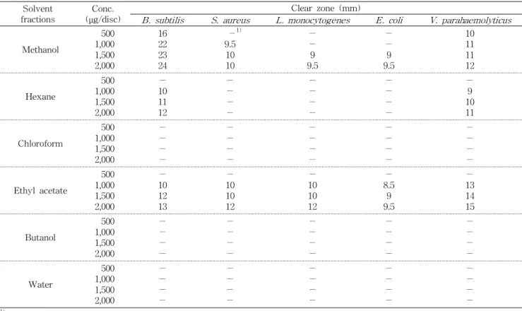
울금 추출물의 분획별 항균성
항균성을 나타낸 울금 methanol 추출물의 항균성 물질을 분리할 목적으로 n-hexane, chloroform, ethyl acetate, n-butanol 및 물 순으로 비극성에서 극성으로 용매를 바꾸 어 순차적으로 분획하여 paper disc 법으로 항균성을 검색한 결과는 Table 2와 같다. 울금의 ethyl acetate 분획 추출물은 낮은 농도인 1,000 μg/disc의 농도에서 식품 부패미생물 5종 모두에 대하여 8.5~13 mm의 clear zone을 형성하였고 2,000 μg/disc 농도에서는 B. subtilis,S. aureus,L. mono- cytogenes,V. parahaemolyticus는 각각 13, 12, 12, 15 mm의 clear zone을 형성하였다. 또한 hexane 분획도B. subtilis와 V. parahaemolyticus균에 대해서는 1,000 μg/disc 농도에서 각각 10, 9 mm의 clear zone을 형성하였다. 그러므로 울금의 항균 효과를 추출 용매별로 살펴보면 ethyl acetate 층이 가 장 우수하고 그 다음이 hexane 층의 순으로 나타났다. 이와 같은 결과는 각 용매 분획 시 용매에 따라 항균성 물질이 용해되어 나타나는 것으로 생각되며 특히, 울금의 항균 물질 은 주로 ethyl acetate 분획에서 모든 균주들에 대해 광범위 한 clear zone을 형성하는 것으로 보아 항균물질은 ethyl
Table 2. Antimicrobial activity of solvent fractions from Curcuma longa L. against various microorganisms Solvent
fractions Conc.
(μg/disc) Clear zone (mm)
B. subtilis S. aureus L. monocytogenes E. coli V. parahaemolyticus
Methanol
1,000500 1,500 2,000
1622 2324
-1) 9.510
10
-- 9.59
-- 9.59
1011 1112
Hexane
1,000500 1,500 2,000
-10 1112
--
--
--
--
--
--
-9 1011
Chloroform
1,000500 1,500 2,000
--
--
--
--
--
--
--
--
--
--
Ethyl acetate
1,000500 1,500 2,000
-10 1213
-10 1012
10- 1012
8.5- 9.59
-13 1415
Butanol
1,000500 1,500 2,000
--
--
--
--
--
--
--
--
--
--
Water
1,000500 1,500 2,000
--
--
--
--
--
--
--
--
--
--
1)No activity.
acetate에 잘 용해되는 물질이고 극성에 가까운 것으로 사료 된다.
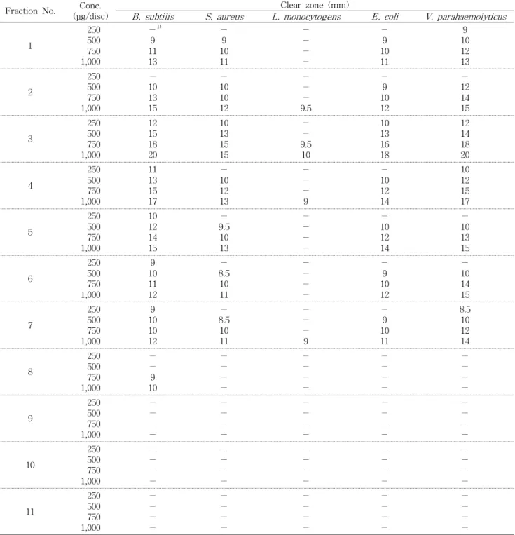
Silica gel chromatography와 TLC에 의한 항균물질 분리
울금의 ethyl acetate층 분획물을 silica gel column chro- matography(10×120 cm)한 후 TLC에 전개하여 Fig. 1과 같이 11개의 sub-fraction을 얻었고 그에 대한 항균성은 Table 3과 같다. 각 분획물의 농도가 250, 500, 750, 1,000 μg/disc가 되도록 paper disc에 첨가한 후 5종의 공시 균주를 대상으로 항균력을 검색하였다. 11개의 분획물중 분획물 3, 4가 모든 공시 균주에 대하여 clear zone이 9.5~20 mm의 clear zone을 형성하여 항균성이 우수하게 나타났다. 500 μg /disc 농도에서 분획물 3과 4에서 B. subtilis의 경우 각각 15, 13 mm, S. aureus의 경우 13, 10 mm, E. coli의 경우 13, 10 mm의 clear zone을 형성하였고,V. parahaemolyticus 는 각각 14, 12 mm의 clear zone을 형성하였다. 그러나 L.
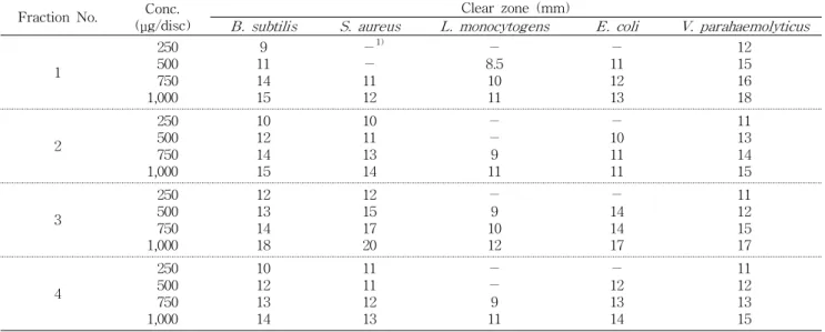
monocytogenes의 경우는 750 μg/disc 농도에서 fraction 3 이 9.5 mm의 clear zone을 형성하여 다소 약한 항균력을 보였다. 따라서 어떤 fraction보다도 높은 항균력을 보인 sub-fraction 3을 다시 silica gel column chromatography (5×75 cm)와 TLC monitoring을 이용하여 4종의 2nd sub-fraction 분획물을 얻었고 이에 대한 항균성 실험을 한 결과는 Table 4와 같다. 이 중에서 항균력이 우수한 2nd
sub-fraction 3은 250 μg/disc 농도에서B. subtilis, S. aur- eus 및V. parahaemolyticus에 대해 각각 12, 12, 11 mm의 clear zone을 형성하였다. 2nd sub-fraction 3을 다시 silica gel column chromatography(2×60 cm)와 TLC monitoring 을 이용하여 항균성물질을 분리하였다.
항균성 물질의 구조 결정
울금의 methanol 추출물로부터 용매별로 계통 분획하여 분리한 ethyl acetate 분획으로부터 silica gel column chro- matography와 TLC monitoring하여 항균성이 가장 우수한 yellow oil의 항균성 물질을 얻었고 이를 다시 GC-MS로 분 석한 결과 spectrum 상에서 m/z 120 mass unit에서 분자 이온 peak가 관측되었다. 이를 다시 확인하기 위하여
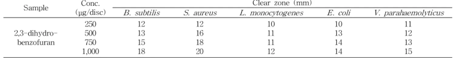
1H-NMR로 분석한 결과 화학식이 C8H8O이며 분자량이 120.154의 2,3-dihydrobenzofuran(coumaran)으로 동정되 었으며(Fig. 2, 3),1H-NMR의 data는 다음과 같다.1H-NMR (CD3OD, δ, 300MHz): 7.59(d, 1H, J=16Hz), 7.50(d, 1H, J=16Hz), 6.86(d, 1H, J=16Hz), 6.61(d, 1H, J=16Hz) 4.87(d, 2H, J=2.7Hz), 3.37(d, 2H, J=2.7Hz). 동정된 2,3-dihydro- benzofuran을 5균주에 대해 항균성을 실험한 결과 모든 균 에 대하여 250 μg/disc 농도에서 10~12 mm의 clear zone을 형성하여 우수한 항균성을 보였다(Table 5). 2,3-dihydro- benzofuran은 다양한 natural product의 근간이 되는 화합물 중의 하나이며 2,3-dihydrobenzofuran 유도체들이 식물로
Table 3. Antimicrobial activity of the first ethyl acetate fractions from methanol extract ofCurcuma longa L. against various microorganisms
Fraction No. Conc.
(μg/disc) Clear zone (mm)
B. subtilis S. aureus L. monocytogens E. coli V. parahaemolyticus
1
250500 1,000750
-1) 119 13
-9 1011
--
--
-9 1011
109 1213
2
250 500750 1,000
- 1013 15
- 1010 12
-
-- 9.5
- 109 12
- 1214 15
3
250500 1,000750
1215 1820
1013 1515
-- 9.510
1013 1618
1214 1820
4
250 500750 1,000
11 1315 17
- 1012 13
-
-- 9
- 1012 14
10 1215 17
5
250500 750 1,000
1012 14 15
9.5- 10 13
--
-
-
-10 12 14
-10 13 15
6
250 500750 1,000
9 1011 12
- 8.510 11
-
--
-
- 109 12
- 1014 15
7
250500 1,000750
109 1012
8.5- 1011
--
-9
-9 1011
8.510 1214
8
250 500750 1,000
-
-9 10
-
--
-
-
--
-
-
--
-
-
--
-
9
250500 1,000750
--
--
--
--
--
--
--
--
--
--
10
250 500750 1,000
-
--
-
-
--
-
-
--
-
-
--
-
-
--
-
11
250500 750 1,000
--
-
-
--
-
-
--
-
-
--
-
-
--
-
-
1)No activity.
부터 추출되어 다양한 약재료 사용되고 있으며 식물로부터 추출된 일부 2,3-dihydrobenzofuran 유도체들은 그 구조식 이 확인되어 화학적으로 합성되어 다양하게 연구되고 있다 (30-34). Ahn과 Obendorf(35)는 Curcuma longa L.에서 GC-MS로 curcumin, curcumene, ferulonylmethane, cou- maran, vanillin, zingiberene 등을 분리하였다고 보고하여 울금에 coumaran이 함유되어 있음을 확인하였고, Bajpai 등
(36)은Silene armeriaL.로부터 essential oil을 추출하여 그 성분을 분리하였는데 methylamine 21.48%, β-butene 17.97%, α-butene 46.40%, coumaran 0.22%, eugenol 0.21%
등 28종류로 구성되어 있었으며, 이 essential oil을B. sub- tilis, L. monocytogenes,S. aureus,Peudomonas aerugi- nosa, Salmonella Typhimurium, E. coli, E. coli-O157:H, Enterobacter aerogenes 등 식품부패 미생물을 대상으로
Table 4. Antimicrobial activity of the second ethyl acetate fractions from methanol extract ofCurcuma longa L. against various microorganisms
Fraction No. Conc.
(μg/disc) Clear zone (mm)
B. subtilis S. aureus L. monocytogens E. coli V. parahaemolyticus
1
250500 1,000750
119 1415
-1)
-11 12
8.5- 1011
-11 1213
1215 1618
2
250 500750 1,000
10 1214 15
10 1113 14
-
-9 11
- 1011 11
11 1314 15
3
250500 1,000750
1213 1418
1215 1720
-9 1012
-14 1417
1112 1517
4
250 500750 1,000
10 1213 14
11 1112 13
-
-9 11
- 1213 14
11 1213 15
1)No activity.
Fig. 2. Scan mass spectrum of antimicrobial compound from Curcuma longa L. in GC/MS.
Fig. 3.1H NMR spectrum of antimicrobial com- pound fromCurcuma longa L. (CD3OD, 300 MHz).
항균효과를 살펴보았는데 MIC가 125~2,000 μg/mL 수준으 로 항균력을 나타내었다고 보고하였으며, Lim과 Kim(37)도 식품부패미생물을 대상으로 쇠비름의 메탄올 추출물의 항 균활성을 살펴보고 성분분석을 한 결과 furan계에서 2,3-di-
hydrobenzofuran이 분리되었고 추출물의 6.13%정도 함유 하여 항균력에 영향을 주었다고 보고하였다. Morimoto 등 (38)도Cyperusspp.로부터 coumaran을 분리하여 살충효과 가 있다고 보고하였다.
Table 5. Effect of antibiotic substances from Curcuma longa L. on the growth of various microorganisms
Sample Conc.
(μg/disc) Clear zone (mm)
B. subtilis S. aureus L. monocytogenes E. coli V. parahaemolyticus 2,3-dihydro-
benzofuran
250500 1,000750
1213 1518
1216 1820
1011 1112
1013 1414
1112 1315
요 약
울금 분말을 methanol로 추출하여 여러 용매로 분획하여 식품부패미생물의 증식억제 효과를 검색하고 그 항균성 물 질을 분리하였다. 울금의 ethyl acetate 분획 추출물은 낮은 농도인 1,000 μg/disc의 농도에서 B. subtilis, S. aureus, L.
monocytogenes,E. coli, V. parahaemolyticus에 대하여 8.5
~13 mm의 clear zone을 형성하여 우수한 항균성을 나타내 었다. 울금의 ethyl acetate 추출물을 silica gel column chro- matography와 TLC로 monitoring 하여 항균성을 실험하였 다. 이 결과 우수한 항균성을 보인 sub fraction을 재차 분리 하여 yellow oil의 항균성물질을 얻었고 이를 GC/MS,
1H-NMR로 구조 분석한 결과 화학식이 C8H8O이며 분자량 이 120.154의 2,3-dihydrobenzofuran(coumaran)으로 동정 되었다.
문 헌
1. Shin DW, Kim MS, Han JS. 1997. Antimicrobial effect of ethanol extracts from some medicinal herbs and their frac- tionates against food-borne bacteria. Korean J Food Sci Tecnol 29: 808-816.
2. 지형준. 1997. 천연식물과 식용색소. 식품과 기술 10: 55-61.
3. Buchanan RL, Shepherd AJ. 1981. Inhibition ofAspergillus parasiticus by thymol. J Food Sci 46: 976-977.
4. Yin MC, Cheng WS. 1998. Inhibition ofAspergillus niger andAspergillus flavusby some herbs and spices.J Food Prot 61: 123-125.
5. Montes-Belmont R, Carvajal M. 1998. Control of Aspergillus flavusin maize with plant essential oil and their components. J Food Prot 61: 616-619.
6. Lee YK. 1995. Identification and antimicrobial activity of cinnamon and clove extracts on food spoilage microorgan- isms. PhD Dissertation. Sookmyung Women's Uniersity, Seoul.
7. Hwang JS, Han YS. 2003. Isolation and identification of an- timicrobial compound from Mordan bark (Paeonia suf- fruticosa ANDR). J Korean Soc Food Sci Nutr 32: 1059- 1065.
8. Choi HY, Han YS. 2003. Isolation and identification of anti- microbial compound from Dansam (Salivia miltiorrhiza Bunge). J Food Sci Nutr 32: 22-28.
9. Sim CJ, Lee GH, Jung JH, Yi SD, Kim YH, Oh MJ. 2004.
Isolation and identification of antimicrobial activity sub- stances from Rhodiola sachlinensis.Kor J Food Preserv 11: 63-70.
10. Park UY, Kim SH, Kim JH, Kim YG, Chang DS. 1995.
Purification of antimicrobial substance for the extract from the root bark ofMorus alba.J Fd Hyf Safety10: 225-230.
11. Yun SM, Jang JH, Lee JS. 2007. Isolation and identification of an antibacterial substance from sea mustard,Undaria pinnatifida, forStreptococcus mutans.J Korean Soc Food Sci Nutr 36: 149-154.
12. Lee SY, Song EJ, Kim KBWR, Yoon SY, Kim SJ, Lee SJ, Hong YK, Lim SM, Ahn DH. 2009. Antimicrobial activity of ethanol extract from Sargassum thunbergii. J Korean Soc Food Sci Nutr 38: 502-508.
13. Kim IH. 1990. The status of Korean food additives pro- duction usage and foreign countries.J Korean Soc Food Nutr 19: 519-528.
14. Lee KA. 1999. Effect of wild plants addition on the shelf-life and characteristics of rice cake. MS Thesis. Sookmyung Women's University, Seoul. p 53-61.
15. Kim SJ. 1998. Inhibitory effect of green laver on the growth of food spoilage microorganism and identification of anti- microbial compounds. MS Thesis. Sookmyung Women's University, Seoul.
16. Kim SI, Han YS. 1997. Isolation and identification of anti- microbial compound from Sancho (Zanthoxylum Schinifol- ium). Korean J Soc Food Sci 13: 56-63.
17. Kim KH. 1999. Isolation and identification of antimicrobial compounds from danelions and plantains and their effects when added to processed foodstuffs. PhD Dissertation. Sookmyung Women's University, Seoul. p 90-98.
18. Kang WS, Kim JH, Park EJ, Yoon KR. 1998. Antioxidative property of turmeric (Curcuma Rhizoma) ethanol extract.
Korean J Food Sci Technol 30: 226-271.
19. Kim KS, Choung MG, Park SH. 2005. Quantitative determi- nation and stability of curcuminoid pigment from turmeric (Curcuma longaL.) root.Korean J Crop Sci50: 211-215.
20. Geoffrey NR, Amitahb C, Muraleedharan GN. 1998. Novel bioactivities of Curcuma longa constituents. J Nat Prod 61: 542-545.
21. Andrew MA, Matthew SM, Ram SM. 2000. Isolation of curcuma from tumeric. J Chem Educ 77: 359-362.
22. An BJ, Lee JY, Park TS, Pyeon JR, Bae JH, Song MA, Baek EJ, Park JM, Son JH, Lee CE, Choi IK. 2006.
Antioxidant activity and whitening effect of extraction con- dition inCurcuma longa L.Korean J Medicinal Crop Sci 14: 168-172.
23. Ryu GY, No KH, Ryu SR, Yang HS. 2005. Study of separa- tion and analysis method an effective component from UlGeum (Curcuma longa) and a contained curcumin as product of national and partial region cultures.Appl Chem 9: 57-60.
24. Masuda T, Isobe T, Jito A, Nakatani N. 1992. Antioxidative curcuminoids from rhizomes of Curcuma xanthorrhiza. J Phytochem 31: 3645-3649.
25. Russell LR. 1988. High performance liquid chromatographic separation and spectral characterization of the pigments in tumeric and anatto. J Food Sci 53: 1823-1826.
26. Negi PS, Jayaprakasha GK, Jagan Mohan Rao L, Sakariah KK. 1999. Antibacterial activity of tumeric oil: A byproduct from curcumin. Agric Food Chem 47: 4297-4301.
27. Ferreira LAF, Henriques OB, Andreoni AAS, Vital GRF, Campos MMC, Harbermehl GG. 1992. Antivenom and bio- logical effects ofar-tumerone isolated fromCurcuma longa zingiberaceae. Toxicon 30: 1211-1218.
28. Speck ML. 1984.Compendium of methods for the micro- biological examination of foods. 2nd ed. American Public Health Association, Washington, DC, USA.
29. Davidson PM, Parish ME. 1989. Methods for testing the efficacy of food antimicrobials.Food Technol43: 148-155.
30. Kokubun T, Harborne JB, Eagles J, Waterman PG. 1995.
Dibenzofuran phytoalexins from the sapwood of coto- neaster acutifolius and five related species. J Phytochem 38: 57-60.
31. Mastelic J, Jerkovic I, Mesic M. 2006. Volatile constituents from flowers, leaves, bark and wood of Prunus mahaleb L. J Flavour Fragr 21: 306-313.
32. Frattini C, Bicchi C, Nano GM. 1977. Volatile flavor compo- nents of licorice. J Agric Food Chem 25: 1238-1242.
33. Lee HJ, Park SY, Lee OK, Jo Hj, Kang HY, Choi DH, Khan M. 2008. Benzofurans and sterol from the seeds ofStyrax
obassia. Chem Nat Comp 44: 435-439.
34. Codagan JIG, John B, MacDonald JF, Rhodes PH. 1996.
Dictionary of organic compounds. 6th ed. Champman &
Hall, New York, USA. Vol 3, p 2216.
35. Ahn CS, Obendorf SK. 2006. GC-MS analysis of dyes ex- tracted from turmaric. Fibers and Polymers 7: 158-163.
36. Bajpai VK, Dung NT, Kwon OJ, Kang SC. 2008. Analysis and the potential applications of essential oil and leaf ex- tracts of Silene armeria L. to control food spoilage and food-borne pathogens.Eur Food Res Technol227: 1613- 1620.
37. Lim MK, Kim LM. 2001. Antimicrobial activity of methanol extract from Soibirhym (Portulace oleracea) against food spoilage or foodborne disease microorganisms and the composition of the extract. Korean J Soc Food Cookery Sci 17: 565-570.
38. Morimoto M, Fujii Y, Komai K. 1999. Antifeedants in Cyperaceas: coumaran and quinones fromCyperusspp.J Phytochem 51: 605-608.
(2009년 6월 29일 접수; 2009년 8월 10일 채택)