대두추출물을 처리한 조골세포 조건배양액은 RANKL에 의해 유도된 파골세포 분화를 억제
박경호1*․주원철1*․여주홍2․이광길2․조윤희1†
1
경희대학교 동서의학대학원 의학영양학과
2
농촌진흥청 농업생물부
Conditioned Medium of Soybean Extract Treated Osteoblasts Inhibits RANKL Induced Differentiation of Osteoclasts
Kyungho Park1*, Won-Chul Ju1*, Joo-Hong Yeo2, Kwang-Gill Lee2, and Yunhi Cho1†
1Dept. of Medical Nutrition, Graduate School of East-West Medical Science, Kyung Hee University, Gyeonggi 446-701, Korea
2Dept. of Agricultural Biology, National Academy of Agricultural Science, Gyeonggi 441-100, Korea
Abstract
Soybean is of particular interest as a food supplement of isoflavones for inhibiting bone resorption in postmenopausal woman. These beneficial effects of isoflavones are caused by functioning as partial agonists or antagonists of estrogen, of which anti-resorptive effect is mediated indirectly through paracrine factors produced by osteoblasts that act on osteoclasts. In this study, the indirect effect of soybean on osteoclastic differentiation of RAW264.7 cells were investigated. The conditioned medium was collected from MC3T3-E1 osbeoblasts treated with 0.001 mg/mL~0.1 mg/mL soybean extracts for 6 days, mixed in 1:1 ratio with osteoclast medium, and then added into RAW264.7 cells with receptor activator of nuclear factor kappa B ligand (RANKL), a differentiation inducer for 3 days. Of paracrine factors in the conditioned medium, the protein expression of osteoprotegerin (OPG) with soybean extract was specifically higher in a dose dependent manner than with 10
-9M~10
-6M of estrogen, genistein or daidzein standards. In RAW264.7 cells, the conditioned medium with soybean inhibited RANKL induced osteoclastic differentiation as total number of multinucleated tartrate- resistant alkaline phosphatase (TRAP)-positive osteoclasts and protein expression of MMP-9 were significantly decreased. Coupled with the low expression of estrogen receptor α and β proteins in RANKL treated RAW264.7 cells, we demonstrate that the conditioned medium of soybean treated osteoblasts inhibits RANKL induced differentiation of osteoclasts with the selective expression of OPG in osteoblasts.
Key words: soybean extract, conditioned medium of osteoblasts, RANKL, OPG, osteoclastic differentiation
*
K. P. and W. J. contributed equally to this study.
†
Corresponding author. E-mail: [email protected]
†
Phone: 82-31-201-3817, Fax: 82-31-201-3817
서 론
골 항상성은 골 흡수에 관여하는 파골세포와 골 형성에 관여하는 조골세포의 긴밀한 상호작용에 의해 유지된다. 파 골전구세포로부터 파골세포로의 분화과정에 조골세포가 관 여함은 여러 보고를 통해 잘 알려져 있다(1). 파골세포분화 는 조골세포에서 발현되는 파골세포 분화유도인자인 re- ceptor activator of nuclear factor-κB lignad(RANKL)와 파 골전구세포에서 발현되는 그 수용체 RANK의 결합에 의해 시작되며, 체내 여러 종류의 전신성 호르몬 및 조골세포에서 생성·분비되는 다양한 국소인자에 의해 조절된다(2,3). 특히, 여성의 골 대사 균형 유지에 중요한 역할을 하는 것으로 알
려진 여성호르몬인 에스트로겐은 세포막 혹은 핵막에 존재 하는 그 수용체(Estrogen Receptor: ER)와 결합 후 파골세 포의 분화를 조절할 수 있는 것으로 보고되어 있다(4). 또한, 조골세포에서 분비되는 국소인자 중 monocyte-macro- phage colony-stimuating factor(M-CSF), tumor necrosis factor(TNF)-α, interleukin(IL)-1 그리고 IL-6 등이 파골세 포의 분화 조절에 관여하는데, 에스트로겐은 이들 인자의 발현감소를 유도하여 파골세포분화를 억제할 수 있다(5,6).
최근 RANKL의 유도인자로서 많은 연구가 되고 있는 os- teoprotegerin(OPG) 역시 에스트로겐에 의해 그 발현증가가 보고되었는데, 이는 RANKL과 직접 결합함으로써 RANKL/
RANK의 결합을 저해하여 파골세포 분화를 강력히 억제하
는 것으로 알려져 있다(7).
상기의 이유로 폐경 후 여성의 에스트로겐 분비 저하에 의한 골량 감소 및 골다공증의 치료는 에스트로겐 투여가 기본적으로 사용되고 있다(8). 이 치료법은 폐경의 기간이나 나이에 관계없이 골량 증가의 효과가 있고 척추 및 고관절 골절의 예방 효과가 뛰어난 것으로 알려져 있으나(9), 장기적 인 에스트로겐의 사용은 오심, 두통, 체중 증가, 유방통, 불규 칙한 자궁 출혈 등의 부작용을 비롯한 자궁내막암 및 유방암 발생을 초래함이 보고되어 있어 장기적인 사용을 기피하는 실정이다(10). 이에 에스트로겐 투여에 의한 위험성을 보완 하기 위해 식품 및 천연물의 활성 성분을 이용한 대체 요법으 로 식물성 에스트로겐에 대한 기능성 연구가 요구되고 있다.
식물성 에스트로겐은 isoflavones, cumestanes, lignans의 3계열로 분류되며 이중 isoflavone은 골 대사와 관련하여 활 발한 연구가 되고 있는 대표적인 식물성 에스트로겐이다 (11). 대두를 비롯한 콩류에 다량함유되어 있는 isoflavone은 비배당체인 genistein, daidzein, glycitin과 이들에 당이 결합 된 배당체 그리고 acetyl 및 malonyl 배당체 각각 3가지 총 12종류가 밝혀져 있다(11,12). 이중 genistein과 daidzein은 에스트로겐과 유사한 화학적 구조에 기인하여 세포내에서 ER과 결합하여 에스트로겐 효과뿐 아니라 항 에스트로겐 효과를 동시에 가지는 선택적 에스트로겐 길항제의 기능을 하는 것으로 알려져 있다(11). 본 연구의 선행연구에서 대두 추출물에 함유된 isoflavone인 genistein 및 daidzein의 함유 량을 분석하였으며 MG-63 조골세포에 대두추출물처리 시 세포증식 및 관련인자의 발현이 증가됨을 확인하였다(13).
본 선행연구를 비롯하여 isoflavone과 이를 다량 함유한 대 두에 대한 연구가 식이 첨가를 통한 임상영양 연구뿐 아니라 난소 절제 동물 모델을 이용하여 활발히 진행되고 있으나 (10,12,13), 폐경 후 골량 감소 및 골다공증의 발병은 조골세 포보다는 파골세포의 상대적인 작용 증가에 기인됨에도 불 구하고 골세포 수준에서의 이소플라본 및 대두 추출물의 연 구는 주로 조골세포에서 이루어져, 파골세포를 이용한 이들 의 골다공증 예방의 효과 및 관련연구는 미흡한 실정이다.
또한 에스트로겐의 영향으로 조골세포에서 분비되는 다양 한 국소인자 역시 파골세포분화에 관여한다는 보고에 근거 하여 파골세포로의 분화관련 연구 시 조골세포에 의한 간접 적인 효과를 살펴보는 것은 매우 중요할 것으로 여겨진다.
이에 본 연구에서는 대두추출물을 처리한 MC3T3-E1 조 골세포 조건배양액에서 다양한 파골세포 분화관련인자의 발현변화를 확인하고, 그 조건배양액을 RAW264.7 파골전 구세포에 처리 시 파골세포 분화를 억제하는 지를 알아보고 자 하였다.
재료 및 방법
재료 및 시약
본 연구에 사용한 RAW264.7 세포와 MC3T3-E1 세포는
American Type Culture Collection(ATCC, Manassas, USA)에서 분양 받아 본 연구실에서 계대 배양하여 사용하 였다. α-Minimal essential medium(α-MEM), penicillin- streptomycin(5000 U/mL penicillin; 5000 μg/mL strepto- mycin) 및 fetal bovine serum(FBS)은 Gibco®사(CA, USA)에서 구입하였다. 파골세포 분화 유도물질인 Recom- binant murine RANKL은 Peprotech Asia Inc.(NJ, USA)로 부터 구입하였고, 표준물질인 17β-estradiol(E
2), genistein, daidzein과 조골세포분화를 위한 비타민 C(ascorbic acid), β -glycerophosphate 및 파골세포 염색시약으로 leukocyte acid phosphatase staining kit 387-A를 Sigma Chemical Co.(MO, USA)로부터 구입하였다. 그리고 1차 항체 (anti-rabbit ER-α, ER-β, IL-1β, IL-6, TNF-α, RANKL, β -actin anti-goat M-CSF, OPG)는 Abcam(MA, USA)에 서 구입하였다. 그 외 다른 시약은 (주)KDR(Seoul, Korea) 에서 구입하여 별다른 정제과정 없이 그대로 사용하였다.
대두 추출물 시료 준비
대두는 경기도 양주산을 구입하여 약 1 kg을 5배가량의 70% 메탄올로 추출하여 여과하고, 여액을 감압 농축한 후 72시간 동결 건조한 것을 대두 메탄올 추출물(0.69% iso- flavone 함유)로 하였다. 대두 메탄올 추출물은 dime- thylsulfoxide(DMSO, Sigma, 최종 농도<0.05%, v/v)에 녹 이고 filter(0.22 mm pore size, Millipore, Bedford, USA)로 여과 멸균하여 사용하였다. 대두 추출물의 농도는 사전 연구 에서 사용한 0.001 및 0.1 mg/mL로 하여 실험하였다. 양성 대조군으로 10
-9~10
-6M 농도의 17-β estradiol(E2), genis- tein, daidzein 단일 표준물질을 DMSO에 녹여 사용하였다.
각 표준물질의 농도는 하루에 50 mg의 isoflavone을 식품으 로 섭취하고 있는 성인 혈중 isflavone 농도인 50~800 ng/
mL(0.2~3×10
-6M)과 성인 여성혈중 에스트로겐 농도인 40~80 pg/mL(0.15~0.3×10
-9M)을 기준으로 하였다.
조골세포와 파골세포의 기본배양 및 분화
MC3T3-E1 조골전구세포와 RAW264.7 파골전구세포는 polystyrene 세포배양접시에 부착시키고 penicillin 및 streptomycin이 함유된 1% antibacterial-antifungal sol- ution(Gibco)과 10% FBS(Gibco)를 첨가한 α-MEM(Gibco) 을 사용하여 5% CO
2와 95% 습도가 유지되는 37
oC in- cubator에서 기본 배양하였다. MC3T3-E1 세포의 조골세포 로의 분화는 이들 세포가 단층을 형성한 후 50 μg/mL 비타 민 C(ascorbic acid)와 10 mM β-glycerophosphate처리에 의해 유도하였으며(14), RAW264.7 세포의 파골세포로의 분 화는 30 ng/mL recombinant murine RANKL 처리에 의해 유도하였다(15).
TRAP(tartrate-resistant acid phosphate) 염색
파골세포의 분화 정도를 Hotokezaka 등(16)의 방법에 따
라 TRAP 염색으로 확인하였다. 각 1×10
5개의 RAW264.7
파골전구세포를 6well plate에 분주하고 30 ng/mL 농도의 RANKL 및 대두 추출물을 비롯한 표준물질을 처리한 조골 세포의 조건 배양액을 처리하여 3일간 배양하였다. 배지를 제거한 후 citrate-acetone 및 formaldehyde 혼합 용액으로 고정한 세포를 leukocyte acid phosphatase staining kit 387-A(Sigma)를 이용하여 염색하였다. 우선, fast garnet GBC base/sodium nitrite(1:1, v/v)를 처리하여 실온에서 3 분간 반응하고, 기질용액(2.5 mM naphthol AS-BI phos- phate, 100 mM acetate solution, pH 5.0 and 50 mM tartrate solution)을 100 μL/well 분주하여 37
oC에서 15~30분간 빛 이 차단된 상태에서 반응시켰다. 이후 TRAP 염색된 거대 다핵의 파골세포는 현미경으로 관찰하였다.
Western blot analysis
조골세포에서 합성된 후 분비되어 파골세포분화 조절에 관여하는 여러 국소 인자들(IL-1β, IL-6, TNF-α, M-CSF, RANKL, OPG) 및 Estrogen Receptor(ER)-α, ER-β의 단백 질 수준의 발현을 western blotting에 의해 확인하였다. 대두 추출물을 비롯한 각 시료를 처리한 조골세포와 조골세포 조 건배양액을 3일간 처리한 파골세포에 1 mL의 RIPA buf- fer[20 mM Tris, pH 8, 150 mM NaCl, 10 mM sodium phos- phate, 100 μM sodium vanadate, 100 μM ammonium mo- lybdate, 10% glycerol, 0.1% nonidet P-40, and 0.1% SDS, and 1×protease inhibitors(Roche, Indianapolis, USA)]를 첨가한 후 초음파 파쇄기를 이용하여 5초 동안 세포벽을 파 괴시킨 다음 16시간 동안 -20
oC에서 보관하였다. 해동 후 그 균질액을 1500×
g에서 20분간 원심분리 하여 상층액을 단백질추출액으로 준비하였다. 또한 조골세포 조건배양액 에서 파골세포관련 국소인자의 발현을 위해 centrifugal fil- ter column(Millipore’s Amicon®, Houston, USA)을 사용하 여 조건배양액을 농축하였다. 단백질 정량은 bovine serum albumin을 표준으로 하여 bio-rad protein assay를 이용하 여 595 nm에서 흡광도를 측정하여 결정하였다. 정량된 단백 질(25 μg/lane)을 10~12% polyacrylamide/SDS gel에서 전 기영동 시킨 후 Hybond ECL nitrocellulose membrane에 흡착시켰다. 5% skim milk으로 blocking한 membrane을 0.05% Tween-20을 함유한 PBST에 각각의 1차 항체를 희 석시켜서 4
oC에서 16시간 동안 반응시켰다. PBST로 5분씩 3차례 세척한 후 PBST에 1:10000배로 희석시킨 perox- idase-conjugated anti-IgG 2차 항체와 실온에서 1시간 동 안 반응시킨 후 PBST를 사용하여 다시 5분씩 3차례 세척하 였다. 발색은 ECL heperfilm으로 확인하였고(17) imaging densitometer(Bioimaging systems ver. 4.6, UVP Inc., Upland, USA)를 사용하여 정량하였다.
통계분석
실험 결과의 자료는 SPSS(Ver 12.0 program, Statistical package for social science)을 이용하여 통계처리 하였다.
결과는 각 군별 평균과 표준오차로 나타내었고 각 실험군 간의 비교는 one way ANOVA로 분석한 후 general linear model(GLM) test로 p<0.05 수준에서 검증하였다.
결과 및 고찰
조골세포 및 파골세포에서 에스트로겐 수용체(Estrogen receptor: ER)의 발현
MC3T3-E1 조골세포와 RAW264.7 파골세포에서 에스트 로겐 수용체(ER-α, ER-β)의 발현을 확인하여 Fig. 1에 제시 하였다. 비타민 C(50 μg/mL)와 β-glycerophosphate(10 mM) 처리에 의해 조골세포로 분화된 MC3T3-E1세포에서 ER-α와 ER-β 모두 발현된 반면(Fig. 1A), RAW264.7 파골 세포에서는 두 수용체 모두 발현되지 않았다(Fig. 1B). 이는 MC3T3-E1, MG-63, SaOS-2 등의 다양한 조골세포에서 ER이 발현되나, 파골세포에는 발현되지 않음을 보고한 여러 사전연구와 일치하는 결과로서(18,19), 에스트로겐 및 식물 성 에스트로겐의 그 수용체와 결합 후 파골세포분화 조절은 주로 조골세포를 통해 간접적으로 이루어지는 것으로 여겨 진다. Srivastava 등(20)의 연구를 비롯한 일부 연구에서 파 골세포에서 ER의 발현됨을 보고하였으나 그 발현 정도가 극히 미미한 수준으로 에스트로겐 및 식물성 에스트로겐의 파골세포 분화억제 효과는 에스트로겐 수용체와의 결합에 의한 효과보다는 tyrosine kinase 등의 신호 전달체계에서 mitogenic activity와 연관된 다양한 매개체들의 활성 및 발 현 억제 그리고 성숙한 파골세포의 세포사멸기전에 의한 것 으로 보고하였다(20,21).
A. +C -C E9 E6 G9 G6 D9 D6 S0.001 S0.1 ER-α
ER-β β-actin
B. +R -R E9 E6 G9 G6 D9 D6 S0.001 S0.1 ER-α
ER-β β-actin
Fig. 1. Expression of ERα and ERβ in MC3T3-E1 cells treat- ed with E
2, isoflavones or soybean and RAW264.7 cells with the conditioned medium of MC3T3-E1 cells. A. Protein extract (25 μg/lane) from control (-C: negative control, +C: positive con- trol), E
2(E9: 10
-9M, E6: 10
-6M), daidzein (D9: 10
-9M, D6: 10
-6M), genistein (G9: 10
-9M, G6: 10
-6M), or soybean (S0.001: 0.001 mg/mL, S0.1: 0.1 mg/mL) treated MC3T3-E1 cells were sub- jected to 10~12% SDS-PAGE and immunoblotting with ERα, ER β or β-actin. B. RANKL-nonstimulated (-R) or stimulated (+R) RAW264.7 cells with conditioned medium of MC3T3-E1 osteo- blasts treated with E
2(E9: 10
-9M, E6: 10
-6M), daidzein (D9: 10
-9M, D6: 10
-6M), genistein (G9: 10
-9M, G6: 10
-6M), or soybean (S0.001: 0.001 mg/mL, S0.1: 0.1 mg/mL) were subjected to 10~
12% SDS-PAGE and immunoblotting with ERα, ERβ or β-actin.
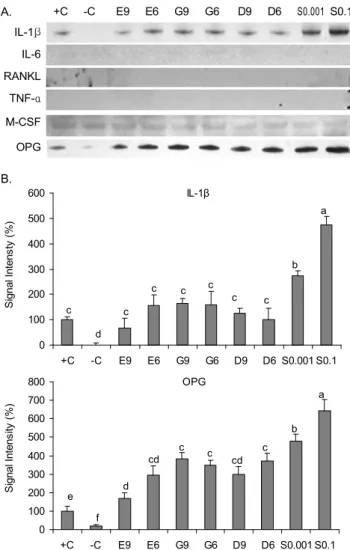
대두추출물을 처리한 조골세포 조건배양액에서 파골세포 분화관련인자의 발현
조골세포에서 생성, 분비되어 파골세포 분화조절에 관여 하는 국소인자인 IL-1β, IL-6, RANKL, TNF-α, M-CSF 및 OPG의 단백질수준의 발현을 조골세포로 분화된 MC3T3-E1세포(양성대조군, +C)를 기준하여 % control로 나타내었다(Fig. 2). IL-6, RANKL 및 TNF-α는 본 연구에 서 발현되지 않았으며, M-CSF발현 역시 모든 처리 군에서
A. +C -C E9 E6 G9 G6 D9 D6 S0.001 S0.1 IL-1β
IL-6 RANKL
TNF-α M-CSF
OPG
B.
IL-1β
0 100 200 300 400 500 600
+C -C E9 E6 G9 G6 D9 D6 S0.001 S0.1
S ig n a l In te n s ty ( % )
OPG
0 100 200 300 400 500 600 700 800
+C -C E9 E6 G9 G6 D9 D6 S0.001 S0.1
S ig n a l In te n s it y ( % )
e f
d cd
c c cd
c b
a c
d c
c c c
c c b
a
Fig. 2. Expression of osteoclastic paracrine factors in the conditioned medium of MC3T3-E1 cells treated with E
2, iso- flavones or soybean. A: Protein extract (25 μg/lane) from con- trol (-C: negative control, +C: positive control), E
2(E9: 10
-9M, E6: 10
-6M), daidzein (D9: 10
-9M, D6: 10
-6M), genistein (G9:
10
-9M, G6: 10
-6M), or soybean (S0.001: 0.001 mg/mL, S0.1: 0.1 mg/mL) treated MC3T3-E1 cells were subjected to 10~12%
SDS-PAGE and immunoblotting with IL-1β, IL-6, TNF-α, M-CSF, RANKL or OPG specific antibodies (abcam Inc.) The region of each gel shown was 31 kDa (IL-1β), 24 kDa (IL-6), 35 kDa (RANKL), 17 kDa (TNF-α), 60 kDa (M-CSF), or 48 kDa (OPG) prestained molecular markers. B: The signal intensities from multiple experiments of A were quantified and the integrated areas were first normalized to the corresponding value of β-actin and then to the signal observed in positive control cells (+C).
Values are mean±SD (n=3). Means with different letters differ at p<0.05.
유사하였다(Fig. 2A). 여러 국소인자 중 파골세포분화를 강 력히 촉진하는 것으로 알려진(22) IL-1β의 발현은 조골세포 로 분화되지 않은 MC3T3-E1세포(음성대조군, -C)에서 거 의 발현되지 않은 반면 양성대조군(+C)에서는 발현되었다.
이는 Khosla 등(23)이 조골 전구세포에서 IL-1β, IL-6, TNF-α, M-CSF, RANKL 등의 파골세포 분화관련 국소인 자의 미비한 수준의 발현이 조골세포로 분화됨에 따라 증가 함을 보고한 연구결과와 상응하는 결과이다. 조골세포로의 분화유도와 함께 10
-9~10
-6M 농도의 E
2, genistein, daid- zein 등의 표준단일물질 처리 시 ~163.8%까지 IL-1β의 발 현을 증가시켰다. 0.001 mg/mL(~273.8%)~0.1 mg/mL(~
475.5%)의 대두추출물 처리 시 농도 의존적으로 그 발현이 증가하였으며, 증가 수준은 E
2를 비롯한 다른 표준단일물질 에 비해 현저하였다(Fig. 2). 조골세포에서 생성, 분비되는 다양한 파골세포 분화관련인자는 파골세포의 분화를 촉진 하는 paracrine 효과뿐 아니라 조골세포의 분화를 촉진하는 autocrine 효과를 동시에 지니고 있다(22). 다른 처리에 비해 대두추출물 처리 시 농도 의존적으로 증가된 IL-1β의 분비 는 조골세포의 분화를 촉진하여 어느 정도 골 형성을 자극할 수 있으나, 다른 국소인자에 비해 강력히 파골세포분화를 자극한다는 보고(6,24)에 기인하여 파골세포 분화촉진에 더 욱 관여할 것으로 여겨진다. OPG는 조골세포에서 생성, 분 비되는 RANKL의 수용성 유도인자로서, RANKL의 수용체 인 RANK와 직접 결합함으로써 RANKL/RANK 결합을 저 해하여 조골세포에서 파골세포로의 분화를 억제하는 주요 인자로 알려져 있다(6,7,22). IL-1β의 발현양상과 유사하게 조골세포로 분화하지 않은 음성 대조군(-C)에서 OPG 발현 은 조골세포로 분화 유도된 양성대조군(+C)의 17.2% 수준 으로 현저히 낮았다. 조골세포로의 분화 유도와 함께 E
2를 비롯한 다른 표준단일물질 처리 시 그 발현을 ~382.3%까지 증가하였고, 0.001 mg/mL 및 0.1 mg/mL의 대두출물처리 시 그 발현은 각 ~479.0%, ~643.4%로 농도 의존적으로 현 저히 증가하였다(Fig. 2). 이는 대두에 다량 함유되어 있는 genistein, daidzein 등을 비롯한 여러 isoflavone 혼합체 간 의 시너지효과에 의한 것으로 생각되어진다.
Tartrate-resistant acid phosphatase staining(TRAP) 염색에 의한 파골세포 분화정도 확인
RAW264.7 파골전구세포에 기존의 분화유도인자(25)인
RANKL 및 대두추출물을 처리한 조골세포조건배양액
(conditioned medium: CM)이 파골세포 분화에 미치는 효과
를 TRAP 염색을 통해 확인하였다 (Fig. 3). TRAP 효소의
활성은 파골세포가 골 흡수 작용을 할 때 증가하는 것으로
보고(26)되었는데, 파골세포는 골 내막에 위치하며 골 조직
에 존재하는 유일한 다핵세포로서 RANKL에 의한 분화초
기 단계에는 단핵의 파골전구세포를 형성하지만 이후 세포
가 유합되어 다핵의 성숙 파골세포로 분화되면 골 표면에
+R -R E9 E6
G9 G6 D9 D6
S0.001 S0.1
Fig. 3. Soybean suppresses the formation of TRAP-positive giant multinucleated cells in RANKL-stimulated RAW264.7 cells. RANKL-nonstimulated (-R) or stimulated (+R) RAW264.7 cells with conditioned medium of MC3T3-E1 osteoblasts treated with E
2(E9: 10
-9M, E6: 10
-6M), daidzein (D9: 10
-9M, D6: 10
-6M), genistein (G9: 10
-9M, G6: 10
-6M), or soybean (S0.001: 0.001 mg/mL, S0.1: 0.1 mg/mL) for 3 days were fixed and stained with TRAP staining kit.
부착되어 주름 막을 형성하여 TRAP 염색 시 보라색으로 염색되는 특징을 가진다(27). 예비실험을 통해 결정된 농도 인 30 ng/mL의 RANKL 처리에 의해 파골세포로 분화된 RAW264.7세포(양성대조군: +R, Fig. 3)에서 파골세포의 특 징인 다핵이 관찰되었으며, 주름 막을 형성하여 파골세포의 화학적 표지효소인 TRAP 염색 시 보라색으로 염색되었다.
그러나 RANKL을 처리하지 않은 일반 RAW264.7 세포(음 성대조군: -R, Fig. 3)는 파골세포의 특징인 다핵과 주름 막 을 형성하지 않았다. 이는 여러 사전연구(25-27)와 일치하는 결과로서 RANKL은 파골세포 분화에 필수물질임을 재확인 하였다. RANKL처리에 의한 파골세포 분화와 함께 10
-9~ 10
-6M 농도의 E
2, genistein, daidzein 등의 표준단일물질 및 저 농도의 대두추출물(0.001 mg/mL)을 처리한 CM처리 시 일부 단핵의 파골전구세포는 확인되었으나, 다핵의 파골 세포와 주름 막은 형성되지 않았다. 그러나 0.1 mg/mL의 고농도 대두추출물 처리 시 양성대조군(+R)의 수준은 아니 지만 일부 다핵의 파골세포와 주름막이 관찰되었다. 이는 저 농도 대두추출물에 의한 파골세포분화 억제효과가 고농 도 대두추출물에 비해 현저함을 의미한다.
RANKL 처리에 의해 파골세포로 유도된 RAW264.7세 포에서 파골세포분화 관련지표의 발현
대두추출물의 파골세포분화 억제정도를 RANKL처리한 RAW264.7세포에서 파골세포분화 관련지표로 알려진 ma- trix metalloproteinase(MMP)-9, cathepsin(Cat)-K 및 cal- citonin receptor(CTR)의 발현을 통해 확인하였다(22)(Fig.
4). Cat-K와 CTR의 발현은 처리 군 간의 차이를 보이지 않아 본 연구에 적절한 분화 maker로서 사용되지 못하였다.
반면, E
2, genistein, daidzein 등의 표준단일물질 및 대두추
출물을 처리한 CM처리 시 MMP-9(upper band: pro-form,
bottom band: active-form)의 발현은 양성대조군에 비해 현
저히 감소하였다. 그러나 대두추출물을 처리한 CM처리 시
MMP-9 발현(pro-form: active-form)의 감소된 수준은 고
농도(26.7%: 16.4%)가 저농도(17.2%: 5.0%)에 미치지 못하
였다. 이 결과는 표준단일물질 및 저농도의 대두추출물을
처리한 CM처리 시 TRAP염색된 파골세포형성이 고농도의
대두추출물에 비해 현저히 감소한 것(Fig. 3)과 상응하는 결
과로서, 고농도의 대두추출물(S0.1)을 조골세포에 처리 시
파골세포 분화억제에 관여하는 OPG의 발현(S0.1: 643.4%)
+R -R E9 E6 G9 G6 D9 D6 S0.001 S0.1 MMP-9
Cat-K CTR β-actin
←pro-form
←active-form (100, 100) (17.2, 5.0) (26.7, 16.4)
Fig. 4. Expression of osteoclastic differentiation markers in RANKL-stimulated RAW264.7 cells. Protein extract (25 μg/lane) from RANKL-stimulated (+R) or non-stimulated (-R) RAW264.7 cells with conditioned medium of MC3T3-E1 osteoblasts treated with E
2(E9: 10
-9M, E6: 10
-6M), genistein (G9: 10
-9M, G6: 10
-6M), daidzein (D9: 10
-9M, D6: 10
-6M), or soybean (S0.001: 0.001 mg/mL, S0.1: 0.1 mg/mL) were subjected to 10~12% SDS-PAGE and immunoblotting with MMP-9, Cat-K, CTR or β-actin specific antibodies (abcam Inc.). The region of each gel shown was 88~92 kDa (pro-form or active-form of MMP-9) between the 27 and 39 kDa (Cat-K), 56.9 kDa (CTR) or the 45 kDa (β-actin) prestained molecular markers. The numerical values below each lane represent relative signal intensities of MMP-9 (pro-form and active-form, respectively), which were quantified and the integrated areas were first normalized to the corresponding value of β-actin and then to the signal observed in positive control cells (+R: 100, 100). The experiments were performed in triplicate.
을 유의적으로 증가시켰으나 동시에 파골세포 분화를 강력 히 촉진하는 IL-1β의 발현 증가수준(S0.1: 475.5%)이 저 농 도(S0.001: 273.8%)의 약 1.7배로 OPG(저농도의 1.3배) 발현 증가수준 이상으로 그 발현이 높았기 때문으로 사료된다.
반면, 저 농도의 대두추출물(S0.001)을 처리한 CM 처리 시 TRAP 염색된 파골세포(Fig. 3) 및 MMP-9의 발현(Fig. 4) 이 표준단일물질 수준으로 억제된 것은 표준단일물질 수준 이상으로 증가된 OPG의 발현과 고농도의 대두추출물 수준 에 크게 미치지 못하는 IL-1β의 발현양상에 의한 것으로 여겨진다. 본 연구의 조골세포조건배양액에서 IL-1β 및 OPG의 발현결과는 양성대조군(+C)에 대비한 상대적인 발 현양상을 확인한 것으로서 추후 각 인자의 절대적인 수치를 확인 및 어느 수준 이상의 IL-1β 및 OPG가 파골세포분화를 각각 촉진, 억제할 수 있는지를 검증할 필요가 있다고 여겨 진다. 다만 저 농도의 식물성에스트로겐에 함유되어 있는 isoflavone은 에스트로겐 효과를 나타내지만 고농도의 식물 성에스트로겐에 함유되어 있는 isoflavone은 항 에스트로겐 효과를 나타내는 등의 biphasic한 효과를 보고한 여러 사전 연구(13,28)와 유사하게 본 연구에서도 저 농도의 대두추출 물을 처리한 조골세포 조건배양액이 파골세포분화억제에 고농도의 대두추출물에 비해 효과적임을 제시한다. 또한 본 연구결과를 기초로 대두추출물의 간접적인 처리에 의한 파 골세포분화억제 관련기전을 세포수준에서의 확인이 추후 연구를 통해 심도 있게 이루어져야 할 것으로 사료된다.
요 약
본 연구는 MC3T3-E1 조골 전구세포에 대두추출물을 처 리 후 그 조건배양액에서 파골세포분화 관련 국소인자 중 파골세포분화 촉진인자(IL-β, IL-6, RANKL, TNF-α, M-CSF) 및 파골세포로의 분화억제에 관여하는 국소인자 인 OPG의 발현변화를 조골세포 조건배양액에서 살펴보았
으며, 이 조골세포 조건배양액을 RAW264.7 파골전구세포 에 처리 시 파골세포로의 분화를 억제정도를 TRAP 염색 및 관련 분화 지표의 발현을 통해 알아보았다. 대두추출물 처리한 조골세포 조건배양액에서 OPG의 발현이 농도 의존 적으로 현저히 증가하였다. 그러나 강력한 파골세포 분화촉 진인자로 알려진 IL-1β의 발현 역시 고농도의 대두추출물 처리 시 현저히 증가하여 고농도의 대두추출물을 처리한 CM 처리 시 TRAP 염색된 일부 파골세포를 확인하였고, 파골세포분화 관련지표인 MMP-9의 발현 또한 저농도 대두 추출물을 처리한 CM 처리에 비해 증가하였다. 이는 저농도 의 대두추출물이 고농도의 대두추출물에 비해 파골세포분 화억제에 효과적임을 의미하나, 추후 파골세포수준에서 분 화억제 관련 기전연구가 필요할 것으로 생각되어진다.
감사의 글
이 연구는 2006년도 경희대학교 연구비지원에 의한 결과 임(KHU-20060554, 공모과제).
문 헌