한국어병학회지 제23 권 제1 호(2010) J. Fish Pathol., 23(1)
:
47 ~ 55 (2010)47
치어기 넙치 사료내 Seleno-yeast 수준에 따른 조직학적 변화
배승철
�∙김영철∙이준호∙김재원
�부경대학교 해양바이오신소재학과 / 사료영양연구소,
�강원도립대학 해양생명과학과
Effects of dietary seleno-yeast levels on histological responses in juvenile olive flounder Paralichthys olivaceus
Sungchul C. Bai�, Young-Chul Kim, Jun-Ho Lee and Jae-Won Kim�
Dept. of Marine Bio-Materials and Aquaculture/FFNRC, Pukyong National University, Busan, 608-737, Korea
�Dept. of Marine life-Science, Gangwon Provincial College, Gangneung, Gangwondo, 210-804, Korea
This study was conducted to investigate the effects of dietary seleno-yeast on histological response in juvenile olive flounder Paralichthys olivaceus. Fish averaging 4.0 ± 0.1 g (mean ± SD) were fed one of the four semi-purified diets containing 0.56, 1.07, 2.86 and 4.56 mg Selenoyeast/kg diet (Se0.56, Se1.07, Se2.86
and Se4.56, respectively) in triplicates for 12 weeks. Swelling of blood cell in glomerulus, the rupture of some epithelial cell in the renal tubules and enlarged macrophage were observed in the Se1.07group. The hepatopancreas had hepatic cell, capillary and zymogen in the pancreas and was normal in the Se0.56group.
Swelling of hepatocyte gradually decreased with increase in selenoyeast supplementation. The gill lamellae showed partial abnormal condition (terminal clubbing) in the Se0.56group, but there were little differences between these and other treatments. These results indicated that the optimum dietary supplementation level of selenoyeast in juvenile olive flounder, Paralichthys olivaceus, could be less than 1.07 mg selenoyeast/kg diet based on histological responses of the fish.
Key words: Seleno-yeast, Olive flounder, Dietary additive, Histological response
셀레늄은 원소 주기율표상 황과 같은 6B족 물질로서 황과 화학적 특성이 매우 비슷한 준금 속으로서 공업적으로 화학촉매, 전기전자, 기계, 유리, 고무산업에 널리 사용되고 있다. 동물의 Se 결핍을 예방 또는 치료할 목적으로 Se 첨가제의 형태로 동물사료에 첨가되고 있으며, 미국 FDA 에서도 셀레늄의 사용을 일부 허용하고 있다. 하 지만 셀레늄은 생물의 항상성이 요구되는 것보 다 조금 높은 농도에서 중독이 되고 축적되는 물질로서 (Bowie et al., 1996) 어류에서 확인되는 셀레늄 영향은 새판 팽창, 간과 신장의 괴사 및
피부병변 등이 알려져 있어 (Lemly, 1997, 2002;
Lohner et al., 2001), 건강한 어류 생산은 모든 국 민의 건강을 위하여 적정 요구량이 필요하다. 현 재까지 연구된 셀레늄 요구량은 무지개 송어 (Hilton et al., 1980)와 채널메기(Gatlin and Wil- son, 1984)에 있어서 각각 0.38 및 0.25 mg Na
2SeO
3kg
-1diet가 적정 요구량으로 알려져 있 으며, 다금바리(Lin and Shiau, 2005)에 있어서 0.78 mg Se-Met kg
-1diet가 적정 요구량으로 보고 되어져 있다.
어류의 독성물질에 대한 민감성을 찾을 수 있
�Corresponding Author : Sungchul C. Bai, Tel : 051-629-5916 E-mail : [email protected]
5...
는 방법으로 생화학적 방법, 생리적 방법 및 조 직병리학적 방법 등이 있다. 생화학적이고 생리 적인 기법이 또한 독성에 대한 병리적인 변화를 결정하는 뚜렷한 방법이지만, 조직병리학적 방 법은 생체 내 실험에서 영향을 받는 특이한 표 적 기관들, 세포, 그리고 세포 소기관들의 변화 를 확인할 수 있다. 더군다나, 조직병리학은 그 들이 현장에서 나타난 것처럼 생화학적이고 생 리적인 변화를 보존하는 유일한 방법이다. 또한 현장 평가에서 조직병리학은 단기간 및 장기간 독성영향 모두 평가하는 가장 쉬운 방법이다. 동 물 조직의 조직병리학적 변화는 이전의 노출된 환경스트레스를 알 수 있는 강력한 지시자이며, 대표적 지시자는 간, 신장, 아가미 및 생식소 등 이다(Hinton and LaurÉn, 1990).
본 연구는 치어기 넙치를 대상으로 사료 내 유기 셀레늄원인 셀레노이스트에 직접적인 반 응을 나타낼 것으로 판단되는 기관계를 대상으 로 사료 내 적정 첨가 농도와 조직학적 반응 양 상을 확인하고자 하였다.
재료 및 방법
실험어 및 사육관리
실험어는 전남 무안에서 운반된 넙치를 부경 대학교 사료영양학실험실내 250 L수조에서 실 험환경에 적응할 수 있도록 2주간 예비 사육하 였다. 예비사육 후 실험어는 평균무게 4.0 ± 0.1 g (mean ± SD) 인 넙치를 30 L 사각수조에 각 실험구 당 각각 20마리씩 3반복으로 무작위 배 치하였다. 각 실험수조는 순환여과식으로 유수 량은 0.8 L/min으로 조절하였으며, 충분한 산소 공급을 위해 에어스톤을 설치하였다. 전 실험기 간 동안 평균 수온은 18 ± 1℃로 하였고 일일 사료공급량은 전 실험기간 동안 어체중의 3~4%(건물량 기준)로 1일 2회 매일 (10시와 16 시) 공급하였으며, 총 사육실험 기간은 12주간 실시하였다.
실험사료 및 실험설계
실험에 사용된 실험사료의 조성표와 일반성분 은 Table 1에 나타내었다. 실험사료의 단백질원 은 카제인과 어분, 탄수화물원은 밀가루, 덱스트 린 및 콘스타치, 지질원으로는 어유를 사용하였 다. 총 4개의 실험사료는 조단백 50%, 조지방 10%로 하였으며, 에너지는 15.8 kJ/g이었다. 그리
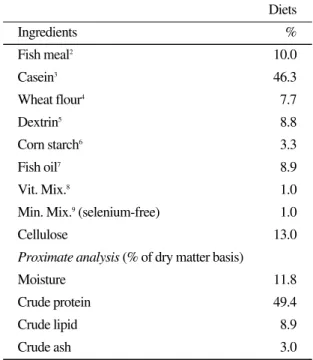
Table 1. Composition and proximate analysis of the basal diet (% of dry matter basis)1
Diets
Ingredients %
Fish meal2 10.0
Casein3 46.3
Wheat flour4 7.7
Dextrin5 8.8
Corn starch6 3.3
Fish oil7 8.9
Vit. Mix.8 1.0
Min. Mix.9(selenium-free) 1.0
Cellulose 13.0
Proximate analysis (% of dry matter basis)
Moisture 11.8
Crude protein 49.4
Crude lipid 8.9
Crude ash 3.0
1Feed stuffs not mentioned here are the same feed stuffs as the domestic aquaculture feed companies are using cur- rently.
2,3,4,5Suhyup feed Co. Uiryeong, Korea
6,7Jeil feed Co. Haman, Korea
8Contains (as mg/kg in diets) : Ascorbic acid, 300; dl-Calci- um pantothenate, 150 ; Choline bitatrate, 3000; Inositol, 150; Menadione, 6; Niacin, 150; Pyridoxine∙HCl, 15;
Riboflavin, 30; Thiamine mononitrate, 15; dl-α-Toco- pherol acetate, 201; Retinyl acetate, 6; Biotin, 1.5; Folic acid, 5.4; B12, 0.06
9Contains (as mg/kg in diets) : NaCl, 437.4; MgSO4∙ 7H2O. 1379.8; NaH2P4∙2H2O, 877.8; Ca(H2PO4)2∙ 2H2O, 1366.7; KH2PO4, 2414; ZnSO4∙7H2O, 226.4; Fe- Citrate, 299; Ca-lactate, 3004; MnSO4, 0.016; FeSO4, 0.0378; CuSO4, 0.00033; Calcium iodate, 0.0006; MgO, 0.00135
고 셀레늄원으로 Selenoyeast를 첨가하여 사료 내 셀레늄을 각각 0ppm, 1ppm, 3ppm, and 5 ppm 으로 설정하였으며, ICP-MS (Perkin-Elmer, Waltham, MA, USA)를 통한 실험사료 내 셀레 늄 함량 분석결과 각각 0.56, 1.07, 2.86 및 4.56 Se/kg으로 나타났다(Se
0.56, Se
1.07, Se
2.86, 및 Se
4.56).
실험사료는 모든 원료를 혼합한 후 펠렛제조기 로 압출∙성형하였으며, 입자크기는 sieve로 고 르게 친 후, 밀봉하여 -20℃에 냉동 보관하면서 사용하였다.
조직학적 연구
어체에서 신장, 간 및 아가미를 고정 포매한 후, Bouin、 s solution에 일정시간 고정 후 수세와 탈수과정을 거쳐 파라핀에 포매 후 4~6 ㎛ 두께 로 연속절편하여 조직표본을 만들었다. 각 기관 계를 광학현미경으로 관찰하기 위하여 파라핀 포매법을 이용하여 표본을 제작한 후 Mayer-
hematoxylin & Eosin 염색을 실시하였다. 조직학 적 변화양상의 기재는 조사된 개체 가운데 50%
이상의 개체에서 나타나는 조직상을 기준으로 하였다.
결 과
사육 실험 종료 후 사료내 셀레노이스트의 농 도가 0.56, 1.07, 2.86 및 4.56 mg/kg사이에 있어 서 증체율, 일간성장율, 사료효율, 단백질전환효 율 및 혈액 측정 항목에 있어서 유의적인 차이 가 없었다(P>0.05). 하지만 사료내 셀레노이스트 43.2 mg/kg 이상의 농도에서 폐사율이 90% 이상 의 증가되었다. 또한 아가미, 근육, 신장 및 간에 있어서 사료내 셀레노이스트 농도가 증가 될수 록 셀레늄의 축적양이 증가되는 경향을 확인할 수 있었다(Lee et al., 2009).
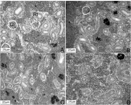
Fig. 1. Histological changes of the kidney of the olive flounder, Paralichthys olivaceus in juvenile stage. A: Se0.56group.
Note glomerulus, renal tubule and some of macrophage. B: Se1.07group. Note the weak swelling of hemocyte of glomeru- lus and macrophage. C: Se2.86group. Note the the some of abnormal renal tubule and glomerulus. D: Se4.56group. Note the degenerating renal tubules. Abbreviations: Gl, glomerulus; Rt, renal tubule; Mp, macrophage.
Fig. 2. Histological changes of the hepatopancreas of the olive flounder, Paralichthys olivaceus in juvenile stage. A: Se0.56
group. Hepatic cell and pancreas with numerous zymogen granules in the cytoplasm. B: Se1.07group. Note the degenerat- ing zymogen and the swelling of the capillary in some of organism. C: Se2.86group. Note the some of degenerated zymo- gen and the swelling hepatocytes. D: Se4.56group. Note fatty change, the irregular hepatocyte and degenerated hepato- cytes. E: Magnitude of degenerated zymogen Abbreviations: Cp, capillary; Hc, hepatocyte; Nu, nuculeus P, pancreas;
Zg, zymogen granules.
신장
Se
0.56개체들의 신장에서 일부 사구체 내 혈구 세포가 팽창하고 작은 점 형태의 흑색 대식세포 (macrophage)들이 관찰되었으며 (Fig. 1-A), 세뇨 관을 구성하는 상피세포들의 팽창들이 일부 보 였다. Se
1.07개체들에서 사구체의 혈구세포가 점 진적인 팽창을 보여주고, 일부 세뇨관의 상피세 포들의 파열도 나타나기 시작하였으며 대식세 포도 점점 커지는 것으로 관찰되었다(Fig. 1-B).
Se
2.86개체들에서 세뇨관의 상피세포들이 일부 탈락되는 것도 관찰되었으나 Se
1.07의 개체보다 병변의 진행이 더 빠른 것으로 나타났다(Fig. 1- C). Se
4.56의 개체는 세뇨관이 나타나기 시작하였 다(Fig. 1-D).
간췌장
Se
0.56개체의 간체장은 간세포, 모세혈관, 췌장
내 효소원 과립들이 정상적인 형태를 갖추고 있 었다(Fig. 2, A). Se
1.07개체의 간췌장 조직은 일부 모세 혈관 내 혈구의 팽창을 뚜렷하게 볼 수 있 었다(Fig. 2-B). Se
2.86개체는 모세혈관 팽창이 Se
1.07개체보다 더 많이 나타나고 일부 췌장 효 소원 감소가 뚜렷하게 관찰되며, 간세포가 일부 팽창되는 것을 볼 수 있었다(Fig. 2-C). Se
4.56개체 는 일부 불규칙적으로 생긴 간세포와 퇴화된 간 세포가 나타났으며 췌장 효소원 과립의 감소가 점차 빠르게 진행되고 있었다(Fig. 2-D).
아가미
Se
0.56의 개체는 새엽의 혈관이 팽창되었으나 새판은 거의 정상적으로 나타났다(Fig. 3-A).
Se
1.07개체의 아가미는 일부 비정상적인 새판을
가지는 것으로 관찰되었으나 Se
1.07개체와 차이
가 크게 나지 않았다 (Fig. 3-B). Se
2.86개체는 일
부 새판 말단의 곤봉화 현상이 나타나며, 새엽 기저의 팽창을 일부 관찰 할 수 있었다(Fig. 3- C). Se
4.56는 새판 말단의 곤봉화 현상이 빠르게 진행되며 새판의 상피세포 이상증식을 일부 관 찰 할 수 있었다(Fig. 3-D).
고 찰
셀레늄은 필요량 이상이 공급 되어졌을 경우 필수 미량영양소인 철이나 구리가 과량 공급되 었을 경우 유해한 활성산소종을 발생시키는 것 처럼 독성 증상을 보이게 된다(Stohs and Bagchi, 1995). 그리고 1987년 미국 FDA에서는 모든 돼 지 사료에 0.3ppm의 무기 셀레늄의 첨가를 허 용 하였으나 일부 환경 단체를 중심으로 호수 및 토양 중에 축적되는 불용성 셀레늄의 문제점 과 이로 인한 야생동물의 번식력 저하 현상을 제기하고 있다(Kim, 2000). 이처럼 셀레늄의 필
요성은 인정되고 있지만 가축에게 공급하였을 경우 발생되는 문제점은 비단 축산뿐 아니라 양 식에 있어서도 고려되어져야할 것이며 양어 사 료의 셀레늄 첨가는 면역 상승의 수준과 독성 수준에 대한 연구가 반드시 선행되어진 후 실행 되어져야 한다.
수서환경에서 독성물질 또는 물리적인 스트레 스로 인한 수서동물 기관계의 형태 및 구조적 변화는 비특이적인 현상이지만, 스트레스에 대 한 노출 조건과 무관한 것이 아니다(Mallatt, 1985). 어류 신장, 간 및 아가미의 조직학적 변화 에 미치는 영향에 관한 연구들에서 You et al.
(1978)은 카드뮴에 노출된 붕어, Carassius caras-
sius의 신장 세뇨관 상피층의 기저막 비후, 간세포의 공포화 및 아가미 새판상피세포의 요해소 체 증가와 기저막의 비후 등을 관찰하였다.
경골어류의 신장은 체내외의 염분과 수분의 적절한 균형을 유지하기 위하여 일차적으로 작
Fig. 3. Histological changes of gill of the olive flounder, Paralichthys olivaceus in juvenile stage. A: Se0.56group. Gill consists of gill filament and gill lamellae. B: Se1.07group. Note the some of the abnormal gill lamellae. C: Se2.86group.Note a few teminal clubbing the lamellae and swelling of the base of lamellae(↓). D: Se4.56group. the hyperplasia of epithelial cells of gill lamellar. Abbreviations: Ec, epithelial cell; Gf, gill filament; Gl, gill lamellae; Hc, hemocyte; Pc, pillar cell.
용하는 기관으로 알려져 있다(Yasutake and Wales, 1983). Kim et al. (2003)은 PCBs 노출된 넙치, Paralichthys olivaceus의 신장에서 흑색 대 식세포의 출현, 세뇨관 상피세포의 비대와 세뇨 관 내부의 공포체와 괴사 등을 관찰할 수 있었 고, Lee et al. (2001)은 구리에 노출된 넙치, Par-
alichthys olivaceus의 신장에서 흑색 대식세포의출현, 세뇨관 내강상피의 alcian blue에 양성반응 및 세뇨관 상피층의 기저막 비후를 보고하였으 며, HCBD (hexachlorobutadiene)에 노출된 금붕 어, Carassius auratus의 세뇨관 상피세포의 공포 화 및 괴사가 뚜렷하게 나타난다고 하였다 (Reimschuessel et al., 1989). 본 연구에서도 사구 체 내 혈구세포가 팽창, 흑색 대식세포 출현, 세 뇨관의 상피세포들의 파열 및 공포화, 세뇨관 상 피세포들이 일부 탈락되는 것을 볼 수 있었다.
흑색 대식세포군의 출현 및 증가는 자체방어 기 작이며(Lee et al., 2001), 세뇨관 상피층 기저막의 비후는 재흡수 장애로 인한 물질의 기저막 투과 에 영향을 주며, 계속적인 기저막의 비후는 간질 조직으로부터 세뇨관 상피층의 탈락으로 진행 될 수 있다고 하였다(Lee et al., 2001; You et al., 1978).
대사의 중심역할과 환경오염의 민감성 때문 에, 간은 다른 어류 종의 유기와 무기화합물의 독성지시자로서 대표적인 기관으로 알려져 있 다 (Baker, 1969; Segner, 1987; Ortiz et al., 1999).
또한, Hinton et al. (1978)과 Braunbeck et al.
(1990)은 연안지역에서 수서생태계 오염물들에 의한 피해가 발생되기 때문에 생체약물 화합물 (xenobiotic componds)에 대한 어류의 조직∙세 포학적인 반응들이 구명화 되어야 한다고 보고 하였으며, 어류의 간구조와 미세구조가 독성물 피해의 민감한 지표로서 평가받고 있다(Hinton
et al., 1978; Braunbeck and Volkl, 1991). PCBs에노출된 넙치, Paralichthys olivaceus의 모세 혈관 내 혈구의 팽창, 간세포의 팽창과 췌장 효소원 과립의 감소 현상을 볼 수 있었고(Kim et al., 2003), Lee et al. (2001)은 구리에 노출된 넙치 간
세포의 팽창 및 췌장 효소원 과립의 감소를 보 고하였다. 본 연구에서도 모세 혈관 내 혈구의 팽창, 췌장 효소원 감소, 간세포의 팽창을 관찰 할 수 있었다. 섭취한 사료를 통하여 흡수된 셀 레늄은 혈액을 통하여 체내 여러 조직으로 운송 되어 축적되는데 신장과 간에서 가장 높은 농도 를 보이게 된다 (Jenkin and Winter, 1973). Lee et
al. (2009)은 조직 내 셀레늄 축적량에 있어서 신장과 간이 사료 내 증가하는 셀레늄의 농도에 따라 축적률이 증가되는 것으로 나타났고, 축적 량은 사료 내 셀레늄의 양이 증가함에 따라 증 가하는 경향을 나타낸다고 보고하였다. 그리고 넙치와 습성이 유사한 어종인 흰철갑상어 (white sturgeon)에 있어서도 신장과 간이 아가미 와 근육 조직 보다 셀레늄 축적률이 더 높은 것 으로 나타났다(Tashjian et al., 2006). 일반적으로 동물의 혈액 중 셀레늄의 농도는 단기간 동안의 셀레늄 영향 상태를 나타내지만, 셀레늄이 조직 중에 축적되는 경우는 상당한 시간이 소요되므 로 조직내의 셀레늄 농도를 통하여 장기간의 셀 레늄에 대한 노출 상태를 확인할 수 있다. 그리 고 간에서의 셀레늄 농도를 통하여 셀레늄 과잉 또는 결핍 유무를 확인할 수 있는 지표로 사용 하고 있다(Ammerman and Miller, 1975).
일반적으로 아가미의 조직병변에 대한 것으로 아가미 상피의 박리 (lifting), 괴사 (necrosis), 이 상비대 (hyperplasia), 이상증식 (hypertropy), 새판 의 융합 등을 들 수 있다(Mallatt, 1985). PCBs 노 출된 넙치, Paralichthys olivaceus의 아가미에서 모세혈관의 팽창, 새판 상피세포의 팽창, 새판 말단의 곤봉화 현상, 새엽 기저의 팽창, 새판의 상피세포 이상증식 등이 관찰하였고(Kim et al., 2003), PCBs에 축적된 배스류, Lepomis auratus 아가미에서 다양한 상피세포의 이상증식과 새 판융합이 나타난다고 하였다(Teh et al., 1997).
Grinwis et al. (1998)은 가자미류, Platichthys fle-
sus에 TBTO (bis(tri-n-butyltin)oxide)를 처리시켰을 때 새엽과 새판 상피세포의 팽창, 새엽의 융
합과 새판상피세포의 이상증식이 생기기 시작
했고 그 정도가 심해짐에 따라 새판의 융합이 일어났다고 보고하였다. 본 연구에서도 넙치 새 엽의 혈관 팽창, 새판 말단의 곤봉화 현상, 새엽 기저의 팽창, 새판의 상피세포 이상증식 등이 관 찰되었다. 이와 같이 셀레늄은 넙치 아가미에 손 상을 유발시킨 후, 아가미 조직을 다양하게 손상 키는 것을 알 수 있었다.
이상 세 기관의 조직학적 연구에서 치어기 넙 치 사료내 첨가제로서 셀레노이스트의 적정 첨 가량은 1.07 mg/kg 이하 수준이 적당한 것으로 사료된다.
요 약
본 실험은 치어기 넙치 사료 내 셀레노이스트 첨가에 따른 영향을 평가하기 위하여 조직학적 반응을 살펴보았다. 사료 내 셀레늄은 각각 0ppm, 1ppm, 3ppm 및 5ppm의 총 4개 실험구로 설정하였으나 실험사료 분석을 통하여 사료내 셀레늄의 함량은 각각 0.56, 1.07, 2.86 및 4.56 mg Se/kg으로 나타났다. (Se
0.56, Se
1.07, Se
2.86, 및 Se
4.56). Se
1.07실험구의 신장에서 사구체의 혈구세 포가 점진적인 팽창을 보여주고, 일부 세뇨관의 상피세포들의 파열도 나타나기 시작하였으며 대식세포도 점차적으로 커지는 것을 알 수 있었 다. Se
0.56실험구의 간에서 정상적인 형태를 갖추 고 있다. 농도가 증가될수록 모세 혈관 내 혈구 의 팽창, 췌장 효소원 과립의 감소, 간세포의 팽 창이 점진적으로 진행되는 볼 수 있었다. 그리고 Se
1.07실험구에서 일부 비정상적인 새판을 가지 는 것으로 나타났으나 Se
0.56실험구와 크게 차이 가 없는 것으로 관찰되었다. 이상의 세 기관의 조직학적 연구에서 치어기 넙치 사료내 첨가제 로서 셀레노이스트의 적정 첨가량은 1.07 mg/kg 이하 수준이 적당한 것으로 사료된다.
사 사
이 논문은 2007학년도 부경대학교 연구년 교
수지원 사업에 의하여 연구되었음(PS-2007-030).
참 고 문 헌
Ammerman, C.B. and Miller, S.M.: Selenium in ruminant nutrition: A review. J. Dairy Sci., 58:1561-1577, 1975.
Baker, J.T.P.: Histological and electron microscopi- cal observations on copper poisoing in the winter flounder (Pseudopleuronectes ameri-
can-us). J. Fish. Res. Bd. Can., 26:2785-2893, 1969.
Bowie, G.L., Sanders, J.G., Riedel, G.F., Gilmour, C.C., Breitburg, D.L., Cutter, G,A. and Por- cella, D.B.: Assessing selenium cycling and accumulation in aquatic ecosystems. Water Air Soil Poll., 90:93-104, 1996.
Braunbeck, T. and Volkl, A.: Induction of biotrans- formation in the liver of eel, Anguilla anguil-
la L. by sublethal exposure to dinitro-o-cresol: An ultrastructural and biochemical study. Ecotoxicol. Environ. Saf., 21:109- 127, 1991.
Braunbeck, T., Gorge, T., Stoach, V. and Nagel, R.:
Hepatic steatosis in zebra fish, Brachydanio
rerio induced by long-term exposure to γhexachlorocyclohexane. Ecotoxicol. Envi- ron. Saf., 19:355-374, 1990.
Gatlin, D.M. III and Wilson, R.P.: Dietary selenium requirement of fingerling channel catfish. J.
Nutr., 114:627-633, 1984.
Grinwis, G.C.M., Boonstra, A., Brandhof, E.J., Dormans, J.A.M.A., Engelsma, M., Kuiper, R.V., Loveren, H, Wester, P.W., Vaal, M.A., Vethaak, A.D. and Vos, J.G.: Short-term tox- icity of bis (tri-n-butyltin) oxide in flounder,
Platichthys flesus : Pathology and immunefunction. Aquat. Toxicol., 42:15-36, 1998.
Hilton, J.W., Hodson, P.V. and Slinger, S.J.: The
requirement and toxicity of selenium in rain- bow trout (Salmo gairdneri). J. Nutr., 110:
2527-2535, 1980.
Hinton, D.E. and LaurÉn, D.J.: Integrative Histopathological Approaches to Detecting Effects of Environmental Stressors on Fishes.
Ameri. Fisher. Soc. Symp., 8:51-66, 1990.
Hinton, D.E., Klaunig, J.E. and Lipsky, M.M.:
PCB-induced alterations in teleost liver. A model for enviromental disease in fish. Mar.
Fish Rev., 40:47-50, 1978.
Jenkin, K.J. and Winter, K.A.: Effects of selenium supplementation of naturally high selenium swine rations on tissue levels of the element.
Can. J. Anim. Sci., 53:561-567, 1973.
Kim, J.W., Jee, J.H., Kang, J.C., Lee, J.S. and Chin, P.: Histological Response of kidney, Gill and Hepatopancreas of the Juvenile Olive Floun- der, Paralichthys olivaceus after PCBs Expo- sure. J. Kor. Fish. Soc., 36:283-289, 2003.
Kim, Y.Y.: Differences in biological activity and methbolism of selenium due to its chemical form. J. Anim. Sci. Technol., 42:835-848, 2000.
Lee J.C., Kang, J.C., Shin, Y.K., Ma, K.H. and Chin, P.: Histological responses of the floun- der, Paralichthys olivaceus exposed to cop- per. J. Fish Pathol., 14:81-90, 2001.
Lee, J.H., Kim, Y.C., Park, S.I., and Bai, S.C.: Eval- uation of the optimum dietary selemium (Se) level to improve immune responses in juve- nile olive flounder, Paralichthys olivaceus.
J. Kor. Fish. Soc., 42(1):26-33, 2009 Lemly, A.D.: Symptoms and implications of seleni-
um toxicity in fish: the Belews Lake case example. Aquat. Toxicol., 57:39-49, 2002.
Lemly, A.D.: A teratogenic deformity index for evaluating impacts of selenium on fish pop- ulations. Ecotox. Environ. Safe, 37:259-266,
1997.
Lin, Y.H. and Shiau, S.Y.: Dietary selenium require- ment of grouper, Epinephelus malabaricus.
Aquaculture, 250:356-363, 2005.
Lohner. T.W., Reash, R.J., Willet, V.E. and Rose, L.A.: Assessment of tolerant sunfish popula- tions (Lepomis sp.) inhabiting selenium- laden coal ash efflu-ents 1. Hematological and population level assessment. Ecotox.
Environ. Safe, 50:203-216, 2001.
Mallatt, J.; Fish gill structural changes induced by toxicants and other irritants: a statixtical review. Can. J. Fish. Aquat. Sci., 42:630- 648, 1985.
Ortiz, J.B., Gonzalez de Canales, M.L. and Saras- quete, C.: Quantifi-cation and histopatholog- ical alterations produced by sublethal copper concentrations in Fundulus heteroclitus.
Ciencias Marinas, 25:119-143, 1999.
Reimschuessel, R., Richard, O.B., Eric, B.M. and Michael, M.L.: Renal histopathological changes in the goldfish, Carassius auratus after sublethal exposure to hexachlorobuta- diene. Aquat. Toxicol., 15:169-180, 1989.
Segner, H.: Response of fed and starved roach.
Rutilus rutilus, to sublethal copper contami-
nation. J. Fish Biol., 30:423-437, 1987.
Stohs, S.J. and Bagchi, D.: Oxidative mechanisms in the toxicity of metal ions. Free Radic.
Biol. Med., 18:321-336, 1995.
Tashjian, D.H., Teh, S.J., Sogomonyan A. and Hung, S.: Bioaccumulation and chronic toxi- city of dietary l-selenomethionine in juvenile white sturgeon, Acipenser transmontanus.
Aquatic Toxicology, 79:401-409, 2006.
Teh, S.J., Adams, S.M. and Hinton, D.E.:
Histopathologic biomarkers in feral freshwa-
ter fish populations exosed to different types
of contaminant stress. Aquat. Toxicol.,
37:51-70, 1997.
Yasutake W.T. and Wales., J.H.: Microscopic anato- my of salmonids: An atals. Department of the interior fish and wildlife service wash- ington D.C. U.S.A., pp 97, 1983.
You, K.H., Choi, C.H., Choe, R.S. and Deung, Y.K.: Ultrastructural studies on cadmium
poisoning in the liver, kidney and gills of
Carassius carassius. Korean J. ElectronMicroscopy, 8:33-48, 1978.
Manuscript Received : December 29, 2009 Revised : April 15, 2010 Accepted : April 21, 2010