Antihyperlipidemic Effect of Ginsenoside Rg1 in Type 2 Diabetic Mice
Jae Hong Park
†, Jiyoun Lee
†, Jiyoung Yeo, Jeong Su Nam and Myeong Ho Jung*
School of Korean Medicine, Pusan National University, Yangsan 626-770, Korea
Received April 14, 2011 /Revised May 11, 2011 /Accepted May 13, 2011Ginsenoside Rg1 is a pharmacologically active component isolated from ginseng. The goal of this study was to clarify the beneficial effects of Rg1 on glucose and lipid metabolism in diabetic animals (db/db mice). To accomplish this, ten week old db/db mice were administered 10 mg/kg of Rg1 for 15 days. Rg1 did not influence the weight of db/db mice when compared with vehicle-treated db/db mice. The administration of Rg1 lowered fasting plasma glucose, and improved glucose tolerance.
Importantly, Rg1 markedly reduced both plasma triglyceride and free fatty acids, and increased high-density lipoprotein cholesterol (HDL-C) concentrations in db/db mice. Rg1 activated promoter ac- tivity of chimeric GAL4-PPARα reporter and increased expression of peroxisome proliferator-activated receptor alpha (PPARα) target genes such as carnitine palmitoyltransferase-1 (CPT-1) and acyl-CoA ox- idase (ACO), which are involved in fatty acid oxidation. These findings indicated that improvement of lipid profiles by Rg1 may be associated with increased fatty acid oxidation via PPARα activation.
Taken together, these results suggest that Rg1 could have beneficial effects for controlling hyper- glycemia and hyperlipidemia associated with type 2 diabetes.
Key words : Ginsenoside Rg1, type 2 diabetes, hyperlipidemia, db/db mice
†Equal contributor
*Corresponding author
*Tel:+82-51-510-8432, Fax:+82-51-510-8437
*E-mail : [email protected]
Introduction
Ginseng (Panax ginseng C.A. Meyer) is a well-known Chinese medicinal herb that has been widely used for thou- sands of years throughout Asia owing to its various pharma- cological actions [9,11,21]. Ginseng is now one of the most popular herbal medicines used nutraceutically, with annual sales of over USD 200 million [29]. Most pharmacological actions of ginseng are attributed to one type of its con- stituents, namely the ginsenosides. Ginsenosides, the major bioactive components of ginseng, have been shown to ac- count for the pharmacological activities of ginseng, such as anti-inflammatory properties [11], anti-platelet action [27], inhibition of lipoxygenase [5] and 15-hydroxyprostaglandin dehydrogenase [6]. With the development of modern tech- nology, more than 40 different ginsenosides have been iso- lated and purified from ginseng to date [13,17,22]. These compounds can be divided into two classes, proto- panaxatriols (Rg1, Rg2, Re, Rf, and Rh1) and proto- panaxadials (Rb1, Rb2, Rc, Rd, Rg3, Rh2, and Rh3), which have slightly different molecular structures [23]. Among the 40 ginsenosides, Rg1, which is the phytosterol from Panax
ginseng and the major active molecule from the total sap- onins of Panax notoginseng (PNS) [19], has received a great deal of attention because it has been shown to possess neuro- trophic and neuroprotective effects with cognitive enhance- ment in many in vitro studies [20,24] as well as anti-aging effects [8]. However, despite these reports, Rg1 has rarely been studied in the field of type 2 diabetes.
Diabetes mellitus is a group of metabolic diseases, and two types (type 1 & 2) of diabetes have a distinct patho- genesis; however, hyperglycemia and diverse life-threat- ening complications including atherosclerosis resulting from long-term hyperglycemia are their most common features.
More than 90% of diabetic patients have type 2 diabetes and suffer from severe insulin resistance [15]. Major complica- tions of type 2 diabetes, such as cardiovascular disease, are partly due to associated abnormalities of plasma lipid and lipoprotein metabolism. Apart from glycemic control, the regulation of lipid metabolism may be an important ther- apeutic target against the complications associated with type 2 diabetes [18].
Accordingly, this study was designed to clarify the possi-
bility of Rg1 as an effective candidate to ameliorate hyper-
glycemia and hyperlipidemia in a diabetic animal model
(db/db mice).
Materials and Methods Animals and diets
Twenty one male C57Bl/KsJ-db/db mice (8 weeks old) were obtained from Jackson Laboratory (Bar Harbor, ME, USA). The mice were all individually housed in poly- carbonate cages under a 12-h light-dark cycle at, 21-23℃ and 40-60% humidity. After a 2-week adaptation period, the 10-week-old mice were randomly divided into three groups (n=7), a diabetic control, acarbose, and Rg1. All groups were fed a standard AIN-76 semi-synthetic diet [2,3]. The mice had free access to food and water, and their body weight was measured once every five days throughout the experiment. The diabetic control group was orally ad- ministered deionized water and two experimental groups (acarbose, and Rg1) were orally administered acarbose (50 mg/kg BW) or Rg1 (10 mg/kg BW) for 15 days. The mice were then starved for 12 hrs, after which they were anes- thetized with ether and their blood samples were taken from the inferior vena cava to measure the plasma biomarkers (HbA
1c, insulin, glucagon, etc.). The mice were handled in strict accordance with the Pusan National University guide- lines for the care and use of laboratory animals.
Plasma glucose, insulin, glucagon levels and lipids contents
The blood glycated hemoglobin (HbA
1c) of each sample sacrificed mice was measured using a MicroMat™ II Hemoglobin A
1cTest (Bio-Rad Laboratories, Hercules, CA, USA). The plasma insulin and glucagon (Mouse ELISA Kit, ALPCO Diagnostics, Salem, NH, USA) levels were de- termined using a quantitative sandwich enzyme immuno- assay kit. In addition, the plasma concentrations of glucose, total cholesterol, triglyceride, and HDL-cholesterol (Asan Diagnostics, Seoul, Korea) were determined using an enzy- matic method. The plasma free fatty acids (FFAs) concen- trations were determined using an enzymatic colorimetric method (Wako Pure Chemical Industries, Japan). All blood samples obtained were centrifuged at 1,000× g for 15 min at 4℃ for biochemical analysis.
Intraperitoneal glucose tolerance test (IPGTT)
One day before the sacrifice, an intraperitoneal glucose tolerance test (IPGTT) was conducted on all of the db/db mice after a 12-h overnight fast. To determine the glucose toler-
ance, the mice were intraperitoneally injected with glucose (0.5 g/kg BW) and the glucose concentrations of blood drawn from the tail vein were determined immediately upon collection at 30, 60, and 120 min after glucose injection using a Glucometer (GlucoDr, Allmedicus, Anyang, Korea).
GAL4/PPARα chimera assay
The PPARα ligand-binding activity was measured using a GAL4/PPARα chimera. COS-7 monkey kidney cells were transfected with pFA-hPPARα, pFR-Gal4 (UAS-Gal4-luciferase) and pFR-galactosidase (Stratagene, La Jolla, CA, USA) using Genejuice (Novagen, Madison, WI, USA). After transfection for 24 hr, the cells were incubated with Rg1 or WY14643 (Sigma, St. Louis, MO, USA) at 5 μM or 2 μM for 24 hr.
The luciferase activities were then determined with a lucifer- ase assay system kit (Promega, Madison, WI, USA).
Real-time RT-PCR
For the quantification of mRNA expression of CPT-1 and ACO, total RNA was extracted using TRIzol reagent (Invitrogen, Carlsbad, CA, USA) and subjected to reverse transcription using reverse transcriptase (Promega). The re- sulted cDNA was then subsequently amplified with gene-specific primers by PCR using a fluorescence temper- ature cycler (Chromo4, Real-Time PCR System; Bio-Rad, Hercules, CA, USA).
Statistical analysis
All data are presented as the means±SE. The data were evaluated by one-way ANOVA and the differences between means were determined using Tukey’s multiple-range test.
All analyses were conducted using GraphPad prism ver.
5.01. Significant difference between two groups was de- termined using Student’s t-test. Values were considered stat- istically significant at p<0.05.
Results Effect of Rg1 on body weight
The body weight of db/db mice in the vehicle-treated
group and Rg1-treated group showed a slight decrease from
Day 0 to Day 15. However, there was no significant differ-
ence in body weight between the vehicle group and the
Rg1-treated group (Fig. 1), suggesting that Rg1 does not af-
fect the bodyweight of db/db mice.
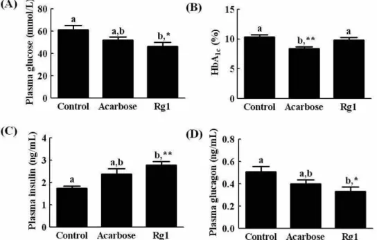
Fig. 2. Effects of Rg1 on plasma glucose (A), the glycated hemogluobin (HbA1c) (B), insulin (C), and glucagon (D) in
db/db
mice over 15 days. Values are expressed as the means±SE, n=7.abmeans not sharing a common letter are significantly different (p
<0.05).Fig. 1. Changes in the body weight of Rg1-treated C57BL/
KsJ-
db/db
mice.Effect of Rg1 on plasma glucose, glycated hemoglobin, insulin and glucagon
We investigated the effect of Rg1 on biochemical parame- ters in the blood of db/db mice after fasting for 12 hr. As shown in Fig. 2, the plasma glucose level of Rg1-treated db/db mice was reduced when compared with vehicle-treated db/db mice (Fig. 2A). The glycated hemoglobin levels were also slightly lower in the Rg1-treated mice, but this difference was not significant (Fig. 2B). The plasma insulin level of the Rg1-treated mice was significantly higher than that of ve-
hicle-treated db/db mice, whereas the plasma glucagon levels were markedly lower (Fig. 2C and 2D).
Effect of Rg1 on IPGTT
We conducted an IPGTT assay to determine the effects of Rg1 on glucose homeostasis in db/db mice, and the results were presented as a percentage of the value at the time of the glucose injection (Fig. 3). The blood glucose was in- creased for up to 30 min after glucose injection in vehicle or treated db/db mice. However, the blood glucose level be- gan to decrease in the Rg1 and acarbose-treated mice at 60 min after glucose injection when compared to the vehicle control db/db mice. The blood glucose level failed to return to the baseline after 120 min in all db/db mice, but the glucose level in the Rg1-treated mice was much lower than that in the vehicle-treated db/db mice, suggesting that Rg1 sig- nificantly improves glucose homeostasis in db/db mice.
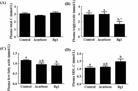
Effect of Rg1 on plasma lipid contents
We examined the effects of Rg1 on plasma lipid contents in db/db mice. The plasma cholesterol level was not sig- nificantly influenced by Rg1 or acarbose treatment (Fig. 4A).
However, Rg1 significantly reduced both the plasma trigly-
ceride and free fatty acid concentrations when compared
with those of vehicle or acarbose-treated mice (Fig. 4B &
Fig. 4. The effects of Rg1 on plasma total cholesterol (A), triglyceride (B), free fatty acids (C), and HDL-cholesterol (D) in
db/db
mice over 15 days. Values are expressed as the means±SE, n=7.abmeans not sharing a common letter are significantly different (p
<0.05).Fig. 3. Effects of Rg1 on the intraperitoneal glucose tolerance test (IPGTT) in C57BL/KsJ-
db/db
mice. The blood glu- cose concentration was measured at the indicated times and presented as a percentage of the level at the time of glucose injection. Values are the means±SE, n=7. ab means not sharing a common letter are significantly dif- ferent (p
<0.05). NS: no significant4C). Furthermore, Rg1 increased the HDL-cholesterol (Fig.
4D), suggesting that Rg1 can ameliorate lipid metabolism.
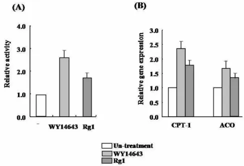
To clarify the mechanism underlying the effect of Rg1 on lipid improvement, we investigated whether Rg1 activates PPARα using cell-based GAL4/PPARα chimera trans-
activation assays. As shown in Fig. 5A, Rg1 increased the PPARα-dependent luciferase activity similar to WY14643, an agonist of PPARα, and stimulated PPARα target genes in- cluding CPT-1, ACO in SK-Hep1 cells (Fig. 5B). Taken to- gether, these findings suggest that Rg1 activates fatty acid oxidation and then improves hyperlipidemia.
Discussion
The role of Rg1 in the severe hyperglycemic state has not yet been fully established; therefore, the effect of Rg1 on hy- perglycemia and hyperlipidemia in the db/db mice was inves- tigated by comparison with acarbose, which is widely used as a prescription drug for the treatment of type 2 diabetes.
In general, hyperglycemia is a modifiable risk factor that
can have a deleterious effect on the development and pro-
gression of microvascular and macrovascular complications
in patients with type 2 diabetes [12]. Hence, many studies
suggest that diabetic treatment may be required to reach hy-
perglycemic control targets in patients who have early-stage
type 2 diabetes [25]. When we measured fasting blood glu-
cose of mice before sacrifice, the fasting blood glucose level
was lower in Rg1-treated mice. Furthermore, Rg1 improved
glucose tolerance, suggesting that Rg1 improves glucose
Fig. 5. The effect of Rg1 on PPARα activation. (A) The PPARα ligand-binding activity was measured using a GAL4/PPARα chimera assay in Rg1-treated cells. (B) Real time PCR was conducted to determine the mRNA expression of CPT-1 and ACO in Rg1-treated Sk-Hep1.
homeostasis. The recent study showed that Rg1 suppressed hepatic glucose production via liver kinase B1 (LKB1)-AMP- activated protein kinase (AMPK)-forkhead box class O1 (FoxO1) pathway [16]. We also found that Rg1 activated AMPK in SK-Hep1 cells (data not shown). Therefore, the suppression of hepatic glucose production by stimulation of AMPK may be responsible for antihyperglycemic effect of Rg1. Additionally, the plasma insulin levels were sig- nificantly higher in the Rg1-treated and acarbose mice than in diabetic control mice, whereas the glucagon levels were reduced by Rg1 treatment. These results appeared to in- dicate that Rg1 positively affects regulation of hyper- glycemia by removing glucose from the blood via increased insulin secretion and decreased glucagon secretion.
The lipotoxic theory states that the accumulation of lipids in target organs such as skeletal muscle, kidneys, the pan- creas and the heart may contribute to the development of obesity-related disorders [10]. Changes in the plasma lipid concentration are a frequent complication in patients with diabetes mellitus [14,28], which certainly contributes to the development of vascular disease. Diabetic dyslipidemia is characterized by high triglyceride (TG) levels, low HDL-C and increased low density lipoprotein cholesterol (LDL) [1,4]. This atherogenic lipid profile contributes to an excess risk of cardiovascular disease (CVD) in individuals with type 2 diabetes. The dyslipidemia is partially corrected by control of the hyperglycemia, but abnormalities persist, in
part due to the effects of insulin resistance on lipoprotein metabolism [7,26]. Low levels of HDL-C and apo A-I are also characteristic of type 2 diabetes. Accordingly, the effects of Rg1 on the plasma lipid concentrations were also investigated. While the plasma lipid concentrations, total cholesterol, and triglyceride were significantly increased in the diabetic control mice, the Rg1- treated mice showed low levels of these compounds, suggesting that Rg1 improved hyperlipidemia. We also found that Rg1 activates PPARα and increases CPT-1 and ACO expression in SK-Hep1 cells.
Thus, these effects may lead to reduced synthesis and secre- tion of triglycerides, thereby decreasing the liberation of fat- ty acids, resulting in antihyperlipidemia.
In conclusion, Rg1 lowered plasma glucose, which ap- peared to be partly mediated via AMPK activation and an increase in plasma insulin, and improved plasma lipids such as free fatty acid and triglycride in the db/db mice via PPARα activation. Therefore, these results suggest that the anti- hyperglycemic and antihyperlipidemic properties of Rg1 could be beneficial for the prevention of diabetic complica- tions such as cardiovascular diseases and mild management of type 2 diabetes.
Acknowledgement
This work was supported by a 2-Year Research Grant of
Pusan National University.
References
1. Aasum, E., A. D. Hafstad, D. L. Severson, and T. S. Larsen.
2003. Age-dependent changes in metabolism, contractile function, and ischemic sensitivity in hearts from db/db mice.
Diabetes
52, 434-441.2. American Institute of Nutrition. 1980. Report of Ad Hoc committee on standards for nutritional studies.
J. Nutr.
110, 1717-1726.3. American Institute of Nutrition. 1977. Report of the American institute of nutrition ad hoc committee on stand- ards for nutritional studies.
J. Nutr.
107, 1340-1348.4. Andallu, B., A. V. Vinay Kumar, and NCh. Varadacharyulu.
2009. Lipid abnormalities in streptozotocin-diabetes:
Amelioration by Morus indica L. cv Suguna leaves.
Int. J.
Diabetes Dev. Ctries.
29, 123-128.5. Anhäuser, M. 2003. Pharmacists seek the solution of a shaman.
Drug Discov. Today
8, 868-869.6. Atta, Ur R. and K. Zaman. 1989. Medicinal plants with hypo- glycemic activity.
J. Ethnopharmacol.
26, 1-55.7. Chen, X., X. Bai, Y. Liu, L. Tian, J. Zhou, Q. Zhou, J. Fang, and J. Chenl. 2009. Anti-diabetic effects of water extract and crude polysaccharides from tuberous root of Liriope spicata var. prolifera in mice.
J. Ethnopharmacol.
122, 205-209.8. Cheng, Y., L. H. Shen, and J. T. Zhang. 2005. Anti-amnestic and anti-aging effects of ginsenoside Rg1 and Rb1 and its mechanism of action.
Acta. Pharmacol. Sin.
26, 143-149.9. DeFronzo, R. A. 1999. Pharmacologic therapy for type 2 dia- betes mellitus.
Ann. Intern. Med.
131, 281-303.10. De Sotillo, D. V. R. and M. Hadley. 2002. Chlorogenic acid modifies plasma and liver concentrations of: cholesterol, tri- acylglycerol, and minerals in (fa/fa) Zucker rats.
J. Nutr.
Biochem.
13, 717-726.11. Gaster, B. and I. B. Hirsch. 1998. The effects of improved glycemic control on complications in type 2 diabetes.
Arch.
Intern. Med.
158, 134-140.12. Hanefeld, M. and T. Temelkova-Kurktschiev. 2002. Control of post-prandial hyperglycemia- an essential part of good diabetes treatment and prevention of cardiovascular complications.
Nutr. Metab. Cardiovasc. Dis.
12, 98-107.13. Hou, J. P. 1977. The chemical constituents of ginseng plants.
Comp. Med. East West
5, 123-145.14. Jang, Y. J., J. K. Kim, M. S. Lee, I. H. Ham, W. K. Whang, K. H. Kim, and H. J. Kim. 2001. Hypoglycemic and hypolipi- demic effects of crude saponin fractions from
Panax ginseng
and gynostemma pentaphyllum.Yakhak Hoechi
45, 545-556.15. Kim, M. J., K. H. Leem, and H. K. Kim. 2009. Hydrangea dulcis folium preserves b-cell mass in diabetic db/db mice.
Food Chem. Toxicol.
47, 1685-1688.16. Kim, S. J., H. D. Yuan, and S. H. Chung. 2010. Ginsenoside Rg1 suppresses hepatic glucose production via AMP-acti- vated protein kinase in HepG2 cells.
Biol. Pharm. Bull.
33, 325-328.17. Lee, S. M., H. J. Shon, C. S. Choi, T. M. Hung, B. S. Min, and K. Bae. 2009. Ginsenosides from heat processed ginseng.
Chem. Pharm. Bull. (Tokyo)
57, 92-94.18. Liu, G., B. Wang, J. Zhang, H. Jiang, and F. Liu. 2009. Total
panax notoginsenosides
prevent atherosclerosis in apolipopro- tein E-knockout mice: Role of downregulation of CD40 and MMP-9 expression.J. Ethnopharmacol.
126, 350-354.19. Ng, T. B. 2006. Pharmacological activity of sanchi ginseng (
Panax notoginseng
).J. Pharm. Pharmacol.
58, 1007-1019.20. Nishijo, H., T. Uwano, Y. M. Zhong, and T. Ono. 2004. Proof of the mysterious efficacy of ginseng: basic and clinical tri- als: effects of red ginseng on learning and memory deficits in an animal model of amnesia.
J. Pharmacol. Sci.
95, 145-152.21. Rosenbloom, A. L., J. R. Joe, R. S. Young, and W. E. Winter.
1999. Emerging epidemic of type 2 diabetes in youth.
Diabetes Care
22, 345-354.22. Sugimoto, S., S. Nakamura, H. Matsuda, N. Kitagawa, and M. Yoshikawa. 2009. Chemical constituents from seeds of Panax ginseng: structure of new dammarane-type triterpene ketone, panaxadione, and HPLC comparisons of seeds and flesh.
Chem. Pharm. Bull.
57, 283-287.23. Tawab, M. A., U. Bahr, M. Karas, M. Wurglics, and M.
Schubert-Zsilavecz. 2003. Degradation of ginsenosides in humans after oral administration.
Drug Metab. Dispos.
31, 1065-1071.24. Tohda, C., N. Matsumoto, K. Zou, M. R. Meselhy, and K.
Komatsu. 2004. Abeta (25–35)-induced memory impair- ment, axonal atrophy, and synaptic loss are ameliorated by M1, A metabolite of protopanaxadiol-type saponins.
Neuropsychopharmacology
29, 860-868.25. Van Gaal, L. F. and I. H. De Leeuw. 2003. Rationale and options for combination therapy in the treatment of type 2 diabetes.
Diabetologia
46, 44-50.26. Verreth, W., J. Ganame, A. Mertens, H. Bernar, M. C.
Herregods, and P. Holvoet. 2006. Peroxisome pro- liferator-activated receptor-alpha,gamma-agonist improves insulin sensitivity and prevents loss of left ventricular func- tion in obese dyslipidemic mice.
Arterioscler. Thromb. Vasc.
Biol.
26, 922-928.27. Xie, J. T., H. H. Aung, J. A. Wu, A. S. Attele, and C. S.
Yuan. 2002. Effects of American ginseng berry extract on blood glucose levels in
ob
/ob
mice.Am. J. Chin. Med.
30, 187-194.28. Xie, J. T., Y. P. Zhou, L. Dey, A. S. Attele, J. A. Wu, M.
Gu, K. S. Polonsky, and C. S. Yuan. 2002. Ginseng berry reduces blood glucose and body weight in db/db mice.
Phytomedicine
9, 254-258.29. Yue, P. Y., N. K. Mak, Y. K. Cheng, K. W. Leung, T. B.
Ng, D. T. Fan, H. W. Yeung, and R. N. Wong. 2007.
Pharmacogenomics and the Yin/Yang actions of ginseng:
anti-tumor, angiomodulating and steroid-like activities of ginsenosides.