연구논문
Abstract:
1)Anti-diabetic effect of the exopolysaccharides (EPS) produced from submerged mycelial culture of Cordyceps sinensis (Cs) was studiedin a type II diabetic animal model (C57BL/6J ob/ob). This study was designed to determine whether Cs-EPS improves clinical symptoms of type II diabetes in ob/ob mice. After Cs-EPS treatment at doses of 200 mg/kg body weight, the fasting blood glucose levels decreased by 47% after 7 weeks compared with those of the control mice. According to the oral glucose tolerance test, the glucose levels recovered its baseline after 120 min in Cs-EPS-treated mice, although the blood glucose levels increased significantly after 30 min. On the other hand, the control group (not-treated) did not recovered its initial level of glucose after 120 min. Furthermore, food intake, body weight, total plasma cholesterol and triglyceride concentrations in ob/ob mice treated with Cs-EPS were significantly decreased, compared with those in control ob/ob mice.
Cs-EPS treatment increased significantly the plasma insulin level and the expression of leptin mRNA in adipose tissue of Cs-EPS-treated ob/ob mice. From these results, it is demonstrated that Cs-EPS could be effective for regulating normal blood glucose levels by increasing the amounts of plasma insulin and leptin expression in ob/ob mice,
대구대학교 생명환경대학 바이오산업학과
Department of Bioindustry, College of Life and Environment, Daegu University, Kyungbuk 712-714, Republic of Korea Tel: +82-53-850-6756, Fax: +82-53-850-6769
e-mail: [email protected]
indicating that this compound could be a candidate material as a dietary supplement to control hyperglycemia in patients suffering from type II diabetes.
Keywords: Anti-diabetic activity, Cordyceps sinensis, exopolysaccharide, leptin, ob/ob mice
1. Introduction
Type II diabetes is a progressive disease that generally has an onset in adulthood and accounts for over 90% of all diabetes cases [1]. The prediabetic state in these individuals is characterized by insulin resistance, which means a failure of peripheral tissues (muscle, adipose, liver, and even the pancreas itself) to respond to normal levels of insulin [2,3].
Currently, pharmacological agents available for type II diabetes have a number of limitations, such as adverse side effects and high rates of secondary failure [4,5]. Due to these reasons, diabetic patients and healthcare professionals are increasingly considering complementary and alternative approaches by the use of medicinal herbs with anti- hyperglycemic activities [6-8]. Generally, the anti-diabetic bioactive materials in medicinal plants are known to be polysaccharide, terpenoids, flavonoids, sterols, and alkanoids [9].
In addition, many researchers have endeavored to study the hypoglycemic effect from either the fruiting body or mycelia of various edible/medicinal fungi including Cordyceps sinensis
제 형 당뇨쥐에서 동충하초로부터 생산된 세포외 다당류의 2 항당뇨 효과
최장원*
Anti-diabetic Effect of the Exopolysaccharides (EPS) Produced from Cordyceps sinensis on ob/ob Mice
Jang Won Choi*
접수 : 2010 년 12 월 27 일 / 게재승인 : 2011 년 월 2 15 일
© 2011 The Korean Society for Biotechnology and Bioengineering
and Lentinus edodes [10,11].
C. sinensis parasitizes insects and has been used as a traditional folk medicine in Asia for hundreds of years [12,13].
Spores germinate inside the caterpillars and a stalked fruiting body is produced [14]. Various bioactive compounds are found in Cordyceps spp., which stimulate immune systems [15], inhibit the growth of tumor cells [16,17], enhance hepatic energy [18]
and promote the secretion of adrenal hormones [19]. It also inhibits rejection of organ transplants [20] and has been used in combination with traditional drugs in this field [20,21]. In recent years, much interests have been focused on exopolysaccharide (EPS) produced by fungi due to their various biological and pharmacological activities and many kinds of EPS have been produced from submerged cultures of macrofungi such as mushrooms and entomopathogenic fungi [22-24].
Leptin, a newly-discovered hormonal product of the obesity (ob) gene, is expressed by white adipose tissue [25]. It has been implicated in the regulation of body weight [26,27], glucose metabolism[28,29] and fertility [30,31]. Leptindeficiency produces severe obesity, insulin resistance and impaired glucose tolerance in ob/ob mice and also, congenital leptin deficiency in humans leads to hyperphagia and marked obesity [32]. The decrease in circulating leptin concentrations during energy restriction in human is related closely to the decrease in plasma glucose level [33]. Leptindeficient ob/ob mice are an ideal model to study the metabolic impact of leptin because the action of leptin may be studied without the interference of physiological changes in endogenous leptin levels in these animals.
In this study, we investigate anti-diabetic effect of EPS produced from mycelial culture of C. sinensis on type II diabetes in ob/ob mice, which do not produce functional leptin, and exhibit obesity, hyperphagia, hyperglycaemia, hyperinsulinaemia and insulin resistance. And also, we study the effect of EPS through changes of leptin expression levels and the possibility of clinical treatment in type II diabetes.
2. Materials and Methods
2.1. Microorganism
The microorganism used in this experiment was C. sinensis (CCRC 36421). The stock culture was inoculated on potato dextrose agar (PDA) slants and subcultured every month.
Slants were incubated at 25 ℃ for 7 days and then stored at 4 . ℃
2.2. Inoculum preparation and culture condition
C. sinensis was initiallygrown on a PDA medium (2.4%
potato dextrose broth and 2% agar) in a petri dish, and then transferred to the seed culture medium. The medium used for seed culture contained the followings (g/L): glucose 10, yeast
extract 3, malt extract 3, meat peptone 5. The seed cultures (50 mL) were incubated in 250 mL Erlenmeyer flasks at 25℃
with shaking at150 rpm for 4 days. For EPS production, submerged culture of C. sinensis was performed as described previously [37]. Briefly, batch cultivation was carried out in a 5-L stirred-tank fermenter (KF-250, KoBioTech, Seoul, Korea) with a working volume of 3 L using a medium containing (g/L) sucrose 20, corn steep powder 25, CaCl
20.78, and MgSO
4· 7H
2O 1.73. After inoculation, the culture was agitated at 150 rpm and the air flow-rate was kept at 2 vvm at 20 ℃ without control of pH during the operation.
2.3. Preparation of EPS
After fermentation, culture broths were centrifuged at 12,000 × g for 30 min and the resulting supernatant was filtered through a Whatman filter paper No. 2 (Whatman International, Maidstone, England) to remove any remaining mycelium. The obtained filtrate was mixed with a four volume of absolute ethanol, stirred vigorously, and stand overnight at 4 . The precipitated EPS was collected by centrifugation ℃ (12,000 × g, 30 min) and the supernatant was discarded.
Finally, the precipitates of crude EPS were lyophilized and the weight was estimated. The crude EPS was used for animal experiment without further purification.
2.4. Purification and content determination of EPS
The ethanol precipitates of EPS were dissolved in 0.2 M NaCl solution at a concentration of 5 g/L, and loaded onto a Sepharose CL-6B column (2.4 cm × 100 cm, Sigma Chemical Co., Louis, MO, USA). The column was eluted with the same solution at a flow rate of 0.6 mL/min. The protein moiety in the crude EPS was monitored at 280 nm, whilst the carbohydrate moiety was monitored at 480 nm. Protein concentrations were determined with a Bio-Rad protein assay kit using bovine serum albumin as a standard [34]. The content of EPS produced by the entomopathogenic fungi, C.
sinensis was determined by phenol sulfuric acid method [35]
using glucose as a standard.Sugar composition was analyzed by gas chromatography (Varian Co., Model: Star 3600CX, Lexington, MA, USA) using fused silica capillary column and flame ionization detector. The composition of amino acid was analyzed by amino acid analyzer (Biochrom Ltd., Model: Biochrom 20, Cambridge, UK) with high performance ion exchange column.
2.5. Animals and breeding conditions
The 5 weeks-old male C57BL/6J ob/ob mice were obtained
from Japan SLC (Hamamatsu, Japan) and left to acclimatize
for 2 weeks before experiment. The animals were housed in
individual stainless steel cages in an air conditioned room
(23 ± 2 ℃ with 55 ± 5% humidity) under a 12 h light and
12 h dark cycle. Astandard mouse chow (Sam Yang Co., Seoul, Korea) and water were provided during the experiment period. These experiments were approved by the Committee for Laboratory Animal Care and Use, Daegu University. All procedures were conducted in accordance with the “Guide for the Care and Use of Laboratory Animals” published by the National Institutes of Health.
2.6. Experimental design
After 2 weeks of acclimatization, the mice with a blood glucose level of 300 mg/dl (16.7 mmol/L) were considered to be diabetic, and then randomly divided into two groups with seven animals in each group. The control group received 0.9% NaCl solution; the diabetic group was treated with 200 mg of Cs-EPS per kg of body weight using an oral zoned daily for 7 weeks. The body weight gain and food intake were periodically measured. After fasting for 4 h, the blood glucose levels were measured using one drop of whole blood obtained from cut on the underside of the mouse’s tail by glucometer (Lifescan, Milpitas, CA, USA).
2.7. Oral glucose tolerance test
To evaluate the acute effects of EPS on hyperglycemia, oral glucose tolerance test (OGTT) was performed on day 50.
Briefly, glucose (2 g per kilogram of body weight) was administrated to the fasted mice during 15 h and then blood samples were collected sequentially from the tail vein at 0 (prior to glucose administration), 30, 60, 90, 120 and 180 min after glucose administration. Blood glucose levels were determined as described above.
2.8. Analytical measurements
The plasma was collected by centrifugation at 3,000 × g for 10 min. Plasma samples from mice were frozen at -80℃
until analysis. The plasma glucose, cholesterol, and triglyceride levels were measured by enzymatic colorimetric assay (YD Diagnostics, Seoul, Korea). Insulin concentration in the plasma was measured by commercially available mouse insulin enzyme-linked immunosorbent assay (ELISA) kit (U-type, Shibayagi, Gunma, Japan). All the samples were analyzed in triplicate.
2.9. RNA isolation
Total RNAs were isolated from blood and various tissues using the RNeasy Protect Mini Kit (Qiagen, Stanford, CA).
The samples were resuspended in RNeasy lysis buffer (Qiagen, Stanford, CA, USA) and homogenized by a 20-gauge needle fitted to a syringe. And then, the extracts were processed following the manufacturer’s instructions. In the final step, the RNA was eluted with 50 L of RNase-free μ water by centrifugation at 10,000 rpm for 1 min. RNAintegrity
was analyzed on a 1% agarose gel containing formaldehyde and the concentration was measured by an UVspectrophotometer (Spectronic instruments, NY, USA).
2.10. RT-PCR analysis
RT-PCR was performed using One Step RT-PCR kit (Takara, California, USA) with leptin-specific primers (forward 5'-TGAGTTTGTCCAAGATGGACC-3’ and reverse 5’-GCC ATCCAGGCTCTCTGG-3’) and -actin (forward: 5’-AGC β CATGTACGTAGCCATCC-3’ and reverse: 5’-CTCTCAG CTGTGGTGGTGAA-3’). PCR reactions were performed in a thermal cycler (Techne, NJ, USA) under following conditions: 50 ℃ for 30 min, 94 ℃ for 2 min (1cycle); 94 ℃ for 30 sec, 50 ℃ for 30 sec, and 72 ℃ for 1 min (25 cycles). The PCR products were fractioned on 1% agarose gel containing ethidium bromide (0.5 g μ /mL), were visualized on UV lamp, and documented by ChemiImager™ system (Alpha Innotech Co, San Leandro, CA, USA).
2.11. Western blot analysis
To determine expression and tissue distribution of the differentially regulated proteins, Western blot analysis was carried out as described below. An aliquot of protein (60 g) μ was diluted in 2 × sample buffer (50 mM Tris-HCl, pH 6.8, 2% SDS, 10% glycerol, 0.1% bromophenol blue, and 5% - β mercaptoethanol), heated for 5 min at 95 , and loaded on ℃ SDS-PAGE. After proteins were fractionated on a SDS-PAGE, proteins were then transferred to PVDF membrane using Trans-Blot SD apparatus (Bio-Rad) and incubated overnight with 5% blocking reagent (Amersham Biosciences) in TBST (10 mM Tris-HCl, pH 7.5, 150 mM NaCl, 0.1%
Tween-20) at 4 . After washing with TBST (four times, ℃ 15 min each), the membrane was incubated with rabbit polyclonal anti-ob (A-20): leptin antibody (200:1 dilution, Santa Cruz Biotechnology) for 2 h. After washing the unbound primary antibodies with TBST (three times, 15 min each), the membrane was incubated for 2 h in horseradish peroxidase- conjugated anti-rabbit IgG secondary antibody (1 1000, : Santa Cruz Biotechnology) and developed using an ECL
TMWestern blotting detection system (RPN2109, Amersham Pharmacia Biotech, USA). The blot was analyzed by scanning with a UMAX PowerLook 1120 (Maxium Technologies, Inc.) and the band density was analyzed by using ChemiImager™
(Alpha Innotech Corporation, San Leandro, CA).
2.12. Statistical analysis
The results were analyzed for statistical significance by one-way analysis of variance (ANOVA) test using the Statistical Package of the Social Science (SPSS) program.
All data were expressed as mean ± SD values. In all analyses,
a p value of <0.05 was considered statistically significant.
3. Results and Discussion
3.1. Composition analysis of Cs-EPS
EPS production reached a maximum concentration of 4.15 g/L after 16 days cultivation under optimal culture condition (in g/L : sucrose 20, corn steep powder 25, CaCl
20.78, MgSO
4·7H
2O 1.73; temperature, 20 ; aeration rate, 2 vvm; ℃ agitation speed, 150 rpm; initial pH 4.0) in a 5-L stirred-tank fermenter. The compositional analysis of Cs-EPS revealed that the component consisted of 77.1% neutral sugar, 2.5%
uronic acid, and 20.4% proteins. The sugar of Cs-EPS was composed of mainly mannose, galactose, and glucose (Table 1).
Exopolysaccharide showing anti-diabetic activity contains mannose, galactose, glucose, arabinose, and xylose in a ratio of 6.15:5.4:1.68:1.0:0.45. A variety of different types of polysaccharides in many species of Cordyceps have been reported in the literature. For example, EPS in the hot water extracts of cultured mycelia of C. sinensis consisted of 83.9%
carbohydrate and 11.8% protein, and the main sugar composition was glucose, mannose, galactose and arabinose [36]. And also, the difference in sugar composition or molar ratio was supposed to due to the composition of culture medium and the sugar moiety may affect on different biological activities [37]. In recent years, biotechnological methods in the production of polysaccharides have gained interest in the view of their commercial significance in different industrial processes like food, pharmaceutical, cosmetic, and other industries. The majority of polysaccharides with various physiological activities are frequently derived from fungi, especially mushrooms [38].
Many types of bioactive metabolites from the fruiting body of Cordyceps were found to have a variety of biological activities.
In our previous paper [37], it was reported that hot water extract of Cordyceps mycelium showed potent mitogenic and
Table 1. Composition of exopolysaccharides (EPS) produced from
submerged mycelial culture of C. sinensis in a 5-L stirred-tank bioreactorComposition Content (%)a
neutral sugar uronic acid protein
77.1 2.5 20.4
Component sugarb Molar ratio
arabinose xylose mannose galactose glucose
1.0 0.45 6.15 5.4 1.68 The maximum concentration of EPS from C. sinensis was 4.15 g/L.
aCrude exopolysaccharides produced from C. sinensis.
bSugar composition was analyzed by gas chromatography (Varian Co., Model: Star 3600CX, Lexington, MA, USA) using fused silica capillary column and flame ionization detector.
immune-stimulating activities and an important factor contributing to the immunological activities was turned out to be carbohydrate moiety. A few reports describing extracellular polymeric substances (e.g. EPS) from Cordyceps strains are dealing with antitumor activity, anticomplementary activity, and hypolipidemic effect [10,39-41]. Accordingly, anti-diabetic activity of the EPS produced from Cordyceps sinensis was investigated in this paper.
3.2. Effect of EPS on fasting blood glucose levels
The anti-diabetic activities of Cs-EPS were confirmed by measuring blood glucose levels. As shown in Fig. 1, all the ob/ob mice in control group showed the high blood glucose levels during 49 days growth from the beginning (at day 0).
After treatment of ob/ob mice with EPS of C. sinensis at doses of 200 mg per kg of body weight, the fasting blood glucose levels decreased by 47% after 7 weeks compared to the control mice (vehicle-treated). We observed that Cs-EPS significantly reduces blood glucose levels in ob/ob mice after 7 weeks treatment, which also demonstrates that there is significantly higher rate of glucose disposal, suggesting an improvement of glucose tolerance by Cs-EPS, presumably caused by recovery of insulin sensitivity. Similar results were reported by other exopolysaccharides produced from Tremella fuciformis and Phellinus baumii in ob/ob mice [5].
Judging from these results, Cs-EPS showed a remarkable improvement in overall glucose response in ob/ob mice and it is expected that Cs-EPS might be one of the important candidates for the industrial applications, especially, food and pharmaceutical additives.
Fig. 1. Effect of Cs-EPS on blood glucose levels in ob/ob mice.
Values are mean ± SD for seven mice in each group. Filled circle, Control group; open circle, Cs-EPS treated group.
3.3. Effect of EPS on oral glucose tolerance test
The administrative effect of Cs-EPS on the body weight gain
and food intake in diabetic mice were tested in ob/ob mice.
As shown in Table 2, the average weights of control and Cs-EPS-treated groups were 37.90 g and 34.89 g at day 0.
After 7 weeks, the weights were increased to 49.17 g and 42.24 g, respectively. The amount of food intake and body weight gain in control ob/ob mice increased by 60.8% and 53%, respectively, compared with Cs-EPS treated group.
However, oral treatments with Cs-EPS considerably reduced the body weight gain and food intake (p < 0.05, p < 0.001) in ob/ob mice. Nevertheless, EPS-treated group showed slightly higher food efficiency ratio than the control ob/ob mice. It was demonstrated that the body weight, food intake, and food efficiency ratio were improved by administration of edible mushroom mycelia [42]. Accordingly, there may be a specific compound in EPS of C. sinensis which contribute to the suppression of food intake although it has not yet been elucidated.
Table 2. Effect of C. sinensis (Cs) EPS on body weight gain and
food intake in ob/ob mice after treatments for 7 weeksaGroup Body weight gain (g/day)
Food intake (g/day)
Food efficiency ratiob Control
Cs EPS
0.23 ± 0.06 0.15 ± 0.04
6.61 ± 0.17 4.11 ± 0.31
0.03 ± 0.00 0.04 ± 0.01
aValues are means ± S.D. (n = 7).
bBody weight gain/Food intake.
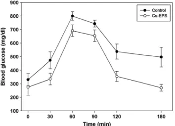
After Cs-EPS treatment for 7 weeks, glucose tolerance was examined using the OGTT methodology. The OGGT data showed the changes in the levels of blood glucose during oral glucose tolerance test (2 g of glucose per kg of body weight) as can be seen in Fig. 2. Compared with the diabetic groups (control mice), Cs-EPS-treated groups showed improvement in overall glucose response. The diabetic groups treated with glucose showed significant increase in the blood glucose level
Fig. 2. Blood glucose levels after the oral glucose tolerance test in
the ob/ob mice. Values are mean ± SD for seven mice in each group.Filled circle, Control group; open circle, Cs-EPS treated group.
after 30 min, and did not recovered its baseline even after 120 min. However, in EPS-treated mice, blood glucose concentration was decreased significantly at 120 min after glucose administration compared to control mice, and returned to the initial glucose level after 180 min, indicating that EPS treatment in ob/ob mice exhibited a significant increase in glucose consumption and there may be due to the presence of some compound in the EPS to largely improve glucose tolerance.
3.4. Effect of Cs-EPS on plasma insulin levels
As shown in Table 3, we studied the effect of Cs-EPS on insulin levels in plasma after its administration in ob/ob mice. After treatment with Cs-EPS for 7 weeks, the insulin levels in plasma were significantly increased in Cs-EPS treated mice (56.40 ± 17.51 ng/mL after 7 weeks) when compared to the initial value (31.84 ± 2.51 ng/mL at day 0).
The plasma insulin levels were measured to be two-fold higher in the Cs-EPS treated group (56.40 ± 17.51 ng/mL) than the control group (28.07 ± 6.34 ng/mL) after 7 weeks, indicating the role of EPS from C. sinensis as an effective insulin-enhancing agent. Similar hypoglycemic effects were reported in ob/ob mice, in which they used exopolysaccharides produced from Tremella fuciformis and Phellinus baumii [5].
They suggested that possible mechanisms by which EPS brings about its hypoglycemic action in diabetic mice may be by creating a potentiating effect of insulin in plasma or by increasing either the pancreatic secretion of insulin from the existing - β cells as an insulin enhancing agent or its release from the bound form. Generally, insulin resistance leads to hyperinsulinemia, resulting high blood glucose levels and type II diabetes. However, our data showed that Cs-EPS treatment improved glucose tolerance and decreased blood glucose level by an increase in insulin sensitivity.
Table 3. Effect of Cs-EPS on plasma insulin levels in ob/ob mice
a Group Plasma insulin level (ng/mL)Day 0 Day 49
Control Cs EPS
22.96 ± 2.94 31.84 ± 2.51
28.07 ± 6.34 56.40 ± 17.51*
aValues are means ± S.D. (n = 7).
*
P < 0.05, compared with that in control mice.
3.5. Effect of EPS on plasma lipids and cholesterol
It was reported that the main features of insulin resistance
include dyslipidemia, which is characterized by high triglyceride,
low high-density lipoprotein (HDL), and dense low-density
lipoprotein (LDL) [43]. Accordingly, we also examined
changes of total plasma cholesterol and triglyceride after
Cs-EPS treatment for 7 weeks. As shown in Table 4, the total
cholesterol levels in EPS-treated group (98 ± 18 mg/dl) was
lower than control ob/ob mice (normal ranges, 130 250 mg/dl) ∼ ,
and the total triglyceride (TG) levels in Cs-EPS-treated groups (73 ± 12 mg/dl) were significantly decreased by approximately 50% when compared to those of the control ob/ob mice (normal ranges, 35~130 mg/dl). Kim et al. [44]
have also reported that plasma levels of total TG and cholesterol were decreased in cinnamon extract-treated ob/ob mice. The mechanism was explained by the adenosine monophosphate-activated protein kinase (AMPK)-enhanced triacylglycerol lipase activity that increases glycogen synthesis in the liver and enhances glucose uptake in skeletal muscle and adipocytes [44]. Our results suggest that dietary EPS improved the composition of blood lipid profiles in ob/ob mice and therefore might has another potential as an anti-obesity ingredient in the application of oriental medicine compounds [45].
Table 4. Effect of Cs-EPS on plasma levels of total cholesterol and
triglyceride in the ob/ob mice after treatments for 7 weeksaGroup Total cholesterol (mg/dl) Triglyceride (mg/dl) Control
Cs EPS
110 ± 20 98 ± 18
153 ± 22 73 ± 12*
aValues are means ± S.D. (n = 7).
*P < 0.05, compared with that ofcontrol mice.
3.6. Effect of Cs-EPS on leptin expression
Leptin, the product of the ob gene and a 16 kDa circulating hormone produced and released by adipocytes, regulates body weight homeostasis through controls of food intake and energy expenditure [46,47]. It was reported that leptin treatment effectively corrects hyperglycemia and hyperinsulinemia in leptin-deficient mice and humans [32,48-51]. Other studies showed the effect of leptin to increase glucose metabolism in the wild-type and ob/ob mice [52,53]. Also, leptin is emerging as a pleiotropic molecule involved in a variety of physiological and pathological conditions such as inflammation, immunity, hematopoiesis, and anti-apoptotic activity [47]. We investigated whether Cs-EPS regulates the expression level of leptin in adipose tissue of ob/ob mice. As can be seen in Fig. 3, the transcriptional level of leptin mRNA in adipose tissue were up-regulated by approximately 69% after treating with Cs-EPS.
Furthermore, western blot analysis showed that the levels of leptin protein in adipose tissue and plasma were also enhanced by approximately 49% and 72%, respectively after treating with Cs-EPS compared to the control ob/ob mice (Fig. 4(a) and 4(b)). Accordingly, some components of Cs-EPS, which had not been elucidated in detail, may be involved in transcriptional regulation of leptin gene (ob) upstream. It appears that increase of leptin expression stimulates its physiological activities in leptin-deficient (ob/ob) mice. This effect is not only attributable to s decreased food intake but also to an increased basal metabolic rate with selective promotion of fat metabolism, resulting in reduction of body
weight, decrease of triglyceride, and stimulation of signaling pathway. It was reviewed that plasma leptin concentrations are correlated with total fat mass, percent body fat and body mass index acting as a sensing hormone in a negative feedback control from adipose tissue to the hypothalamus, the brain center responsible for satiety [28], suggesting that leptin plays an important role in type II diabetes, obesity and metabolic disorders.
Fig. 3. Effects of Cs-EPS on the expression of leptin mRNA of
adipocyte in ob/ob mice. Values are mean ± SD of triple determinations.* Signifies different from control at the P < 0.05 level.
(a)
(b)
Fig. 4. Detection of leptin protein in (A) Adipose tissue and (B)
Plasma of ob/ob mice by Western blotting. Values are mean ± SD of triple determinations.In conclusion, we have demonstrated that the administration
of Cs-EPS caused not only the significant reduction of fasting
blood glucose levels but also the improvement of glucose tolerance in ob/ob mice. In addition, plasma levels of insulin and leptin were increased by treating with Cs-EPS. The increases in leptin expressions may be related to the regulation of hyperglycemia and dyslipidemia in ob/ob mice. Taken these results together, we suggested that Cs-EPS would be an excellent candidate to develop as functional food additives or therapeutic agents in anti-obesity and anti-diabetic through improvement in glucose homeostasis and preservation of insulin reserves. Further study will be processing about elucidating components of Cs-EPS which directly affect on anti-diabetic effect.
Acknowledgement
This study was supported by the Technology Development Program for Food, Ministry for Food, Agriculture, Forestry and Fisheries, Republic of Korea (Project No.: 109144-03-2).
References
1. Zimmet, P., K. G. M. M. Alberti, and J. Shaw (2001) Global and societal implications of the diabetes epidemic. Nature 414: 782-787.
2. Polonsky, K. S., J. Sturis, and G. I. Bell (1996) Non-insulin- dependent diabetes mellitus a genetically programmed failure of the beta cell to compensate for insulin resistance. New Engl.
J. Med. 334: 777-783.
3. Taylor, S. I. (1999) Deconstructing type 2 diabetes. Cell 97:
9-12.
4. Holman, R. R. and R. C. Turner (1991) Oral agents and insulin in the treatment of NIDDM. pp 467-469. In: Pickup, J. and G.
Williams (eds) Text book of Diabetes. Blackwell Scientific Publication, Oxford, UK.
5. Cho, E. J., H. J. Hwang, S. W. Kim, J. Y. Oh, Y. M. Baek, J. W.
Choi, S. H. Bae, and J. W. Yun (2007) Hypoglycemic effects of exopolysaccharides produced by mycelia cultures of two different mushrooms Tremella fuciformis and Phellinus baumii in ob/ob mice. Appl. Micribiol. Biotechnol. 75: 1257-1265.
6. Attele, A. S., Y. P. Zhou, J. T. Xie, J. A. Wu, L. Zhang, and L.
Dey (2002) Antidiabetic effects of Panax ginseng berry extract and the identification of an effective component. Diabetes 51: 1851-1858.
7. Xie, J. T., H. H. Aung, J. A. Wu, A. S. Attele, and C. S. Yuan (2002a) Effects of American ginseng berry extract on blood glucose levels in ob/ob mice. Am. J. Clin. Med. 30: 187-194.
8. Xie, J. T., Y. P. Zhou, L. Dey, A. S. Attele, J. A. Wu, and M. Gu (2002b) Ginseng berry reduces blood glucose and body weight in db/db mice. Phytomedicine 9: 254-258.
9. Li, W. L., H. C. Zheng, J. Bukuru, and N. De Kimpe (2004) Natural medicines used in the traditional Chinese medical system for therapy of Diabetes mellitus. J. Ethanopharm. 92: 1-21.
10. Kiho, T., A. Yamane, J. Hui, S. Usui, and S. Ukai (1996) Polysaccharide in fungi, XXXVI. Hypoglycemic activity of a polysaccharide (CS-F30) from the cultural mycelium Cordyceps
sinensis and its effect onglucose metabolism in mouse liver.
Biol. Pharm. Bull. 19: 294-296.
11. Yang, B. K., D. H. Kim, S. C. Jeong, S. Das, Y. S. Choi, J. S.
Shin, S. C. Lee, and C. H. Song (2002) Hypoglycemic effect of a Lentinus edodes exo-polymer produced from a submerged mycelial culture. Biosci. Biotechnol. Biochem. 66: 937-942.
12. Kobayashi, Y (1982) Key to the taxa of the genera Cordyceps and Torrubiella. Trans Mycol. Soc. Japan 23: 329-364.
13. Spatafora, J. W. and M. Blackwell (1993) Molecular systematic of unitunicate perithecia ascomycetes; the Clavicipitales- Hypocreales connection. Mycologia 85: 912-922.
14. Li, Q. S., W. Zeng, D. H. Yi, and T. F. Huang (1998) Studies on the alternation of generations in Cordyceps sinensis. Chung
Kuo Yao Tsa Chil 23: 210-212.
15. Kuo, Y. C., W. J. Tsai, M. S. Shiao, C. F. Chen, and C. Y. Lin (1996) Cordyceps sinensis as an immunomodulatory agent.
Am. J. Clin. Med. 24: 111-125.
16. Lee, J., C. Chung, H. Jeong, and K. Lee, K. (1990) Anticomplementary and antitumor activities of the alkali extract from the mycelia of Lentinus edodes IY 105. Kor. J. Appl. Microbiol. Biotechnol.
18: 571-577.
17. Bok, J. W., L, Lermer, J. Chilton, H. G. Klingeman, and G. H.
Towers (1999) Antitumor sterols from the mycelia of Cordyceps
sinensis. Phytochem. 51: 891-898.
18. Manabe, N., M. Sugimoto, Y. Azuma, N. Taketomo, A. Yamashita, H. Tsuboi, A. Tsunoo, N. Kinjo, H. Nian-Lai, and H. Miyamoto (1996) Effect of the mycelial extract of cultured Cordyceps
sinensis on in vivo hepatic energy metabolism in the mouse.
Jpn. J. Pharmacol. 70: 23-29.
19. Wang, S. M., L. J. Lee, W. W. Lin, and C. M. Chang (1998) Effect of a water-soluble extract of Cordyceps sinensison steroidogenesis and capsular morphology of lipid droplet in cultured rat adrenocortical cell. J. Cell Biochem. 69: 483-489.
20. Zhu, X. Y. and H. Y. Yu (1990) Immunosuppressive effect of cultured Cordyceps sinensis on cellular immune response.
Chung Hsi I Chieh Ho Tsa Chih 10: 485-487.
21. Zhang, Z. and S. S. Xia (1990) Cordyceps sinensis as an immunosuppressant in heterotopic heart allograft model in rats. J. Tongji. Med. Univ. 10: 100-103.
22. Kim, S. W., H. J. Hwang, C. P. Xu, J. M. Sung, J. W. Choi, and J. W. Yun (2003) Optimization of submerged culture process for the production of mycelia biomass and exopolysaccharide by Cordyceps militaris C738. J. Appl. Microbiol. 94: 120-126.
23. Hwang, H. J., S. W. Kim, C. P. Xu, J. W. Choi, and J. W. Yun (2003) Production and molecular characteristics of four group of exopolysaccharides from submerged culture of Phellinus
gilvus. J. Appl. Microbiol. 94: 708-719.
24. Cho, E. J., J. Y. Oh, H. Y. Chang, and J. W. Yun (2006) Production of exopolysaccharides by submerged mycelial culture of
Tremella fuciformis. J. Biotechnol. 127: 129-140.
25. Zhang, Y., R. Proenca, M. Maffei, M. Barone, L. Leopold, and J. M. Friedman (1994) Positional cloning of the mouse obese gene and its human homologue. Nature 372: 425-432.
26. Baskin, D. G., J. E. Blevins, and M. W. Schwartz (2001) How the brain regulates food intake and body weight: the role of leptin. J. Pediatr. Endocrinol. Metabol. 14: 1417-1429.
27. Mantzoros, C. S. (2001) The role of leptin and hypothalamic neuropeptides in energy homeostasis: update on leptin in obesity. Growth Hormone and IGF Research 11A, S85-S89.
28. Fruhbeck, G. and J. Salvador (2000) Relation between leptin and the regulation of glucose metabolism. Diabetologia 43: 3-12.
29. Ceddia, R. B., H. A. Koistinen, J. R. Zierath, and G. Sweeney (2002) Analysis of paradoxical observations on the association between leptin and insulin resistance. FASEB Journal 16:
1163-1176.
30. Brann, D.W., M. F. Wade, K. M. Dhandapani, V. B. Mahesh, and C. D. Buchanan (2002) Leptin and reproduction. Steroids 67: 95-104.
31. Moschos, S., J. L. Chan, and C. S. Mantzoros (2002) Leptin and reproduction: a review. Fertility and Sterility 77: 433-444.
32. Montague, C. T., I. S. Farroqi, J. P. Whitehead, M. A. Soos, H. Rau, N. J. Wareham, C. P. Sewter, J. E. Digby, S. N.
Mohammed, J. A. Hurst, C. H. Cheetham, A. R. Earley, A. H.
Barnett, J. B. Prins, and S. O’Rahilly (1997) Congenital leptin deficiency is associated with severe early-inset obesity in humans. Nature 387: 903-908.
33. Dubue, G. R., S. D. Phinney, J. S. Stern, and P. J. Havel (1998) Changes of serum leptin and endocrine and metabolic parameters after 7 days of energy restriction in men and women.
Metabol. 47: 429-434.
34. Bradford, M. M. (1976) A rapid and sensitive method for the quantification of microgram quantities of protein utilizing the principle of protein-dye binding. Anal. Biochem. 72:
248-254.
35. Dubois, M., K. A. Gillis, J. K. Hamilton, P. A. Rebers, and F.
Smith (1956) Colorimetric method for determination of sugar and related substance. Anal. Chem. 28: 350-356.
36. Koh, J. H., K. W. Yu, H. J. Suh, Y. M. Choi, and T. S. Ahn (2002) Activation of macrophages and the intestinal immune system by an orally administered decoction from cultured mycelia of Cordyceps sinensis. Biosci. Biotechnol. Biochem.
66: 407-411.
37. Suh, H. J. and J. W. Choi (2010) Morphological changes by submerged culture conditions for the mycelia optimal growth of
Cordyceps sinensisand immunological properties of hot water
extract of mycelium. Kor. Soc. Biotech. Bioeng. J. 25: 47-54.38. Yu, K. W., Y. S. Kim, K. S. Shin, J. M. Kim, and H. J. Suh (2005) Macrophage-stimulating activity of exobiopolymer from cultured rice bran with Monascus pilosus. Appl. Biochem.
Biotechnol. 126: 35-48.
39. Yamada, H. (1984) Structure and antitumor activity of an alkali-soluble polysaccharide from Cordyceps ophioglossoides.
Carbohydr. Res. 125: 107-115.
40. Song, C. H., Y. J. Jeon, B. K. Yang, K. S. Ra, and J. M. Sung (1998) The anti-complementary activity of exo-polymers produced from submerged mycelial cultures of higher fungi with particular reference to Cordyceps militaris. J. Microbiol.
Biotechnol. 8: 536-539.
41. Yang, B. K., J. Y. Ha, S. C. Jeong, S. Das, J. W. Yun, Y. S. Lee, J. W. Choi, and C. H. Song (2000) Production of exo-polymers by submerged mycelial culture of Cordyceps militaris and its hypolipidemic effect. J. Microbiol. Biotechnol. 10: 784-788.
42. Yang, B. K., D. H. Kim, and C. H. Song (2002) Production of Lentinus edodes mycelia in submerged culture and it’s hypoglycemic effect in diabetic rats. Korean J. Mycol. 30:
131-135.
43. Peter, P., S. L. Nuttall, and M. J. Kendall (2003) Insulin resistance- the new goal. J. Clin. Pharm. Ther. 28: 167-174.
44. Kim, S. H., S. H. Hyun, and S. Y. Choung (2006) Anti-diabetic effect of cinnamon extract on blood glucose in db/db mice. J.
Ethnopharmacol. 104: 119-123.
45. Chae, M. H., J. G. No, and D. Y. Jhon (2007) Hangbisan, Sulfur- based Oriental Medicine, Lowers the Blood Cholesterol Level of
ob/ob Obese Mice. J. Korean Soc. Food Sci. Nutr. 36: 27-31.
46. Seekamp, A., G. O. Till, M. S. Mulligan, J. C. Paulson, D. C.
Anderson, M. Miyasaka, and P. A. Ward (1994) Role of selectins in local and remote tissue injury following ischemia and reperfusion. Am. J. Pathol. 144: 592-598.
47. Fantuzzi, G. and R. Faggioni (2000) Leptin in the regulation of immunity, inflammation, and hematopoiesis. J. Leukoc. Biol.
68: 437-446.
48. Farooqi, I. S., S. A. Jebb, G. Langmack, E. Lawrence, C. H.
Cheetham, A. M. Prentice, I. A. Hughes, M. A. McCamish, and S. O’Rahilly (1999) Effects of recombinant leptin therapy in a child with congenital leptin deficiency. New Engl. J. Med.
341: 879-884.
49. Muzzin, P., R. C. Eisensmith, K. C. Copeland, and S. L. Woo (1996) Correction of obesity and diabetes in genetically obese mice by leptin gene therapy. Proc. Natl. Acad. Sci. USA. 93:
14804-14808.
50. Pelleymounter, M. A., M. J. Cullen, M. B. Baker, R. Hecht, D.
Winters, T. Boone, and F. Collins (1995) Effects of the obese gene product on body weight regulation in ob/ob mice. Science 269: 540-543.
51. Yu, Y., S. K. Hu, and H. Yan (2008) The study of insulin resistance and leptin resistance on the model of simplicity obesity rats by curcumin. Zhonghua Yu Fang Yi Xue Za Zhi 42: 818-822.
52. Burcelin, R., S. Kamohara, J. Li, G. S. Tannenbaum, M. J.
Charron, J. M. Friedman (1999) Acute intravenous leptin infusion increases glucose turnover but not skeletal muscle glucose uptake in ob/ob mice. Diabetes 48: 1264-1269.
53. Kamohara, S., R. Burcelin, J. L. Halaas, J. M. Friedman, and M. J. Charron (1997) Acute stimulation of glucose metabolism in mice by leptin treatment. Nature 389: 374-377.