Korean J. Environ. Biol. 36(3) : 299~307(2018) https://doi.org/10.11626/KJEB.2018.36.3.299
INTRODUCTION
Cyanobacteria or blue-green algae are a prokaryote that plays a role of primary producer through photosynthesis since about 3.5 billions years(Graham et al. 2009). The cy- anobacteria are found in various environments even extreme conditions like hot spring, polar region, ultra-oligotrophic or high pH/salinity water(Parker et al. 1981; Wharton et al.
1983; Whitton 2012).
Planktonic cyanobacteria are floating algae suspended in slow moving water such as in a lake. They usually clustered within 30cm of the water surface caused by colonization, mucilaginous sheath and gas vesicles. It often produces bright green or blue-green soup-like scums(Samuel et al.
2014).
Nowadays, freshwater habitats are threatened world- wide by blooming of cyanobacteria, such as Microcystis, Anabaena. Once they are bloomed, they can cause the prob- lems in water environments and drinking water supplies by
producing toxins like microcystin and anatoxin(Codd 1995;
Kim et al. 1995). Therefore, continuous researches of cya- nobacteria, especially Microcystis, Anabaena, Oscillatoria, and Aphanizomenon are being conducted worldwide(Park and Kim 1995; Park 2005; Lee et al. 2017).
The 4,617 taxa of cyanobacteria have been reported to AlgaeBase(Guiry and Guiry 2018), and 377 taxa have been reported in Korea(Kim 2015; Song and Lee 2017; Yim et al. 2017). Microcystis have been reported 51 taxa in world- wide, and 7 species in Korea. Anabaena have been reported 151 taxa in worldwide(Guiry and Guiry 2018), and 10 taxa in Korea.
For this study, we collected unrecorded cyanobacteria from fresh and brackish waters to add the algal flora of Ko- rea.
MATERIALS AND METHODS
We collected planktonic cyanobacteria from nine sites in Korea from August 2016 to May 2018(Fig. 1; Tables 1,
* Corresponding author: Ok Min Lee, Tel. 031-249-9643, Fax. 031-241-0860, E-mail. [email protected]
ⓒ2018. Korean Society of Environmental Biology.
A Study of Nine Unrecorded Species of Planktonic Cyanobacteria (Cyanophyceae, Cyanophyta) in Korea
Byoung Cheol Yim, Hyun Chul Jung, Sung Do Bang and Ok Min Lee*
Department of Life Science, College of Natural Science, Kyonggi University, Suwon 16227, Republic of Korea Abstract - Samples were collected from planktonic habitats of the fresh and brackish waters in Korea from August 2016 to May 2018. As a result, three genera and nine species were newly recorded in Korea. The unrecorded indigenous genera were Anathece, Chondrocystis and Geminocystis, and nine species were Anabaenopsis arnoldii, Anathece smithii, Chondrocystis dermochroa, Coelosphaerium aerugineum, Eucapsis microscopica, Geminocystis herdmanii, Microcystis panniformis, Synechococcus nidulans and Woronichinia karelica. Anathece smithii, Coelosphaerium aerugineum, Eucapsis microscopica, Microcystis panniformis and Synechococcus nidulans had been reported to inhabit freshwater, but these were found in brackish water in this study. Microcystis panniformis, which is a potential genus for causing green-tide, is taxonomically valuable in Korea.
Keywords : brackish water, cyanobacteria, Korean unrecorded species, planktonic
<Original article>
2). They were collected using phytoplankton net with mesh size 20μm and diameter 30 cm(Sournia 1978). Each sam- ple was sealed in sterile bottles and transported to the lab- oratory(Crispim et al. 2004). Measurement of salinity was performed by marine tester(DMT-10, DYS, Korea).
The samples were examined under BX53 light micro- scope at ×400-1,000(Olympus, Tokyo, Japan) and photo- graphed using AxioCam HRC camera(Carl Zeiss, Oberko- chen, Germany). The taxonomic classification system was based on Komárek et al.(2014) and Algaebase(Guiry and Guiry 2018). In this study, identification of cyanobacteia re- ferred to Hirose et al.(1977), Prescott(1982) Chung(1993), Komárek and Anagnostidis(1999, 2005), John et al.(2011) and Komárek(2013).
RESULTS AND DISCUSSION
Three genera and nine species of planktonic cyanobacte- ria were newly recorded in Korea. The newly recorded gen- era were Anathece, Chondrocystis and Geminocystis, and the newly recorded species were Anabaenopsis arnoldii, Anathece smithii, Chondrocystis dermochroa, Coelosphae-
rium aerugineum, Eucapsis microscopica, Geminocystis herdmanii, Microcystis panniformis, Synechococcus nidu- lans and Woronichinia karelica.
Order Chroococcales Schaffner 1922 Family Chroococcaceae Rabenhorst 1863 Genus Chondrocystis Lemmermann 1899
Colonies are free-living and more or less irregular or ir- regular-polygonal. Colonies are surrounded by packet-like outline, gelatinous, laterally usually flattened or(in masses) slimy mucilage envelopes. They are composed of numerous partial subcolonies. Subcolonies are surrounded by more or less cubic or polygonal flattened, widened, colourless, de- limited slime, cells with individual thin, gelatinous, slightly stratified envelopes. Envelopes are partially thickened.
Reproduction is occured by small subcolonies after colony disintegration. This genus is newly recorded in Korea.
Chondrocystis dermochroa(Nägeli ex Kützing) Komárek and Anagnostidis 1995(Fig. 2)
Synonym: Gloeocapsa dermochroa Nägeli ex Kützing 1849 Table 1. Sampling sites in Korea from where planktonic samples were collected from August 2016 to May 2018
Site Local name Latitude (N) Longitude (E)
st.1 629-3, Bangsan-dong, Siheung-si, Gyeonggi-do 37.413256 126.755274
st.2 1169, Jangcheon-ri, Ibang-myeon, Changnyeong-gun, Gyeongsangnam-do 35.610430 128.357497 st.3 1-5, Deokcheon-ri, Byeonggok-myeon, Yeongdeok-gun, G yeongsangbuk-do 36.568571 129.420167 st.4 49, Uljindaege-ro, Hupo-myeon, Uljin-gun, Gyeongsangbuk-do 36.677738 129.446123
st.5 1061-3, Gupo-dong, Buk-gu, Busan 35.203433 128.992730
st.6 1-55, Nonhyeon-dong, Namdong-gu, Incheon 37.412225 126.748987
st.7 150, Yonggok-ri, Jangdong-myeon, Jangheung-gun, Jeollanam-do 34.718328 126.955823 st.8 1108-14, Anseong-ri, Dongjin-myeon, Buan-gun, Jeollabuk-do 35.784821 126.755452
st.9 275-6, Daeeum-ri, Inju-myeon, Asan-si, Chungcheongnam-do 36.854477 126.851721
Table 2. The environment of sampling sites from August 2016 to May 2018
Site Collection date Environment Salinity(‰)
st.1 May 4, 2018 Eutrophic and brackish paddy field 0.6
st.2 Aug. 24, 2016 Midstream of Nakdong river -
st.3 Mar. 30, 2018 Eutrophic and brackish riverside nearby farmlands 1
st.4 Sep. 23, 2017 Eutrophic brackish waterway 5.1
st.5 Jun. 9, 2017 Downstream of Nakdong river -
st.6 May 4, 2018 Mesotrophic and brackish wetland 6.6
st.7 Aug. 7, 2017 Mesotrophic reservoir at mountainside -
st.8 Sep. 26, 2017 Brackish waterway at reclaimed land 0.2
st.9 Aug. 7, 2017 Deep-water of seawall -
Colony size is microscopic or rarely macroscopic. Col- onies are more or less spherical, hemispherical or oval.
Large colonies are composed of very densely packed and small subcolonies. Subcolonies are spherical or polygonal- ly-rounded, and composed of clustered cells. Mucilaginous envelope that surround the subcolony is narrow and firm, but later diffluent. Mucilaginous envelope forms single or slightly lamellated layers. The cell is spherical and blue- green in color. Cell diameter is 1.5-3μm.
Ecology: This species appears in freshwater and subaero- phytic habitat. This species lives usually in limestone areas or on rocks with periodically flowing water, but rarely in the water-level zone of streams(Komárek and Anagnostidis 1999). We collected it from midstream of Nakdong river.
Distribution: Arctic: Svalbard(Matula et al. 2007); North America: Arkansas(Smith 2010).
Site of collection: Yulji-gyo, Changnyeong-gun, Gyeong-
sangnam-do(August 24, 2016).
Specimen Locality: ACKU2018MD03
Genus Geminocystis Korelusová et al. 2009
Cells are solitary, and spherical or slightly oval, after di- vision hemispherical. Cell’s diameter is 3-10μm. Mucilage is abscent or narrow, fine, colorless, and usually diffluent and indistinct mucilaginous envelopes. Cell contents are homogeneous, without separation of centro- and chromato- plasma, sometimes with lengthwise striation(thylakoids sit- uated in cells lengthwise). Color of cells is pale blue-green, bright-green, grey, or pinkish. Cells are divided by binary fission into two morphologically equal, hemispherical daughter cells, which reach the original globular shape be- fore next division. This genus is newly recorded in Korea.
Geminocystis herdmanii Korelusová et al. 2009(Fig. 3) They are solitary, not forming a colony. The cell is spher- ical but hemispherical after cell division by binary fission.
Cell contents are homogeneous or has slightly irregularly keritomic thylakoid content. Thylakoids in cells are ar- ranged irregularly or in parallel. The cell is blue-green in color and diameter is 3-5μm.
Ecology: This species appears in freshwater and planktonic Fig. 1. The map showing the nine sampling sites in Korea from
August 2016 to May 2018.
Fig. 2. Microscopic photographs of Chondrocystis dermochroa (Nägeli ex Kützing) Komárek and Anagnostidis. The pho- tographs show the pellets of cells (A-C) and the shape of cells (D). Scale bar: 10 μm.
A B
C D
habitat. They lives in oligotrophic lakes(Komárek and An- agnostidis 1999). We collected it from mesotrophic reser- voir at mountainside.
Distribution: North America: Wisconsin(Korelusová et al.
2009).
Site of collection: Yonggok-reservoir, Jangdong-myeon, Jangheung-gun, Jeollanam-do(August 7, 2017).
Specimen Locality: ACKU2018MD02
Family Synechococcaceae Komárek and Anagnostidis 1995
Genus Anathece(Komárek and Anagnostidis) Komárek et al. 2011
Colonies are multicellular, irregular, microscopic or rare- ly macroscopic. They are composed of irregularly arranged cells and surrounded by fine, colourless, homogeneous and diffluent mucilage, but sometimes they disintegrat- ed in solitary cells. Colonies fragment into solitary cells.
Mostly planktonic, rarely benthic. Cells are oval to rod- like, straight or slightly arcuate, without own mucilaginous envelopes. Cells are pale-grey, blue-green or olive-green colored, and(0.8)1-2(6)μm long, 0.3-2μm width. Cell division is occured by binary fission, perpendicular to the long axis. This genus is newly recorded in Korea.
Anathece smithii(Komárková-Legnerová and Cron- berg) Komárek et al. 2011(Fig. 4)
Synonym: Aphanothece smithii Komárková-Legnerová and G. Cronberg 1994
Colonies are microscopic and free floating. Colonies are more or less spherical or irregular in outline and sometimes elongated. Cells that form colonies are usually arranged regularly, not very densely. Mucilage is colorless and usual- ly wraps all the cells by a narrow, hyaline. The cell is more or less oval to cylindrical and straight. The cell is pale blue- green or grey-blue in color. The cell’s length is 2-3.5μm and width is 1-1.5μm.
Ecology: This species occurs in freshwater and planktonic habitat. They lives in mesotrophic and slightly eutrophic, usually large water bodies(Komárek and Anagnostidis 1999). In this study, we collected it from eutrophic brackish waterway(salinity 5.1‰).
Distribution: Australia and New Zealand: Queensland (Bostock and Holland 2010).
Site of collection: Eutrophic waterway, Hupo-myeon, Ul- jin-gun, Gyeongsangbuk-do(September 23, 2017).
Specimen Locality: ACKU2017IR10 Genus Synechococcus Nägeli 1849
Synechococcus nidulans(Pringsheim) Komárek in Bourrelly 1970(Fig. 5)
Fig. 4. Microscopic photographs (A, C) and illustration (D) of Anathece smithii (Komárková-Legnerová and Cronberg) Komárek et al. The photographs show the cells enveloped in very diffluent and hyaline mucilaginous sheath. Scale bars: 10 μm.
A B
C D
Fig. 3. Microscopic photographs of Geminocystis herdmanii Kore- lusová et al. The photographs show the shape of cells and arrangement of thylakoids (red point). Scale bar: 10 μm.
A B
C D
Synonym: Lauterbornia nidulans Pringsheim 1968
Cells are solitary and free-floating. The cells are straight or sometimes slightly sigmoid or arcuate and without muci- lage. Cell contents are homogeneous and pale blue-green in color. Cell’s length is 1.5-6.5μm and width is 1-1.5μm.
Ecology: This species appears in freshwater and planktonic habitit. They lives in small water bodies, ditch ponds and large lakes(Komárek and Anagnostidis 2005). We collected it from eutrophic and brackish waterway(salinity 1‰).
Distribution: South America: Brazil(Lopes et al. 2005);
Australia and New Zealand: Queensland(Bostock and Hol- land 2010).
Site of collection: Goraebuldae-gyo, Byeonggok-myeon, Yeongdeok-gun, Gyeongsangbuk-do(March 30, 2018).
Specimen Locality: ACKU2018IR06
Order Nostocales Borzì 1914 Family Aphanizomenonaceae Genus Anabaenopsis Miller 1923
Anabaenopsis arnoldii Aptekar 1926(Fig. 6)
Trichomes are free-floating, solitary or clustered in small,
irregular colonies, irregularly screw-like coiled, with 1-12 coils. The coil width is ±25μm, and the length is 7-30μm, constricted at the cross-walls, with wide, colourless, difflu- ent mucilaginous envelopes. Cells are spherical or widely barrel-shaped, yellow-green. Heterocytes are spherical, 8-10×7-10μm.
Ecology: This species occurs in freshwater ponds and lakes Fig. 5. Microscopic photographs of Synechococcus nidulans (Pringsheim) Komárek in Bourrelly. The photographs show the irregular shape
of cells (red arrows) and the cells surrounded by other cyanobacteria. Scale bar: 5 μm.
A B C
D E F
Fig. 6. Microscopic photographs of Anabaenopsis arnoldii Apteka.
The photographs show the specific coiling arrangement of cells. The red arrows point towards the heterocyst (right) and akinete (left). Scale bars: 10 μm (A, B), 20 μm (C, D).
A B
C D
(Komárek 2013). We collected it from seawall.
Distribution: Russia(Medvedeva and Nikulina 2014); Aus- tralia and New Zealand: Queensland(Bostock and Holland 2010).
Site of collection: Deep-water of seawall, Inju-myeon, Asan-si, Chungcheongnam-do(August 7, 2017).
Specimen Locality: ACKU2017NR17
Family Coelosphaeriaceae Elenkin 1933 Genus Coelosphaerium Nägeli 1849
Coelosphaerium aerugineum Lemmermann 1898(Fig. 7)
Colonies are free floating and spherical. However, some- times they consist of 2-3 hemispherical subcolonies with irregularly and sparsely or densely aggregated cells(where they never touch one another). Cells are spherical or hemi- spherical after division and have a bit of a granule, but without gas vesicles. The cells are usually bright blue-green in color and diameter is 2.2-3.5μm.
Ecology: This species appears in freshwater and mainly planktonic habitit. They lives in eutrophic to mesotrophic water bodies(Komárek and Anagnostidis 1999). We col- lected it from brackish waterway(salinity 0.2‰).
Distribution: North America: Arkansas(Smith 2010).
Site of collection: Okpo-gyo, Dongjin-myeon, Buan-gun,
Jeollabuk-do(September 26, 2017).
Specimen Locality: ACKU2017IR01
Genus Woronichinia Elenkin 1933
Woronichinia karelica Komárek and Komárková- Legnerová 1992(Fig. 8)
Colonies are solitary and irregularly spherical or oval, and they are composed of subcolonies when matured. The colonies are composed of cells that arranged radially and densely packed in the peripheral layer, and which grow up to 50μm in diameter. The cells joined by the faintly visible, colorless and thick stalks radiating from the colonial center.
The cells are more or less elongated and obovated with a homogeneous contents and pale blue-green in color. The cell’s length is 3-3.5μm and width is 1.5-2μm.
Ecology: This species appears in freshwater and planktonic habitat. They lives in oligo- or mesotrophic lakes and ponds of northern part of the whole temperate zone(Komárek and Anagnostidis 1999). We collected it from downstream of Nakdong river.
Distribution: Asia: Russia(Medvedeva and Nikulina 2014).
Site of collection: Gupodae-gyo, Gupo-dong, Buk-gu, Bu- Fig. 7. Microscopic photographs of Coelosphaerium aerugineum
Lemmermann with focus on the surface of the colony (A, B) and absence of stalks (C, D). Scale bar: 10 μm.
A B
C D
Fig. 8. Microscopic photographs of Woronichinia karelica Ko- márek and Komárková-Legnerová with focus on the sur- face (A, B) and the middle part (C, D) of the colonies. The red arrow points towards the mucilaginous sheath. Scale bar: 10 μm.
A B
C D
san(June 9, 2017).
Specimen Locality: ACKU2017NR18
Family Merismopediaceae Elenkin 1933 Genus Eucapsis Clements and Shantz 1909
Eucapsis microscopica(Komárková-Legnerová and G. Cronberg) Komárek and Hindák 2016(Fig. 9) Synonym: Chroococcus microscopicus Komárková-Legn-
erová and G. Cronberg 1994
Colonies are microscopic and free floating. They are composed of numerous cumulus-like densely packed groups of cells. Mucilage is colorless, without structure, delicate and more or less clearly delimited or diffluent.
Cells are spherical and usually 4-8 cells forms subcolonies arranged regularly and surrounded by a mucilage, which is easily recognizable after staining or in phase contrast. The cell contents are homogeneous and pale blue-green in color.
The cell’s diameter is 0.9-1.3μm.
Ecology: This species appears in freshwater and planktonic habitit. They lives in mesotrophic lakes; known only from Sweden, but probably more widely distributed in northern lakes(Komárek and Anagnostidis 1999). We collected it from mesotrophic and brackish wetland(salinity 6.6‰).
Distribution: Europe: Baltic Sea(Hällfors 2004).
Site of collection: Soreapogu-wetland, Nonhyeon-dong, Namdong-gu, Incheon(May 4, 2018).
Specimen Locality: ACKU2018IR01
Family Microcystaceae Elenkin 1933 Genus Microcystis Lemmermann 1907
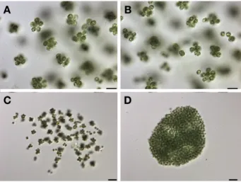
Microcystis panniformis Komárek, Komárková-Legn- erová, Sant’Anna, Azevedo and Senna 2002(Fig. 10) Colonies are microscopic and macroscopic. The young colonies are subsphaerical close to a sphere; as they ma- ture, they get somewhat flattened, growing irregularly into a lobe-shape. Cells that form colonies are arranged homogeneously or slightly clathrate. Mucilage is colorless, homogeneous, diffluent and indistinct of the margin. Repro- duction of cells happen on the margin of matured colonies.
Cells are spherical or hemispherical after division, and they have gas vesicles. The cells are yellow-green or olive brown in color, and their diameter is 3.5-4.5μm.
Ecology: This species are founded in freshwater and plank- tonic habitit. They lives in subtropical eutrophic lakes and ponds(Komárek et al. 2002). We collected it from eutrophic and brackish paddy pond(salinity 0.6‰).
Fig. 9. Microscopic photographs of Eucapsis microscopica(Ko- márková-Legnerová and G. Cronberg) Komárek and Hindák, dyed with India ink. The photographs show the pellets of cells surrounded by very diffluent and hyaline mucilaginous sheath. Scale bar: 10 μm.
A B
C D
Fig. 10. Microscopic photographs of Microcystis panniformis Komárek, Komárková-Legnerová, Sant’Anna, Azevedo and Senna. The photographs show aggregates of young subcolonies (A, C) and old colonies (D). Scale bars: 10 μm (A, B), 20 μm (C, D).
A B
C D
Distribution: Australia and New Zealand: Queensland (Bostock and Holland 2010).
Site of collection: Paddy field nearby Jangsu-stream, Bang- san-dong, Siheung-si, Gyeonggi-do(May 4, 2018).
Specimen Locality: ACKU2018IR02
ACKNOWLEDGEMENTS
This study was supported by a grant from the National Institute of Biological Resources(NIBR201801205) and the Nakdonggang National Institute of Biological Resources (NNIBR2017287), funded by the Ministry of Environment (MOE) of the Republic of Korea.
REFERENCES
Bostock PD and AE Holland. 2010. Census of the Queensland Flora. Brisbane: Queensland Herbarium Biodiversity and Ecosystem Sciences, Department of Environment and Re- source Management, Queensland.
Chung J. 1993. The Microscopic Illustrations of the Freshwater Algae of Korea. Academy Publishing Co., Seoul.
Codd GA. 1995. Cyanobacterial toxins: occurrence, properties and biological significance. Water Sci. Technol. 32:149- 156.
Crispim CA, CC Gaylarde and PM Gaylarde. 2004. Biofilms on church walls in Porto Alegre, RS, Brazil, with special attention to cyanobacteria. Int. Biodeterior. Biodegradation 54:121-124.
Graham LE, JM Graham and LW Wilcox. 2009. Algae. Second edition. Pearson Benjamin Cummings, San Francisco.
Guiry MD and GM Guiry. 2018. AlgaeBase. World-wide elec- tronic publication, National University of Ireland, Galway.
http://www.algaebase.org. accessed on 20 Aug. 2018.
Hällfors G. 2004. Checklist of Baltic Sea phytoplankton spe- cies(including some heterotrophic protistan groups). Baltic Sea Environ. Proceed. 95:1-208.
Hirose HM, T Akiyama, H Imahori, H Kasaki, S Kumano, H Kobayasi, E Takahashi, T Tsumura, M Hirano and T Yamagishi. 1977. Illustrations of the Japanese Freshwater Algae. Uchidarokakugo Publishing Co. Ltd., Tokyo.
John DM, BA Whitton and AJ Brook. 2011. The Freshwater Algal Flora of the British Isles. An Identification Guide to Freshwater and Terrestrial Algae. Second edition. Cam- bridge University Press, Cambridge.
Kim BC, EK Kim, DJ Pyo, HD Park and WM Heo. 1995. Tox- ic cyanobacterial blooms in Korean lakes. Environ. Res.
13:64-70(in Korean).
Kim HS. 2015. National List of Species of Korea: Blue-green Algae. The National Institute of Biological Resources Pub, Incheon.
Komárek J. 2013. Cyanoprokaryota. 3. Heteracystous Genera.
Süsswasserflora von Mitteleuropa 19/3. Spektrum Akade- mischer Verlag, Heidelberg.
Komárek J and K Anagnostidis. 1999. Cyanoprokaryota. 1.
Chroococcales. Süsswasserflora von Mitteleuropa 19/1.
Spektrum Akademischer Verlag, Heidelberg.
Komárek J and K Anagnostidis. 2005. Cyanoprokaryota. 2.
Oscillatoriales. Süsswasserflora von Mitteleuropa 19/2.
Spektrum Akademischer Verlag, Heidelberg.
Komárek J, J Kastovsky, J Mares and JR Johansen. 2014. Tax- onomic classification of cyanoprokaryotes(cyanobacterial genera) 2014, using a polyphasic approach. Preslia 86:295- Komárek J, J Komárková-Legnerová, C Sant’Anna, MTP 335.
Azevedo and PAC Senna. 2002. Two common Microcys- tis species(Chroococcales, Cyanobacteria) from tropical America, including M. panniformis sp. nov. Cryptogam.
Algol. 23:159-177.
Korelusová J, J Kastovský and J Komárek. 2009. Heterogene- ity of the cyanobacterial genus Synechocystis and descrip- tion of a new genus, Geminocystis. J. Phycol. 45:928-937.
Lee EH, KS Cho and AJ Son. 2017. Detection and quantifi- cation of toxin-producing Microcystis aeruginosa strain in Water by NanoGene assay. J. Microbiol. Biotechnol.
27:808-815(in Korean).
Lopes MRM, CEM Bicudo and MC Ferragut. 2005. Short term spatial and temporal variation of phytoplankton in a shallow tropical oligotrophic reservoir, southeast Brazil.
Hydrobiol. 542:235-247.
Matula J, M Pietryka, D Richter and B Wojtun. 2007. Cyano- prokaryota and algae of Arctic terrestrial ecosystems in the Hornsund area, Spitsbergen. Pol. Polar Res. 28:283-315.
Medvedeva LA and TV Nikulina. 2014. Catalogue of Fresh- water Algae of the Southern Part of the Russian Far East.
Dalnauka, Vladivostok.
Park JG. 2005. Developmental characteristic of cyanobacte- rial bloom in lake Daecheong. Korean J. Environ. Biol.
23:304-314(in Korean).
Park SK and JH Kim. 1995. Cross correlation analysis of en- vironmental factors affecting water-bloom of Microcystis aeruginosa(Cyanophyta). Korean J. Limnol. 28:381-391 (in Korean).
Parker BC, GM Simmons, KG Seaburg, DD Cathey and FCT
Allnutt. 1981. Modern stromatolites in Antarctic Dry Val- ley lakes. BioScience 31:656-661.
Prescott GW. 1982. Algae of the Western Great Lakes Area.
WC Brown Company Publishers, USA.
Samuel DC, KA Kaplan, M Modanu, KM Sirianni1, S Annan- dale and I Hewson. 2014. Biogeography of planktonic and benthic cyanobacteria in coastal waters of the Big Island, Hawai’i. FEMS Microbiol. Ecol. 89:80-88.
Smith TE. 2010. Revised list of algae from Arkansas, U.S.A.
and new additions. Int. J. Algae 12:230-256.
Song MA and OM Lee. 2017. A study of six newly recorded species of cyanobacteria(Cyanophyceae, Cyanophyta) in Korea. J. Species Res. 6:154-162.
Sournia A. 1978. Phytoplankton Manual. Muséum National
d’Histoire Naturelle, Paris.
Wharton RA, BC Parker and GM Simmons. 1983. Distribu- tion, species composition and morphology of algal mats in Antarctic dry valley lakes. Phycologia 22:355-365.
Whitton BA. 2012. Ecology of Cyanobacteria II: Their Diver- sity in Space and Time. Springer. Dordrecht.
Yim BC, MA Song, SD Bang, SR Yoon and OM Lee. 2017.
Notes on six unrecorded indigenous species of filamentous cyanobacteria(Cyanophyceae, Cyanophyta) in Korea. Ko- rean J. Environ. Biol. 35:296-304.
Received: 20 August 2018 Revised: 11 September 2018 Revision accepted: 12 September 2018