219
버섯 세균갈색무늬병균(Pseudomonas tolaasii)에 항균활 성을 가지는 미생물 Pseudomonas azotoformans HC5
이찬중
1*·유영미
1·한주연
1·전창성
1·정종천
1·문지원
1·공원식
1·서장선
1·한혜수
2·차재순
31농촌진흥청 국립원예특작과학원 버섯과, 2흙살림, 3충북대학교 식물의학과
Pseudomonas azotoformans HC5 Effective in Antagonistic of Mushrooms Brown Blotch Disease Caused by
Pseudomonas tolaasii
Chan-Jung Lee
1*, Young-Mi Yoo
1, Ju-Yeon Han
1, Chang-Sung Jhune
1, Jong-Chun Cheong
1, Ji-Won Moon
1, Jang-Sun Suh
1, Hye-Su Han
2and Jae-Soon Cha
31Mushroom Research Division, NIHHS, RDA, Eumseong 369-873, Korea
2Heuksalim, Cheongwon 363-885, Korea
3Department of Plant Medicine, Chungbuk National University, Cheongju 361-763, Korea
ABSTRACT : A gram-negative bacterium was isolated from spent substrate of Agaricus bisporus and showed marked antagonistic activity against Pseudomonas tolaasii. The bacterium was identified as Pseudomonas azotoformans by based on the cultural, biochemical and physiological characteristics, and 16S rRNA gene sequence. The isolated bacterium was saprophytic but not parasitic nor pathogenic to cultivation mushroom. The isolated bacterium for P. tolaasii cell was not sufficient for inhibition in vitro. Control efficacy of Pseudomonas azotoformans HC5 to brown blotch of P. tolaasii was 73, 78, and 71% on A. bisporus, Flammulina velutipes, and Pleurotus ostreatus, respectively. In the future, the suppressive bacterium may be useful for development of a biocontrol system.
KEYWORDS : Antagonistic, Control efficacy, Inhibition, Mushrooms, Pseudomonas tolaasii
서 론
버섯에 발생하는 갈색무늬병, 세균성무름병, 미이라병 등 은 농가에 재배되고 있는 다양한 버섯에 발생하여 버섯의 수량감소 및 시장에서 상품가치를 떨어뜨리는 하나의 원인
이 되고 있다. 이들 병원균은 Pseudomonas tolaasii, Pseudo- monas agarici, Pseudomonas gingeri 그리고 병원성 Pseu- domonas reactans 등으로 알려져 있다[1-3]. 세균갈색무늬 병은 1915년 Tolaas에 의하여 처음으로 보고되었으며[4], 1919년 Paine에 의하여 P. tolaasii로 명명되었다[5]. P. tol-
aasii는 느타리, 양송이, 표고 등에 갈색무늬병을 일으키며
[6-9], 분류학적으로 버섯에서 분리되는 여러 종의 형광성 Pseudomonas 종과 매우 유사하여, 생리적인 특성 및 영양 요구성에 의한 방법으로는 동정하기가 쉽지 않다[1, 9]. 그 리고 병징에 있어서는 P. tolaasii에 의한 갈색무늬 증상은 P. gingeri에 의해 발생되는 옅은 갈색(yellow-brown)의 반 점병인 ginger blotch disease와 유사한 특성을 갖는다[2, 10]. 이 병은 병 발생의 예측이 매우 어렵고, 병 발생 후에 는 방제가 거의 불가능하며, 한번 발생하면 재배사 전체로 급격하게 전염되어 심한 경우에는 버섯을 전혀 수확하지 못하게 하는 특성이 있다[7, 11]. P. tolaasii 균체는 버섯이 나 또는 주위에 약 5×10⁷의 높은 밀도로 존재하며 이외에도
*Corresponding author E-mail: [email protected] Received May 7, 2014 Revised June 23, 2014 Accepted June 23, 2014
This is an Open Access article distributed under the terms of the Creative Commons Attribution Non-Commercial License (http://
creativecommons.org/licenses/by-nc/3.0/) which permits unrestricted non-commercial use, distribution, and reproduction in any medium, provided the original work is properly cited.
Kor. J. Mycol. 2014 September, 42(3): 219-224 http://dx.doi.org/10.4489/KJM.2014.42.3.219 pISSN 0253-651X • eISSN 2383-5249
© The Korean Society of Mycology
다른 세균 즉 P. agarici, P. gingeri 등의 병원균과 함께 버섯 조직으로부터 영양분을 흡수하여 생존한다[2, 3]. 이 병원균 은 tolaasin이라는 독소를 생산하여 세포 밖으로 분비하는 데, tolaasin은 아미노산 18개로 구성된 분자량 1,985 Da의 lipodepsipeptide로, N-말단이 -hydoxyoctanoic acid와 acy- lation되어 있으며, C-말단의 lysine은 14번째의 threonine 과 lactone을 형성하여 환상결합을 하고 있는 2차 대사산 물임이 밝혀졌다[12, 13].
최근에는 지속 가능한 친환경농업에 대한 관심이 집중되 면서 화학농약의 대안으로 식물병원균에 대한 길항미생물 의 생물학적 방제에 의한 연구가 활발하게 진행되고 있다 [14]. 버섯의 유해균을 억제하지만 버섯 균의 생장과 생육 에는 영향을 주지 않는 길항미생물을 이용한 방제법은 버 섯에 병이 발생하였을 때 가장 바람직한 방제 방법이라고 할 수 있으나 아직 실용화 된 경우는 많지 않다. 길항균을 이용한 갈색무늬병의 방제를 오스트리아에서 Nair과 Fahy [15]가 처음으로 연구하였고, 프랑스[16]와 타이완[17]에서 시행한 연구를 이용해 한때 상업적으로 생물학적 제제로 이용하였다. P. fluorescens가 P. tolaasii에 대해 길항성을 가 진다고 하였으나 병원균에 비해 80배의 많은 수가 있어야 효과가 있으므로 실용성이 떨어진다고 보고하였다[12]. 세 균갈색무늬병은 그 피해가 큼에도 불구하고 병원세균에 대 한 전염경로 및 생태파악이 제대로 이루어져 있지 않으며, 효과적인 방제법도 아직 개발되어 있지 않다. 일부 항생제 를 이용한 화학적 방제와 길항미생물을 이용한 생물적 방 제법이 계속하여 시도되고 있지만 아직 만족할 만한 효과 를 거두지 못하고 있는 실정이다[18]. 또한 버섯은 화학 처 리에 의한 방제는 안전성문제로 사용이 어렵기 때문에 생 물적 방제법에 의한 새로운 방법의 도입이 요구되고 있다.
따라서 본 연구에서는 세균갈색무늬병의 생물학적 방제 를 위하여 길항미생물을 선발 및 동정하여 화학농약을 대 체하고 친환경적인 버섯 병해 방제를 위하여 수행하였다.
재료 및 방법
미생물 분리
유용한 미생물을 분리하기 위해 재배중인 느타리 폐면배 지와 양송이 퇴비를 농가별 3점씩 채취하여 실험에 사용하 였다. 미생물의 분리는 R2A배지에 단계별로 희석 배양하여 50~60개의 colony를 형성한 plate로부터 독립적으로 분리 하였다. 순수 분리한 미생물은 R2A배지에서 2일 동안 배 양한 후 균체를 모아서 20%(v/v) 글리세롤 용액에 넣어 -70oC에 보존하면서 검정용 시료로 사용하였다.
길항미생물의 선발
배지에서 순수 분리한 미생물로부터 세균갈색무늬병균에 대한 길항균을 선발하기 위하여 paper disk methods를 이 용하여 실험하였다. 병원균을 R2A배지에 48시간 배양한
후 멸균수로 현탁(5×10⁶)하여 페트리디쉬에 도말한 후 분 리 미생물을 접종하여 생육저지환의 정도에 따라 각 균주 에 대한 길항능력을 평가하였다.
DNA 분리 및 16S rRNA 유전자 염기서열의 결정 DNA는 Quiagen Genomic DNA Isolation Kit(Quiagen, USA)을 사용하여 분리하였고, PCR 증폭은 Techne ther- mocycler(Techne LTD, Duxford, Cambridge, U.K.)로 수 행하였다. PCR 반응혼합액은 1 × buffer (10 mM Tris-HCl pH 9.0, 50 mM KCl, 2.5 mM MgCl2, 0.01% gelatin and 0.1% Triton X-100), 최종농도 200 µM의 deoxyribonucleo- tide triphosphates(dATP, dCTP, dTTP), 0.6 U Taq DNA polymerase(Molecular Biochemicals, Mt Wellington, Au- ckland, New Zealand), 최종농도 2 µL의 정, 역방향의 primers [fD1(5'-AGAGTTTGATCCTGGCTCAG-3')와 rP2 (5'-ACGGCT ACCTTGTTACGACTT-3')] 그리고 10 ng template DNA로 이루어졌다. PCR은 94oC에서 1분, 56oC 에서 1분 그리고 72oC에서 2분간 30cycles로 수행하였고, 반응 후 primer와 dNTP는 High Pure PCR Product Puri- fication Kit(Bioneer Co., Chungbuk, Korea)을 사용하여 PCR 산물로부터 제거하였다. 정제된 PCR 산물은 pT7 blue Vector(Novagen Co., Madison, WI, USA)에 클로닝하여 Big Dye Terminator Kit와 ABI Prism 310 Genetic Analy- zer(Perkin Elmer, New Jersey, USA)를 사용하여 염기서열 을 결정하였다. 결정한 16S rRNA 유전자의 염기서열은 GenBank Database에 등록하였다. 종 유사성 결정을 위해 Clustral W 분석프로그램[19]을 사용하여 GenBank에 있 는 다른 염기서열들과 비교하였다. Jukes와 Cantor[20] 방 법을 이용하여 evolutionary distance matrix를 작성하고, MEGA 4의 Neighbor-joining 방법을 이용하여 계통수를 작성하였으며, tree의 안정성은 1000 반복의 bootstrap 분 석으로 조사하였다.
선발균의 생리·생화학적 특성
선발 유용미생물의 생화학적인 특성을 조사하기 위하여 기본배지[21]에 다양한 종류의 탄소원과 질소원, 유기산 등 을 0.1%(w/w)씩 첨가하여 생육정도를 조사하였으며, 부가 적으로 API 20E, API 20NE, 50CH 키트(BioMerieus, Marcy I'Etoile, France)를 사용하였고, Bergey's Manual of Syste- matic Bacteriology[22]에 준하여 실험을 하였다.
Fatty Acid Methyl Esters의 분석
세포의 지방산 조성에 의한 분류동정은 상법에 따라 약 50 mg의 균체로부터 지방산을 추출하여[223 fatty acid me- thyl esters(FAMEs) 분석으로 수행하였다. FAMEs profile 은 25 mm × 0.2 mm의 methyl phenyl silicone fused silica capillary column을 사용하여 MIDI Hewlett-Packard Micro- bial Identification System(MIDI Inc., Newark, DE, USA)
software를 가진 마이크로프로세스를 장착한 Gas Chroma- tography(HP 5890A, Avondale, Pa)로 분석하였다.
길항균의 갈색무늬병 방제효과검정
세균갈색무늬병의 방제효과를 검정하기 위해 병재배된 느타리와 팽이버섯 그리고 폿트(90×30 cm)에 재배된 양송 이를 실험에 사용하였다. 버섯 자실체에 병원균 현탁액을 분무살포하고 30분 후 유용미생물 현탁액을 분무살포하고 온도 15oC, 습도 95%의 생육실에서 재배하면서 병 발생율 을 조사하였다. 발병율 및 방제가는 다음 식으로 계산하였 다.
발병율 = 발병조사개체수/조사개체수 × 100
발병율 = (무처리발병율−처리발병율)/무처리발병율 × 100
결과 및 고찰
길항미생물 HC5의 선발
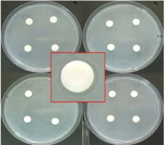
세균갈색무늬병을 일으키는 P. tolaasii에 대해 길항성을 나타내는 미생물을 선발하기 위하여 버섯배지로 부터 약 3,500균주의 미생물을 분리하여 길항력을 조사하였다. 그 결과 미생물 HC5가 가장 높은 길항력을 보였지만 대부분 의 분리 미생물은 길항력이 없었거나 아주 약한 길항력을 보였다(Fig. 1). Kim 등[24]은 Bacteriophages가 P. tolaasii 에 의해 일어나는 세균성갈색무늬병의 발생을 억제하였다 고 보고하였고, 국내에서도 길항미생물로 P. fluorescens가
보고되어 있지만[17], 신속하고 정확한 방제 효과를 기대 하기 어렵고, 병 발생 후의 치료 효과가 매우 낮으며 환경 의 영향을 많이 받기 때문에 처리 효과가 일정하게 나타나 지 않는 등의 단점으로 생물농약으로 실용화되지는 못하 고 있는 실정이다.
길항미생물 HC5의 16S ribosomal DNA 분석
길항세균 HC5균주의 16S rDNA를 PCR 증폭에 의해 약
Fig. 2. Phylogenetic tree of HC5 based on 16S rRNA sequence similarity. Branching values determined using 1000 bootstraps.
Bar, 1 substitution per 100 nucleotides.
Fig. 1. Antimicrobial activity of P. azotoformans HC5 against P. tolaasii.
1.5 kb의 유전자를 확보하였으며, 염기서열을 결정하였다.
이 염기서열을 Ribosomal database project를 이용하여 상 동성을 분석한 결과 P. azotoformans와 97%의 유사성을 보 였다. 또한 Neighbor Joining방법을 이용한 유연관계를 분 석한 결과 길항균 HC5는 P. azotoformans와 같은 그룹을 형성하였다(Fig. 2).
생리·생화학적인 특성
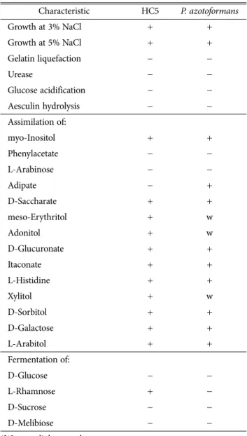
본 연구의 길항세균 HC5균주는 NaCl 3%와 NaCl 5%를 첨가한 배지에서 균생육이 가능하였으며, gelatin을 액화시 키지 못하였다. 그리고 Urease, Glucose acidification, Aes- culin hydrolysis에서 음성이었다. myo-Inositol, D-Saccha- rate, meso-Erythritol, Adonitol, D-Glucuronate, Itaconate, L-Histidine, Xylitol, D-Sorbitol, D-Galactose, L-Arabitol
그리고 L-Rhamnose등과 같은 산과 당을 이용하였고, Phe- nylacetate, L-Arabinose, Adipate, D-Glucose, D-Sucrose 그리고 D-Melibiose 등은 이용하지 못하였다(Table 1). 지방 산 분석 결과 전체 지방산 중에서 Sum in Feature 3(16:1 w7c/16:1 w6c), 16:0, Sum In Feature 8(18:1 w7)가 가장 높 았고, 낮은 양의 10:0 지방산이 존재하였다(Table 2).
생물 검정 효과
느타리에 세균갈색무늬병균을 접종한 후에 HC5을 처리 한 결과 무처리구에서는 93.8%의 이병율을 보였지만 HC5 처리에서는 27.8%의 이병율을 보여 71%의 방제효과가 있 었다. 양송이에서는 무처리에서 56.3%의 이병율을 보였고, HC5처리에서는 15.3%의 이병율을 보여 73%의 방제효과를 보였다. 그리고 팽이버섯에서는 무처리에서 89.8%의 이병 율을 보였고, HC5처리에서는 19.5%의 이병율을 보여 78%
의 방제효과가 있었다. 이상의 결과로 HC5균주는 세균갈 색무늬병을 일으키는 거의 모든 버섯에 높은 병 방제효과 가 관찰되었다(Table 3, Fig. 3). Lee 등[25]은 세균갈색무늬 병원균의 분비독소를 저해는 미생물인 Pseudomonas 속이 느타리, 팽이버섯, 양송이 등에 높은 방제효과가 있었다고 보고하였으나, 현재까지 국내에서는 세균갈색무늬병에 대 Table 1. Phenotypic and biochemical characteristics of Pseu-
domonas azotoformans HC5 and type strain Pseudomonas azotoformans
Characteristic HC5 P. azotoformans
Growth at 3% NaCl + +
Growth at 5% NaCl + +
Gelatin liquefaction − −
Urease − −
Glucose acidification − −
Aesculin hydrolysis − −
Assimilation of:
myo-Inositol + +
Phenylacetate − −
L-Arabinose − −
Adipate − +
D-Saccharate + +
meso-Erythritol + w
Adonitol + w
D-Glucuronate + +
Itaconate + +
L-Histidine + +
Xylitol + w
D-Sorbitol + +
D-Galactose + +
L-Arabitol + +
Fermentation of:
D-Glucose − −
L-Rhamnose + −
D-Sucrose − −
D-Melibiose − −
*W: very slight growth
Table 2. Fatty acid profile of Pseudomonas azotoformans HC5 by MIDI system
Fatty acid HC5
10:0 0.08
10:0 3-OH 3.06
12:0 3.11
12:0 2-OH 3.85
12:1 3-OH 3.51
14:0 0.36
Sum in Feature 3 (16:1 w7c/16:1 w6c) 26.32 Sum in Feature 3 (16:1 w7c/16:1 w7c) 4.98
16:1 w5c 0.11
16:0 25.60
16:0 2-OH 0.34
17:0 iso 0.15
17:0 cyclo 3.56
17:0 0.21
Sum In Feature 8(18:1 w7) 22.75
18:1 w5c 0.16
18:1 w9c 0.22
18:0 1.42
19:0 cyclo w8c 0.21
Summed Feature 3(16:1 w6c/16:1 w7c) 31.30
Summed Feature 8(18:1 w6c) 22.75
한 생물적 제제가 개발되어 상품화된 것은 없다. 따라서 길 항세균 HC5 균주의 항균활성 물질탐색 및 생산의 최적 조 건 검토, 농가 편의성을 고려한 제형화 기술개발과 살포방 법 등을 추가적으로 개발한다면 산업적으로 충분히 가치가 있을 것으로 판단된다.
적 요
P. tolaasii에 의해 발생하는 세균갈색무늬병은 버섯재배 에서 문제가 되는 대표적인 병해이다. 본 연구에서는 세균 갈색무늬병의 생물학적 방제법에 이용할 수 있는 길항미생 물의 항균활성과 선발된 길항미생물에 대해 폿트 수준의 생물검정 실험을 실시하였다. 재배중인 느타리 폐면배지와 양송이 퇴비에서 세균갈색무늬병원균을 강하게 억제하는 길항세균 HC5를 선발하였으며, 생리·생화학적 실험과 유 전적 실험결과 HC5균주는 P. azotoformans로 동정되었다.
P. azotoformans HC5를 양송이, 팽이버섯, 느타리에 처리한 결과 각각 78%, 73%, 71%의 방제효과를 보였다. 따라서 P.
azotoformans HC5가 버섯 세균갈색무늬병 방제를 위해 합 성농약을 대체할 수 있는 친환경 방제제가 될 수 있을 것으 로 생각된다.
감사의 글
본 연구는 농촌진흥청 기관고유 연구과제(PJ0099610120 14)에 의하여 수행된 연구결과입니다.
REFERENCES
1. Wells JM, Sapers GM, Fett WF, Butterfield JE, Jones JB, Bouzar H, Miller FC. Postharvest discolorization of the cultivated mushroom Agaricus bisporus caused by Pseudomo- nas tolaasii, P. 'reactans', and P. 'gingeri'. Phytopathology 1996;
86:1098-104.
2. Wong WC, Fletcher JT, Unsworth BA, Preece TF. A note on ginger blotch, a new bacterial disease of the cultivated mush- room, Agaricus bisporus. J Appl Bacteriol 1982;52:43-8.
3. Young JM. Drippy gill: a bacterial disease of cultivated mush- rooms caused by Pseudomonas agarici n. sp. N Z J Agr Res 1970;13:977-90.
4. Tolaas AG. A Bacterial disease of cultivated mushrooms.
Phytopathology 1915;5:51-4.
5. Paine SG. Studies in bacteriosis . A brown blotch disease of cultivated mushrooms. Ann Appl Biol 1919;5:206-19.
6. Tsuneda A, Suyama K, Muradami S, Ohira I. Occurrence of Pseudomonas tolaasii on fruiting bodies of Lentinula edodes formed on Quercus logs. Mycoscience 1995;36:283-8.
7. Kim JW, Kim KH, Kang HJ. Studies on the pathogenic Pseu- domonas causing bacterial disease of cultivated mushroom in Korea. 1. On the causal organisms of the rots of Agaricus bisporus, Pleurotus ostreatus and Lentinus edodes. Kor J Plant Pathol 1994;10:197-210.
8. Rainey PB, Brodey CL, Johnstone K. Biology of Pseudomonas tolaasii, cause of brown blotch disease of cultivated mush- room. Pages 95-118 in: Advances in Plant Pathology, Vol. 8. J.
H. Andrews and I. Tommerup, eds. Academic Press, Inc., New York. 1992.
9. Goor M, Vantomme R, Swings J, Gillis M, Kersters K, de Ley J. Phenotypic and genotypic diversity of Pseudomonas tolaasii and white line reacting organisms isolated from cultivated Table 3. Control efficacy of brown blotch disease on different
mushrooms by HC5 strain
Mushrooms Treatment Infection rate (%)
Control value (%) Pleurotus
ostreatus
Control 93.8
HC5 27.8 71
Agaricus bisporus
Control 56.3
HC5 15.3 73
Flammulina velutipes
Control 89.8
HC5 19.5 78
Fig. 3. Effect of spraying of HC5 suspension on brown blotch disease development in Flammulina velutipes (A), Pleurotus ostreatus (B) and Agaricus bisporus (C). Left: control treat- ment, Right: HC5 treatment.
mushrooms. J Gen Microbiol 1986;132:2249-64.
10. Cutri SS, Macauley BJ, Roberts WP. Characteristics of patho- genic non-fluorescent (smooth) and non-pathogenic fluore- scent (rough) forms of Pseudomonas tolaasii and Pseudomo- nas 'gingeri'. J Appl Bacteriol 1984;57:291-8.
11. Lee HI, Cha JS. Cloning of a DNA fragment specific to Pseu- domonas tolaasii causing bacterial brown blotch disease of oyster mushroom (Pleurotus ostreatus). Kor J Plant Pathol 1998;14:177-83.
12. Nutkins JC, Mortishire-Smith RJ, Packman LC, Brodey CL, Rainey PB, Johnstone K, Williams DH. Structure determina- tion of tolaasin, an extracellular lipodepsipeptide produced by the mushroom pathogen Pseudomonas tolaasii Paine. J Am Chem Soc 1991;113:2621-7.
13. Jourdan F, Lazzaroni S, Mendes BL, Lo Cantrore P, de Julio M, Amodeo P, Iacobellis NS, Evidente A, Motta A. A left- handed alpha-helix containing both L- and D-amino acids:
the solution structure of the antimicrobial lipodepsipeptide tolaasin. Proteins 2003;52:534-43.
14. Scherwinski K, Grosch R, Berg G. Effect of bacterial antago- nists on lettuce: active biocontrol of Rhizoctonia solani and negligible, short-term effects on nontarget microorganisms.
FEMS Microbiol Ecol 2008;64:106-16.
15. Nair NG, Fahy PC. Bacteria antagonistic to Pseudomonas tola- asii and their control of brown blotch of the cultivated mush- room Agaricus bisporus. J Appl Bacteriol 1972;35:439-42.
16. Olivier JM, Guillaumes J, Martin G. Study of a bacterial disease of mushroom caps. Proceedings of 4th International Conference Plant Pathogenic Bateria, Angers, France. 1978;
903-16.
17. Liao YM, Tu CC, Jeng JJ. Control of bacterial blotch of mushroom. Taiwan Mushrooms 1980;4:34-41.
18. Park BS, Cho NC, Chun UH. Identification of Pseudomonas fluorescens antagonistic to Pseudomonas tolaasii and its culti- vation. Kor J Biotechnol Bioeng 1992;7:296-301.
19. Thompson JD, Higgins DG, Gibson TJ. Clustal W: Improving the sensitivity of progressive multiple sequence alignment through sequence weighing position-specific gap penalties and weight matrix choice. Nucleic Acids Res 1994;34-637.
20. Jukes TH, Cantor CR. Evolution of protein molecules, In:
Munro HN, editor. Mammalian Protein Metabolism. NY:
Academic Press; 1969. p. 21-132.
21. Stainer RY, Palleroni NJ, Doudoroff M. The aerobic pseudo- monads: a taxonomic study. J Gen Microbiol 1966;43:159- 271.
22. Palleroni NJ. Genus. Pseudomonas. In: Krieg NR, Hotr JG, editors. Bergey's manual of systematic bacteriology. Vol. I, Baltmore: Williams and Wilkins; 1984. p. 141-219.
23. Sasser MJ. Identification of bacteria by gas chromatography of cellular fatty acids. Technical note 101. Newark, DE:
Microbial ID Inc. 1990.
24. Kim MH, Park SW, Kim YK. Bacteriophages of Pseudomonas tolaasii for the biological control of brown blotch disease. J Kor Appl Biol Chem 2011;54:99-104.
25. Lee CJ, Yoo YM, Han JY, Jhune CS, Cheong JC, Moon JW, Suh JS, Han HS, Cha JS.Isolation of the bacterium Pseudo- monas sp. HC1 effective in inactivation of tolaasin produced by Pseudomonas tolaasii. Kor J Mycol 2013;41:248-54.