녹용대보탕 열수 추출물의 실험적으로 유발된 랫트 아급성 출혈성 빈혈에 대한 효과
1 부산대학교 한방병원 여성의학과, 2 대구한의대학교 한의과대학 부인과교실
김정아 1 , 김동철 2
ABSTRACT
Ameliorating Effects of Nokyongdaebo-tang on Experimental Subacute Hemorrhagic Anemia in Rats
Jung-Ah Kim 1 , Dong-Chul Kim 2
1 Dep. of Korean Ob & Gy, Pusan National University Korean Medicine Hospital
2 Dept. of Korean Obstetrics & Gynecology, College of Korean Medicine, Daegu Haany University
Objectives: The object of this study is to observe the possible ameliorating effects of Nokyongdaebo-tang (NYDBT) on the experimental subacute hemorrhagic anemia (SHA) in rats.
Methods: In the present study, SHA in rats was induced by exsanguinations from orbital plexus, and ameliorating effects of NYDBT was observed based on the changes of body and hematopoietic organ (spleen, liver and femur) weights, red blood cell (RBC) related hematological values, smear cytology, histopathological changes and immunohistochemistrical analysis of hematopoietic stem cells in the femur bone marrow, liver and spleen. In addition, the gastrointestinal motility and the surface mucosa thicknesses of remnant fecal pellets in the colon lumen, mucosa thicknesses and the mucous producing cell numbers in the colonic mucosa were analyzed to observe the digestive disorders, especially on the constipation, the major discomfort problems in iron supplement.
Results: SHA related abnormal anemic signs were markedly and dose-dependently inhibited by oral administration of NYDBT 500, 250 and 125 mg/kg in a condition of this experiment. In addition, no meaningful changes on the gastrointestinal motilities and mucous component on the colon and remnant feces were noticed in all three different dosages of NYDBT treated rats as compared with intact vehicle and SHA control rats in this study.
Conclusions: It, therefore, is expected that NYDBT will be promising as a novel alternative hematopoietic and therapeutic agent for anemia.
Key Words: Nokyongdaebo-tang, Ferromax, Subacute Hemorrhagic Anemia, Hematopoietic Effect
1)
Corresponding author(Dong-Chul Kim) : Department of Obstetrics & Gynecology Korean Medicine, Pohang Hospital of Korean Medicine, Daegu Haany University, 411, Saecheonnyeon-daero, Nam-gu,
Pohang-si, Gyeongsangbuk-do, Korea
Tel : +82-54-271-8002 Fax : +82-54-281-0055 E-mail : [email protected]
I. Introduction
Anemia is a decrease in number of red blood cells (RBCs, erythrocytes) or less than the normal quantity of hemoglobin in the blood 1) . Acute blood loss anemia, also called hemorrhagic anemia, is specific type of anemia that sufficient decrease in RBC due to acute hemorrhage 1) . Although hemorrhagic shock was induced in a case of acute blood losses, over 20% in mammalian body, chronic or subacute blood losses can be induced the regenerative and iron deficient anemia 2) .
Until now, hemorrhage related regenerative iron deficient anemia has been treated by iron supplements and they have been showed potent anti-anemic effects. However, they induced various side effects including digestive discomfort, especially constipation 4) .
Therefore, new strategies highlighted as a modern concern to search hematopoietic potent anti-anemic agents have less toxic effects rather than simple alternative iron supplements, especially concerned on natural herbs or their mixtures 5) .
As traditional medicine, anemia is belong to blood deficiency 6) . Relationship of qi and blood is explained as ‘Qi can make blood, qi can move blood 7) '. Therefore, supplement with qi and blood is treatment as traditional medicine for anemia 7) .
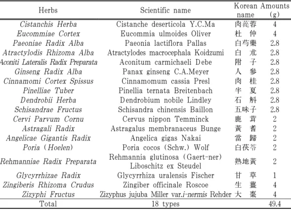
Nokyongdaebo-tang 8) (NYDBT) is a traditional medicine and aqueous polyherbal formula that has been used for several hundred years, predominantly to tonic purpose in a weak constitution and
supplement with qi and blood 8) (Table 1).
Studies have been reported the specific bioactivities of NYDBT, which include neuroprotective activities to Alzheimer's disease model 9) , hypolipidemic effects 10) , antithrombotic activities 11) , anti-oxidative effects 12) and anti-aging activities 13) . However, there are no reports dealing anti-anemic activities of NYDBT, upon our knowledge, especially on the experimentally induced subacute hemorrhagic anemia (SHA).
Therefore, in this study, we examined the ameliorating effects of NYDBT on experimental subacute hemorrhagic anemia in rats and compared with commercial iron supplements, Ferromax TM (7.14%
ferric hydroxide polymaltose complex; FM, Hanmi Pharm. Co. Ltd., Seoul, Korea).
Ⅱ. Materials and methods
1. Animals and husbandry
Total sixty healthy female SPF/VAF Crl : CD1 [Sprague-Dawley] rats (6 weeks old upon receipt; Orient Bio, Seungnam, Korea; Body weight ranged in 120~150 g upon receipt), were used after acclimatization for 8 days. Animals were assigned four to five per polycarbonate cage in a temperature (20~25℃) and humidity (50~55%) controlled room. Light : dark cycle was 12 hr : 12 hr, and standard rodent chow (Samyang, Seoul, Korea) and water were supplied free to access.
After 8 days after acclimatization, eight
rats in each group were selected based
on the body weight deviations (Average
201.52±8.13 g, ranged in 183~217 g) as follows. All laboratory animals were treated as the national regulations of the welfare and usage of laboratory animals, admitted by the Institutional Animal Care and Use Committee in Daegu Haany University (Gyeongsan, Gyeongbuk, Korea) before animal experiment (Approval No DHU2015-029, April 13, 2015).
Experimental groups (Six groups, 8 rats in each group were used)
1. Intact vehicle control : Distilled water administered intact rats
2. SHA control : SHA-induced and distilled water administered rats
3. FM : SHA-induced and FM 5 ml/kg administered rats
4. NYDBT 500 : SHA-induced and NYDBT 500 mg/kg administered rats
5. NYDBT 250 : SHA-induced and NYDBT 250 mg/kg administered rats
6. NYDBT 125 : SHA-induced and NYDBT 125 mg/kg administered rats
2. Preparations and administrations of test materials
Aqueous extracts of NYDBT (Table 1) (yield=15.17%) as brown powders were prepared by routine methods using rotary vacuum evaporator (N-1110, Eyela, Tokyo, Japan) and programmable freeze dryer (FDB-5503, Operon, Kimpo, Korea), which were purchase from local voucher (Jecheon Hanbang Yakcho, Jecheon, Korea) after confirm the morphology under microscopy.
Total 494 g of NYDBT were boiled in 5 L of distilled water for 4 hrs, three times
at 60℃ and evaporated using automated round flasked evaporator (Eyela N-1110, Tokyo, Japan), then lyophilized completely.
Total 74.94 g (yield=15.17%) of lyophilized aqueous NYDBT extracts were acquired.
NYDBT extracts were stored at -20℃
in a refrigerator to protect from light and humidity until used. The voucher specimens documenting this purchase and some specimens of lyophilized aqueous extracts of NYDBT were deposited in the herbarium of the Medical Research center for Globalization of Herbal Formulation, Daegu Haany University (Code NYDBT2015KDC).
Deep brown solutions of FM (Hanmi Pharm. Co. Ltd., Seoul, Korea) were also stored at 4℃ in a refrigerator to safeguard from light and humidity until used. FM was well diluted as 1:4 (v/v) indistilled water, and NYDBT extracts were well dissolved upto 100 mg/ml in distilled water, at least in a condition of this experiment. Test articles were orally administered 5 ml/kg, once a day for 7 days at 1 hr after exsanguination.
NYDBT extracts were dissolved in
distilled water as 100, 50 and 25 mg/ml
concentration, and orally administered
in a volume of 5 ml/kg, equivalence to
500, 250 and 125 mg/kg, respectively. FM
solution was diluted by distilled water
as 1:4 (v/v), and orally administrated
in a volume of 5 ml/kg, once a day for
7 days at 1 hr after exsanguination. In
intact and SHA control rats, distilled
water 5 ml/kg was orally administered,
instead of test substance.
Herbs Scientific name Korean name
Amounts (g) Cistanchis Herba Cistanche deserticola Y.C.Ma 肉蓯蓉 4
Eucommiae Cortex Eucommia ulmoides Oliver 杜 仲 4
Paeoniae Radix Alba Paeonia lactiflora Pallas 白芍藥 2.8 Atractylodis Rhizoma Alba Atractylodes macrocephala Koidzumi 白 朮 2.8 Aconiti Lateralis Radix Preparata Aconitum carmichaeli Debe 附 子 2.8 Ginseng Radix Alba Panax ginseng C.A.Meyer 人 參 2.8 Cinnamomi Cortex Spissus Cinnamomum cassia Presl 肉 桂 2.8 Pinelliae Tuber Pinellia ternata Breitenbach 半 夏 2.8 Dendrobii Herba Dendrobium nobile Lindley 石 斛 2.8 Schisandrae Fructus Schisandra chinensis Baillon 五味子 2.8
Cervi Parvum Cornu Cervus nippon Temminck 鹿 茸 2
Astragali Radix Astragalus membranaceus Bunge 黃 耆 2 Angelicae Gigantis Radix Angelica gigas Nakai 當 歸 2
Poria (Hoelen) Poria cocos (Schw.) Wolf 白茯苓 2
Rehmanniae Radix Preparata Rehmannia glutinosa (Gaert-ner)
Liboschitz ex Steudel 熟地黃 2
Glycyrrhizae Radix Glycyrrhiza uralensis Fischer 甘 草 1 Zingiberis Rhizoma Crudus Zingiber officinale Roscoe 生 薑 4 Zizyphi Fructus Zizyphus jujuba Miller var.i-nermis Rehder 大 棗 4
Total 18 types 49.4
Table 1. Composition of NYDBT
3. Induction of SHA
SHA in rats was induced according to the previous studies 14) with some modifications.
Briefly, animals were anesthetized with 2 to 3% isoflurane (Hana Pharm. Co., Hwasung, Korea) in the mixture of 70%
N 2 O and 28.5% O 2 , exsanguinations of whole bloods 1 ml/head, match to about 0.5% of body weights, were continuously executed from orbital plexus, once a day for 7 days in each rats, at 1 hr before test material administration. In intact vehicle control rats, no exsanguinations were conducted but system inhalation anesthesia was also conducted in this experiment.
4. Changes in body weights
Changes of body weight were checked at once a day from 1 day before initial exsanguinations and test material throughout all periods of experiment using an automatic electronic balance (Precisa Instrument, Zuerich, Switzland). To reduce the differences of individuals, the body weight gains after 7 days of administrations were calculated as follow Equation [1].
Equation [1]. Body Weight Gains (g) during 7 days of test article treatment from initiation of test article administrations to end of 7 days of test article administrations
=Body weights at sacrifice (24 hrs after
last administrations and exsanguinations)
-body weights at start of administrations
or exsanguinations
5. Organ weight measurements
At sacrifice, the weights of spleen, liver and left femur were measured at g levels as absolute wet-weights and to reduce the differences of individual body weights, the relative weights (% of body weights) were also computed using body weight at sacrifice and absolute weight as follow Equation [2].
Equation [2]. Relative Organ Weights (% of body weight)
=(Absolute spleen, liver or femur weights /Body weight at sacrifice)×100
6. Hematology
Blood samples were drawn from posterior vena cava using a syringe with a 23 gauge needle under 2 to 3% isoflurane inhalation anesthesia in the mixture of 70% N 2 O and 28.5% O 2 . The animals had been 18 hrs overnight fasted (water was not restricted) prior to sacrifice and blood collecting. The blood samples were collected into CBC bottles containing EDTA-2K (1.8 mg/ml of blood). Items for hematology measurement are listed below and the detecting methods and unit measured are presented as follow items, respectively.
All hematological measurements were conducted in Veterinary Teaching Hospital, College of Veterinary Medicine, Kyungpook National University (Daegu, Korea) using automated hematology cell counter (MS9-5V;
Melet Schloesing Lab., Paris, France).
7. Measurement of intestinal charcoal transit ratio
Assessment of gastrointestinal propulsion of charcoal meal was determined as Sagar et al 15) with minor modifications 16) . Test animals were starved for 18 hrs before the experiment, but consumed water ad libitum. Ten minutes after last 7th test material administration, animals from each group were fed on 1 ml of charcoal meal (3% suspension of activated charcoal in 0.5% aqueous methylcellulose (Sigma -Aldrich, St. Louise, MO, USA)). After thirty minutes of administrating of charcoal meals, the animals of each groups were killed by cervical dislocation. And then, total small intestine length (pyloric sphincter to caecum) and the distance which the charcoal moved as a fraction of that length was measured. The intestinal charcoal transit ratio was calculated as the difference between the total small intestinal length and length of charcoal meal transferred as Equation [3] 17) .
Equation [3]. Charcoal Transit Ratio (%)
=(Length of charcoal meal transferred /Total small intestine length)×100
8. Smear cytology
At sacrifice, 0.5 ml of whole bloods
were collected from orbital plexus under
inhalation anesthesia, and directly smeared
on slide. Preparations were dried, and
fixed by submerging in absolute methanol
(for 30 minutes). Fixed slides were stained
by 1:6 diluted Giemsa solutions (Sigma
-Aldrich, St. Louise, MO, USA) for 10
minutes. Slides were randomly coded and examined under 400 times magnification by two different experts. Mean RBC diameters (μm/cell) and mean PCEs (PCE/1000 cells) were observed using a computer-assisted image analysis program (iSolution FL ver 9.1, IMT i-solution Inc., Vancouver, Quebec, Canada) to decide whether microcytic or macrocytic anemia, regenerative or degenerative anemia 1,18) .
9. Histopathology
The left femur, left lateral lobes of liver and spleen were sampled at 7 days after first exsanguinations and after measurement of charcoal transfer, and fixed in 10% neutral buffered formalin (NBF). After fixation, femur samples were decalcified using decalcifying solution [24.4% formic acid, and 0.5N sodium hydroxide] for 5 days (mixed decalcifying solution was exchanges once a day for 5 days), not in liver and spleen samples.
After that, they were lengthways trimmed and embedded in paraffin, sectioned (3~4 μm) and stained with Hematoxylin &
Eosin (H&E). The histological profiles of the femur bone marrow regions, hepatic parenchyma has focused on the hematopoietic spots, spleen red pulps were observed as compared with the SHA and intact vehicle control. The mean numbers of total femur bone marrow cells (×10 3 cells/mm 2 ), area of hematopoietic spots in the liver parenchyma (%/mm 2 ) and numbers of total spleen red pulp cells (×10 3 cells/mm 2 ) were measured as histomorphometrical
analyses at prepared longitudinally trimmed samples using a computer-assisted image analysis program. In addition, assessment of histological observations of colon mucosa and fecal pellets remnant in the colon lumen were determined according to Wu et al 19) with minor modifications 16) . Briefly, the segments of rat distal colon containing one fecal pellet were seperated by ligatures, removed, and immediately fixed with 10%
NBF at intestinal charcoal transit ratio measurement. The fixed tissue segments were embedded in paraffin and serially cut into 3 μm thick cross sections. The sections were stained with alcian blue at pH of 2.5. Eight tissue segments per group were prepared and, the histological profiles were interpreted as mean thickness of mucosal layers at the fecal surface (μm/fecal pellets), mucous-producing goblet cell (alcian blue positive cell) numbers (cells/mm 2 of colonic mucosa) and colonic mucosa thicknesses (μm/colon) using a computer-assisted image analysis program.
The histopathologist was blinds to group distribution when this analysis was made.
10. Immunohistochemistry
The changes of CD34 and CD45-
immunoreactive cells, the hematopoietic
stem cells, were observed by
immunohistochemistrical methods using
purified rat anti-mouse CD34 and CD45
antibodies (BD Biosciences, San Jose, CA,
USA) with avidin-biotin-peroxidase (ABC)
and peroxidase substrate kit (Vector Labs,
Burlingame, CA, USA). Briefly, endogenous
peroxidase activity was cut off by incubated in methanol and 0.3% H 2 O 2 for 30 minutes.
Non-specific binding of immunoglobulin was cut off with normal horse serum blocking solution for 1 hr in humidity chamber after heating (95~100℃) based epitope retrievals in 10 mM citrate buffers (pH 6.0) in spleen and liver samples or by pretreatment of trypsin (Sigma-Aldrich, St. Louise, MO, USA) and 2NHCl in femur samples 20) . Primary antisera was treated for overnight at 4℃ in humidity chamber, and then incubated with biotinylated universal secondary antibody and ABC reagents for 1 hr at room temperature in humidity chamber. Finally, reacted with peroxidase substrate kit for 3 minutes at room temperature. All sections were rinse in 0.01 M PBS for 3 times, between each step. The cells taken by over 20% of immunoreactivities, the density of CD34 and CD45, were regarded as positive. The mean numbers of CD34 and CD45-immunoreactive cells dispersed in the liver, spleen and femur bone marrow (mm 2 ) were counted using a computer -assisted image analysis program, respectively.
The histopathologist was blinded to the group distribution when performing the analysis.
11. Statistical analyses
All Data was expressed as mean±
standard deviations (SD) of eight rats.
Multiple comparison tests for all dose groups were treated. Variance homogeneity was examined using the Levene test 21) .
If the Levene test signified no significant deviations from variance homogeneity, the obtain data was analyzed by one way ANOVA test followed by least-significant differences multi-comparison (LSD) test to determine which pairs of group comparison were significantly different.
In case of significant deviations from variance homogeneity were observed at Levene test, a non-parametric comparison test, Kruskal-Wallis H test was conducted.
When a significant difference is observed in the Kruskal-Wallis H test, the Mann -Whitney U (MW) test was conducted to determine the specific pairs of group comparison, which are significantly different.
Statistical analyses were conducted using SPSS for Windows (Release 14.0K, IBM SPSS Inc., Armonk, NY, USA). In addition, the percent changes between intact vehicle and SHA control rats were calculated to observe the severities of SHA induced by continuous exsanguinations in this study, and the percent changes as compared with SHA control and test material treated rats were also calculated to help the understanding of the anti-anemic effects of test substances as follow Equation [4] and [5], according to previous method described by Kang et al 22) , respectively.
Equation [4]. Percent Changes as Compared with Intact Vehicle Control (%)
={(Data of SHA control-Data of intact vehicle control rats)/Data of intact vehicle control rats}×100
Equation [5]. Percent Changes as
Compared with SHA Control (%)
={(Data of test material treated rats -Data of SHA control)/Data of SHA control}×100
Ⅲ. Results
1. Effects on body weights
Significant (p<0.01) decreases of body weights were detected in SHA control from 3 days after exsanguinations as compared with intact vehicle control, consequently, the body weight gains during 7 days of treatment period were also significantly (p<0.01) decreased. No meaningful or
significant changes on the body weight and gains were observed in FM treated rats as compared with SHA control rats, throughout the whole experimental periods, but NYDBT 500, 250 and 125 mg/kg treated rats showed significant (p<0.01 or p<0.05) increased body weights from 4, 5 and 6 days after administration, and accordingly, the body weight gains during 7 days of administration periods were also significantly (p<0.01) increased in all three different dosages of NYDBT administered rats as compared with SHA control rats, respectively (Table 2).
Items Groups
Body weights (g)
Body weight gains [B-A]
Before treatment
At initial treatment [A]*
At last treatment [B]*
Controls
Intact 201.25±6.94 181.13±8.95 211.25±4.98 30.13±6.79 SHA 201.63±10.13 180.25±10.14 186.38±7.33 a 6.13±6.69 a FM 5 ml/kg 201.50±10.30 179.38±10.39 187.00±8.91 a 7.63±3.07 a NYDBT
500 mg/kg 201.38±7.23 180.63±9.30 208.00±6.00 c 27.38±6.95 c 250 mg/kg 201.50±8.55 181.50±10.41 202.88±9.99 bc 21.38±3.54 ac 125 mg/kg 201.88±7.86 181.88±9.78 198.88±3.91 ac 17.00±6.39 ac
Values are expressed mean±SD of eight rats.* : All animals were overnight fasted.
a : p<0.01 and b : p<0.05 as compared with intact vehicle control by LSD test c : p<0.01 as compared with SHA control by LSD test
Table 2. Changes on the Body Weights
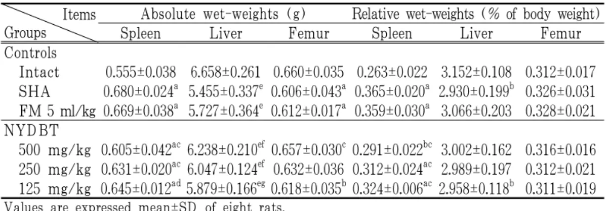
2. Changes on the spleen weights Significant (p<0.01) increases of absolute and relative spleen weights were demonstrated in SHA control rats as compared with intact vehicle control rats. However, significant (p<0.01) decreases of spleen weights were noticed in NYDBT 500, 250 and 125 mg/kg treated rats as compared
with SHA control rats, but not in FM treated rats, in this experiment (Table 3).
3. Changes on the liver weights
Significant (p<0.01 or p<0.05) decreases
of absolute and relative liver weights were
demonstrated in SHA control rats as compared
with intact vehicle control, respectively. FM
treatment did not influenced on the hepatic weights as compared with SHA control rats.
Significant (p<0.01 or p<0.05) increases of absolute liver weights were demonstrated in all three different dosages of NYDBT 500, 250 and 125 mg/kg treated rats as compared to those of SHA control rats, but not in relative liver weights (Table 3).
4. Changes on the femur weights Significant (p<0.01) decreases of absolute
femur weights were demonstrated in SHA control rats as compared with intact control. Anyway, no meaningful changes on the femur weights were demonstrated in all test substance treated rats as compared with SHA control rats, except for significant (p<0.01) increases of absolute femur weights detected in NYDBT 500 mg/kg treated rats as compared with SHA control rats, respectively (Table 3).
Items Groups
Absolute wet-weights (g) Relative wet-weights (% of body weight)
Spleen Liver Femur Spleen Liver Femur
Controls
Intact 0.555±0.038 6.658±0.261 0.660±0.035 0.263±0.022 3.152±0.108 0.312±0.017 SHA 0.680±0.024 a 5.455±0.337 e 0.606±0.043 a 0.365±0.020 a 2.930±0.199 b 0.326±0.031 FM 5 ml/kg 0.669±0.038 a 5.727±0.364 e 0.612±0.017 a 0.359±0.030 a 3.066±0.203 0.328±0.021 NYDBT
500 mg/kg 0.605±0.042 ac 6.238±0.210 ef 0.657±0.030 c 0.291±0.022 bc 3.002±0.162 0.316±0.016 250 mg/kg 0.631±0.020 ac 6.047±0.124 ef 0.632±0.036 0.312±0.024 ac 2.989±0.197 0.312±0.021 125 mg/kg 0.645±0.012 ad 5.879±0.166 eg 0.618±0.035 b 0.324±0.006 ac 2.958±0.118 b 0.311±0.019
Values are expressed mean±SD of eight rats.a : p<0.01 and b : p<0.05 as compared with intact vehicle control by LSD test c : p<0.01 and d : p<0.05 as compared with SHA control by LSD test
e : p<0.01 as compared with intact vehicle control by MW test f : p<0.01 and g : p<0.05 as compared with SHA control by MW test
Table 3. Changes on the Spleen, Liver and Femur Weights
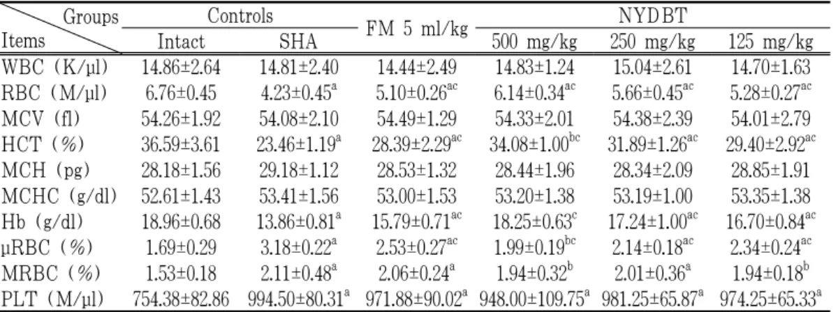
5. Changes on the hematology
Significant (p<0.01) decreases of RBC numbers, Hct and Hb and increases of PLT numbers, μRBC and MRBC ratios were observed in SHA control rats as compared with intact vehicle control rats, respectively. However, these abnormal changes on the hematological inspections were significantly (p<0.01) normalized by
treatment of all test substances except
for PLT numbers. No meaningful changes
on the WBC numbers, MCV, MCH and
MCHC were noticed in all SHA-induced
rats, and also no significant changes on
the MRBC ratios were noticed in FM 5
ml/kg, NYDBT 500, 250 and 125 mg/kg
administered rats as compared with SHA
control rats, in the present study (Table 4).
Groups Items
Controls
FM 5 ml/kg NYDBT
Intact SHA 500 mg/kg 250 mg/kg 125 mg/kg
WBC (K/μl) 14.86±2.64 14.81±2.40 14.44±2.49 14.83±1.24 15.04±2.61 14.70±1.63 RBC (M/μl) 6.76±0.45 4.23±0.45 a 5.10±0.26 ac 6.14±0.34 ac 5.66±0.45 ac 5.28±0.27 ac MCV (fl) 54.26±1.92 54.08±2.10 54.49±1.29 54.33±2.01 54.38±2.39 54.01±2.79 HCT (%) 36.59±3.61 23.46±1.19 a 28.39±2.29 ac 34.08±1.00 bc 31.89±1.26 ac 29.40±2.92 ac MCH (pg) 28.18±1.56 29.18±1.12 28.53±1.32 28.44±1.96 28.34±2.09 28.85±1.91 MCHC (g/dl) 52.61±1.43 53.41±1.56 53.00±1.53 53.20±1.38 53.19±1.00 53.35±1.38 Hb (g/dl) 18.96±0.68 13.86±0.81 a 15.79±0.71 ac 18.25±0.63 c 17.24±1.00 ac 16.70±0.84 ac μRBC (%) 1.69±0.29 3.18±0.22 a 2.53±0.27 ac 1.99±0.19 bc 2.14±0.18 ac 2.34±0.24 ac MRBC (%) 1.53±0.18 2.11±0.48 a 2.06±0.24 a 1.94±0.32 b 2.01±0.36 a 1.94±0.18 b PLT (M/μl) 754.38±82.86 994.50±80.31 a 971.88±90.02 a 948.00±109.75 a 981.25±65.87 a 974.25±65.33 a
Values are expressed mean±SD of eight rats.a : p<0.01 and b : p<0.05 as compared with intact vehicle control by LSD test c : p<0.01 as compared with SHA control by LSD test
Table 4. Changes on the Hematological Values
6. Changes on the gastrointestinal motility
Although no significant changes on the charcoal transfer rates, the digestive motility, were detected in SHA control rats and all three different dosages of NYDBT administered rats as compared with intact
vehicle control rats, significant (p<0.01) decreases of charcoal transfer rates in gastrointestinal tract were observed in FM 5 ml/kg treated rats as compared with intact and SHA vehicle control rats, in this study (Table 5).
Items Groups
Gastrointestinal motility Total small intestine
length (cm) [A]
Length of charcoal meal transferred (cm) [B]
Charcoal transfer rates (%) [B/A×100]
Controls
Intact 108.25±8.14 81.88±5.08 75.94±6.47
SHA 107.38±7.82 79.75±8.40 74.63±9.40
FM 5 ml/kg 105.88±9.11 52.75±10.39 ab 49.89±9.32 ab NYDBT
500 mg/kg 107.25±4.74 80.75±9.10 75.37±8.74
250 mg/kg 110.50±5.48 83.38±6.99 75.46±5.45
125 mg/kg 108.13±2.80 82.88±7.47 76.60±5.90
Values are expressed mean±SD of eight rats.
a : p<0.01 as compared with intact vehicle control by LSD test b : p<0.01 as compared with SHA control by LSD test
Table 5. Changes on the Gastrointestinal Motility
7. Changes on the blood smear cytology Significant (p<0.01) decreases of mean RBC diameters and increases of PCEs were observed in SHA control rats as compared with intact vehicle control rats, respectively. However, these abnormal changes on the smear cytological inspections were significantly (p<0.01) normalized by treatment of all test substances including FM 5 ml/kg, in this result.
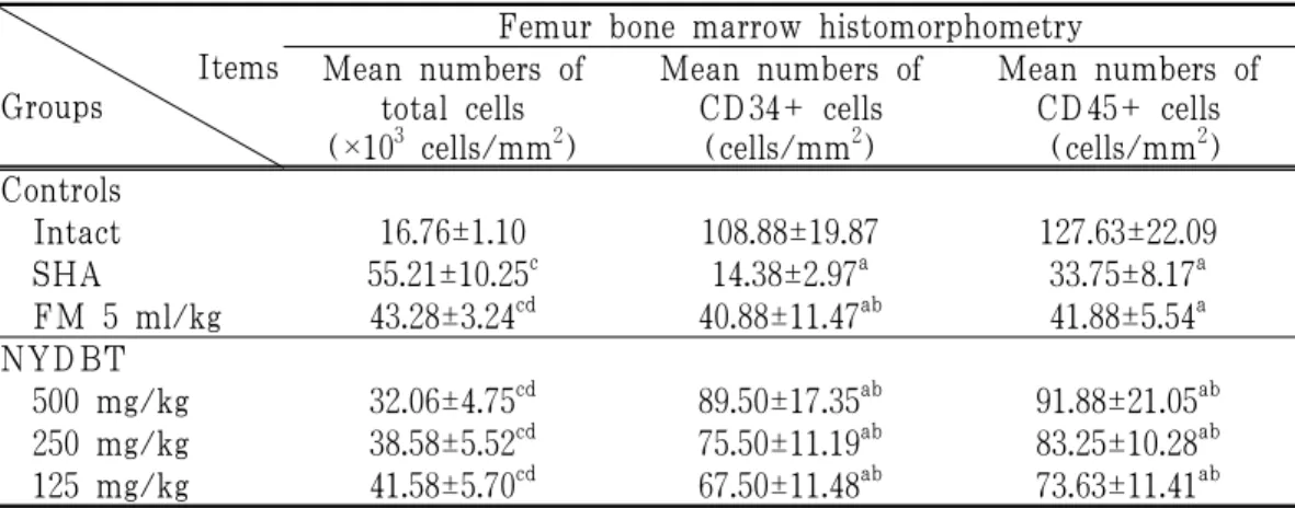
8. Changes on the femur bone marrow histopathology
Significant (p<0.01) increases of total femur bone marrow cell numbers and decreases of CD34 and CD45 immunolabeled cells were observed in SHA control rats as compared with intact vehicle control rats, respectively. Significant (p<0.01) decreases of total femur bone marrow cell numbers and marked increases of CD34 and CD45 immunoreactive cells were observed in all test substance treated rats as compared with SHA control rats, in the current study (Table 6).
Items Groups
Femur bone marrow histomorphometry Mean numbers of
total cells (×10 3 cells/mm 2 )
Mean numbers of CD34+ cells
(cells/mm 2 )
Mean numbers of CD45+ cells (cells/mm 2 ) Controls
Intact 16.76±1.10 108.88±19.87 127.63±22.09
SHA 55.21±10.25 c 14.38±2.97 a 33.75±8.17 a
FM 5 ml/kg 43.28±3.24 cd 40.88±11.47 ab 41.88±5.54 a NYDBT
500 mg/kg 32.06±4.75 cd 89.50±17.35 ab 91.88±21.05 ab 250 mg/kg 38.58±5.52 cd 75.50±11.19 ab 83.25±10.28 ab 125 mg/kg 41.58±5.70 cd 67.50±11.48 ab 73.63±11.41 ab
Values are expressed mean±SD of eight rats.a : p<0.01 as compared with intact vehicle control by LSD test b : p<0.01 as compared with SHA control by LSD test
c : p<0.01 as compared with intact vehicle control by MW test d : p<0.01 as compared with SHA control by MW test
Table 6. Changes on the Femur Bone Marrow Histomorphometrical Analysis
9. Changes on the liver histopathology Significant (p<0.01) increases of the area occupied by hematopoietic spots and decreases of CD34 and CD45 immunolabeled cells were observed in SHA control rats as compared with intact vehicle control rats, respectively. Significant (p<0.01 or p<0.05)
decreases of the area percentages occupied
by hematopoietic spots and increases of
portal triad CD34 and CD45 immunoreactive
cells were observed in all test substance
treated rats as compared with SHA
control rats, in the current experiment
(Table 7).
Items Groups
Liver histomorphometry Mean area of
hematopoietic spots (%/mm 2 )
Mean numbers of CD34+ cells
(cells/mm 2 )
Mean numbers of CD45+ cells
(cells/mm 2 ) Controls
Intact 1.63±0.90 173.75±16.65 260.75±44.68
SHA 29.00±5.81 d 68.00±12.28 d 104.38±20.30 a
FM 5 ml/kg 16.98±2.35 df 93.63±11.25 df 145.88±10.38 ac NYDBT
500 mg/kg 6.93±2.82 df 253.63±53.13 df 280.25±45.56 b 250 mg/kg 12.58±3.14 df 215.88±42.96 ef 234.38±41.69 b 125 mg/kg 15.41±1.91 df 165.25±22.50 f 193.88±51.20 ab
Values are expressed mean±SD of eight rats.a : p<0.01 as compared with intact vehicle control by LSD test b : p<0.01 and c: p<0.05 as compared with SHA control by LSD test
d : p<0.01 and e: p<0.05 as compared with intact vehicle control by MW test f : p<0.01 as compared with SHA control by MW test
Table 7. Changes on the Liver Histomorphometrical Analysis
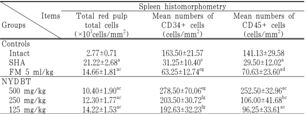
10. Changes on the spleen histopathology Significant (p<0.01) increases of the total red pulp cell numbers and decreases of CD34 and CD45 immunolabeled cells were observed in the spleen of SHA control rats as compared with intact vehicle control rats, respectively. However, significant
(p<0.01 or p<0.05) decreases of the total red pulp cells and increases of red pulp CD34 and CD45 immunoreactive cells were observed in all test substance treated rats including NYDBT 125 mg/kg administered rats as compared with SHA control rats, in this study (Table 8).
Items Groups
Spleen histomorphometry Total red pulp
total cells (×10 3 cells/mm 2 )
Mean numbers of CD34+ cells
(cells/mm 2 )
Mean numbers of CD45+ cells
(cells/mm 2 ) Controls
Intact 2.77±0.71 163.50±21.57 141.13±29.58
SHA 21.22±2.68 a 31.25±10.40 e 29.50±12.02 a
FM 5 ml/kg 14.66±1.81 ac 63.25±12.74 eg 70.63±23.60 ad NYDBT
500 mg/kg 10.40±1.90 ac 278.50±70.06 eg 252.50±32.96 ac 250 mg/kg 12.30±1.77 ac 203.50±30.72 fg 106.00±41.68 bc 125 mg/kg 14.22±1.53 ac 192.63±32.23 fg 96.25±33.61 ac
Values are expressed mean±SD of eight rats.a : p<0.01 and b : p<0.05 as compared with intact vehicle control by LSD test c : p<0.01 and d : p<0.05 as compared with SHA control by LSD test
e : p<0.01 and f : p<0.05 as compared with intact vehicle control by MW test g : p<0.01 as compared with SHA control by MW test
Table 8. Changes on the Spleen Histomorphometrical Analysis
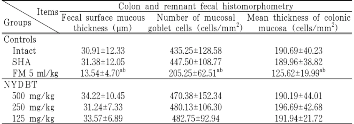
11. Changes on the histopathology of colon and remnant feces
No meaningful or significant changes on the histopathological profiles of the colon and remnant feces in situ, the surface mucosa thicknesses of remnant fecal pellets in the colon lumen, mucosa thicknesses and the mucous producing cell numbers in the colonic mucosa were demonstrated in SHA control and all three different
dosages of NYDBT treated rats as compared with intact vehicle control, respectively.
However, significant (p<0.01) decreases of the surface mucous thicknesses of remnant fecal pellets in the colon lumen, the mucosa thicknesses and mucous producing cell numbers in the colon were detected in FM 5 ml/kg treated rats as compared with intact vehicle and SHA control rats, in the present study (Table 9).
Items Groups
Colon and remnant fecal histomorphometry Fecal surface mucous
thickness (μm)
Number of mucosal goblet cells (cells/mm 2 )
Mean thickness of colonic mucosa (cells/mm 2 ) Controls
Intact 30.91±12.33 435.25±128.58 190.69±40.23
SHA 31.38±12.05 447.50±108.77 189.96±38.82
FM 5 ml/kg 13.54±4.70 ab 205.25±62.51 ab 125.62±19.99 ab NYDBT
500 mg/kg 34.22±10.45 470.38±152.34 190.19±44.01
250 mg/kg 31.24±7.33 480.13±106.30 196.69±42.68
125 mg/kg 33.57±6.89 482.75±92.94 191.94±21.72
Values are expressed mean±SD of eight rats.
a : p<0.01 as compared with intact vehicle control by LSD test b : p<0.01 as compared with SHA control by LSD test
Table 9. Changes on the Histomorphometrical Analysis of the Colon and Remnant Fecal Pellets in situ
Ⅳ. Discussion
Iron deficiency anemia is a common anemia because of chronic or subacute blood losses including in case of women with menorrhagia or menometrorrhagia.
It has been treated by iron supplements, but can induce digestive discomfort, especially constipation. Therefore, new treatments have to be concerned on natural herbs less toxic effects than iron supplements.
We can consider a traditional medicine prescription used in blood deficiency 7) . NYDBT is a traditional medicine that has been used for several hundred years, predominantly to tonic purpose in a weak constitution as qi and blood deficiency or ameliorating loss of blood of women 8) .
Considerable amounts of bleeding leads
hypovolemic situations in mammals, and
accordingly marked decreases of body
weights were accompanied 23) . In the present
study, noticeable decreases of body weights were also induced by continuous exsanguinations of 1 ml/head (about 0.5%
body weights), once a day, from forth bleedings in SHA control rats, with significant decreases of body weight gains after seven repeated exsanguinations as secondary results from hypovolemic situations considering the facts that indicated by other investigators 23) . In the present study, NYDBT dose-dependently and significantly inhibited the hypovolemic related decreases of body weights, but not in FM 5 ml/kg treatment. These results are suggested that NYDBT can be ameliorating the body weight losses induced by SHA through their hematopoietic activities.
Iron deficient and regenerative anemia have been showed marked decreases of RBC, Hct and Hb with increases of μ RBC and MRBC and peculiarly noticeable increases of PLT were also accompanied in subacute or chronic iron deficient and regenerative anemia on the hematological observations 24) . In the present study, rats exsanguinations of 1 ml/head, once a day for 7 days, also showed classic SHA related hematological changes including decreases of RBC, Hct and Hb with increases of PLT, μRBC and MRBC, respectively. These SHA related hematological changes were effectively inhibited by oral treatment of commercial iron supplements, FM and also by oral administrations of all three different dosages of NYDBT, dose-dependently, except for PLT, MRBC numbers.
On the smear cytological aspects, classic microcytic and hypochromatic anemia were observed in hemorrhage related regenerative anemia 1,18) , and also decreases of RBC diameters and increases of PCEs were detected in SHA control rats of the present study, as typical regenerative and iron deficient anemia. These smear cytological changes were also favorably inhibited by oral treatment of FM 5 ml/kg, and also dose-dependently in NYDBT 500, 250 and 125 mg/kg administered rats, respectively.
Continuous bleeding related hemorrhagic anemia induced excessive erythropoiesis in the bone marrow with start of extramedullary ectopic hematopoiesis in fetal hematopoietic organs, liver and spleen 25) . Marked and significant increases of total femur bone marrow cells, increases of hematopoietic spots in liver and increases of splenic red pulp cells, may be indicated ectopic hematopoiesis were also observed in SHA control rats, in the present study, with noticeable increases of spleen weights.
Once again, these ectopic hematopoiesis and bone marrow hyperplasia were meaningfully inhibited by treatment of all test substances used in this study.
In chronic anemia, depletion of
hematopoietic stem cells, the decreases
of absolute cell numbers, have been
observed related to over differentiation
into erythrocyte precursor cells or migration
into peripheral bloods with premature
erythrocytes like MRBC or PCEs 26) . In
the present study, CD34+ and CD45+
cells were regarded as hematopoietic stem cells on the basis of the previous reports 24) , marked decreases of hematopoietic stem cells were detected in the femur bone marrow, splenic red pulp and hepatic portal triad regions after immunohistochemistrical analysis using CD34 and CD45 antibodies, may be related to the over differentiations or migration considering the report of other investigators 26) . However, these SHA- related decreases of hematopoietic stem cells were marked and favorably inhibited by treatment of FM and all three different dosages of NYDBT. Obvious dose-dependently inhibitions of the CD34+ and CD45+ cells were observed in NYDBT treated rats in all three observed hematopoietic organs - the femur bone marrow, spleen and liver.
However, the possibilities that NYDBT also potentially inhibited the migration of stem cells, were also could not completely exclude in the present study. Therefore, more fundamental mechanism studies should be tested in future.
The transit process of the entire gastrointestinal tract reflected the overall gastrointestinal motor activity, and measuring gastrointestinal charcoal transit ratio is useful in diagnosis of constipation 27) . Since the decrease of gastrointestinal charcoal transit ratio means constipation 15,16) , the decreased gastrointestinal charcoal transits induced by treatment of FM 5 ml/kg, detected in this study, were considered as one of direct evidences that commercial iron supplement used in this experiment induced the digestive discomfort, constipation,
well corresponded to the previous reports 4) . In addition, reduces of mucous production on the colonic mucosa are directly related with constipation 15,16) , and marked decreases of colonic mucosa layer thicknesses and mucous producing cells has been detected in animals suffering from constipation at histopathology 16) . Moreover, FM 5 ml/kg also induced noticeable and significant decreases of mucous components in the colon and remnant feces in situ under alcian blue staining as direct evidences that they induced constipation. However, no meaningful changes on the charcoal transfers, mucous components in the colonic mucosa and also in remnant feces were demonstrated in all three different dosages of NYDBT as compared to those of intact vehicle and SHA control in this experiment.
These are direct evidences that NYDBT has potent favorable anti-anemic effects on SHA through proliferating effects on hematopoietic stem cells with less digestive discomfort, constipations, one of major problems in iron supplements 4) .
The results obtained in this study suggest
that oral administration of NYDBT 500,
250 and 125 mg/kg has clear dose-dependently
favorable anti-anemic in SHA rats through
proliferating effects on hematopoietic stem
cells with less digestive discomfort at least,
in a condition of this experiment. It,
therefore, is expected that NYDBT will
be promising as a novel alternative
hematopoietic and therapeutic agent for
anemia. Since NYDBT consisted of 18
herbs and each herb has various active
ingredients, the screening of the biological active compounds should be conducted in future with more detail mechanism studies.
Ⅴ. Conclusion
In this study, we reached the following results:
1. NYDBT dose-dependently and significantly inhibited the hypovolemic related decreases of body weights, but not in FM 5 ml/kg treatment.
2. Significant decreases of spleen weights, increases of absolute liver weights were noticed in NYDBT treated rats, but not in FM treated rats. In case of femur weights, significant increases of absolute femur weights were only demonstrated in NYDBT 500 mg/kg.
3. More favorable and significant increases of RBC numbers, Hct and Hb and decreases of μRBC ratios were demonstrated in FM 5 ml/kg and NYDBT dose-
dependently.
And also, more favorable and significant increases of RBC diameters and decreases of PCEs were demonstrated in FM 5 ml/kg and NYDBT dose-dependently.
4. Obvious increases of the CD34+ and CD45+ cells were observed in FM 5 ml/kg and NYDBT dose-dependently treated rats in all three observed hematopoietic organs - the femur bone marrow, spleen and liver.
5. Significant decreases of gastrointestinal charcoal transits, mucous components in the colon and remnant feces in situ were induced by treatment of FM 5 ml/kg only. However, no meaningful changes on all three different dosages of NYDBT.
According to these results, oral administrations of NYDBT 500, 250 and 125 mg/kg has clear dose-dependently anti-anemic effect on SHA rats.
□ Received : Jul 25, 2017
□ Revised : Jul 29, 2017
□ Accepted : Aug 16, 2017
국문초록