Anti-oxidant and anti-inflammatory activities of the various kinds of herbal tea
Jin Wook Lee
1#, Hyun Ji Eo
1, Gwang Hun Park
1, Hun Min Song
1, So Hee Woo
1, Mi Kyoung Kim
1, Jung Hye Eom
2, Man Hyo Lee
3, Jeong Rak Lee
3,
Jin Suk Koo
1,2, Jin Boo Jeong
1,2*1 : Department of Bioresource Sciences, Andong National University, Andong, 760749, Korea 2 : Department of Medicinal Plant Resources, Andong National University, Andong, 760749, Korea
3 : Gyeongbuk Institute for Bio-industry, Andong, 760380, Korea
ABSTRACT
Objectives : Reactive oxygen species (ROS) are involved in a wide spectrum of diseases including chronic inflammation and cancer. In this study, we investigated the antioxidant activities and anti-inflammatory effects of the extracts from the herbal teas such as Lonicera japonica Thunberg ( L. japonica ), Chrysanthemum morifolium Ramat ( C. morifolium ), Mentha arvensis L. ( M. arvensis ), and P.rhizoma.
Methods : Anti-oxidant activity was evaluated using DPPH radical scavenging assay and Fe
2+chelating assay.
And DNA cleavage assay was performed to evaluate an anti-oxidative effect. Anti-inflammatory effect was performed using NO generation assay and western blot in LPS-stimulated RAW264.7 cell line.
Results : L. japonica scavenged DPPH radical by 9.8% at 12.5 µg/ml, 24.8% at 25 µg/ml, 34.3% at 50 µg/ml, 61.1% at 100 µg/ml and 75.8% at 200 µg/ml, respectively. In addition, C. morifolium and M. arvensis removed DPPH radical by 15.6% and 10.4% at 12.5 µg/ml, 34.8% and 22.8% at 25 µg/ml, 66.9% and 43.3% at 50 µg/ml, 87.4% and 69.1% at 100 µg/ml, and 92.1% and 73.2% at 200 µg/ml, respectively. However, P. rhizoma did not affect on DPPH radical scavenging. The Fe
2+chelating activity was highest in L. japonica , but lowest in P.
rhizoma among the herbal teas. In addition, the extracts from L. japonica , C. morifolium and M. arvensis inhibited oxidative DNA damage via its anti-oxidant activity. In anti-inflammatory effect, the extracts from C.
morifolium inhibited NO production. In addition, it suppressed the NF-κ B signaling pathway in LPS-stimulated RAW 264.7 cells.
Conclusions : Together, this study indicates that L. japonica , M. arvensis and C. morifolium possess the protective effect against the oxidative DNA damage. Furthermore, C. morifolium exerts an anti-inflammatory effect.
Key words : Herbal teas; Anti-oxidant; Oxidative damage; Anti-inflammation
Introduction 1)
Reactive oxygen species (ROS) produced by cellular aerobic respiration have been regarded as a inducer of oxidative stress including damage of cell matrices such as lipids, proteins and DNA, which is associated with human diseases such as cancer and chronic inflammation
1-3). Thus, antioxidant activity can be
defined as a suppression of oxidative damage of organic molecule including lipids, proteins, DNA and other molecules
4). Antioxidants can be divided to two types;
primary antioxidants directly remove the generated ROS and second antioxidants indirectly inhibits the ROS generation by Fenton's reaction. In generally, herbal teas have been reported to have these two type capacities
5).
*Corresponding author : Jin Boo Jeong, Department of Medicinal Plant Resources, Andong National University, Andong, Korea, 760-749.
·Tel : +82-54-820-7757 ·Fax : +82-54-820-6252 ·E-mail : [email protected]
#First author : Jin Wook Lee, Department of Medicinal Plant Resources, Andong National University, Andong, Korea, 760-749.
·Tel : +82-54-820-7753 ·E-mail : [email protected]
·접수:2014년 2월 21일 ·수정:2014년 3월 12일 ·채택:2014년 3월 13일
In addition, ROS have been regarded as a mediator of chronic inflammation by activating proinflammatory cytokines, which has been regarded as a major mechanism for inflammation injury
6). Especially, ROS stimulates nitric oxide (NO), one of the inflammation mediators by which inflammatory processes can be provoked or sustained. Thus, free radicals are important mediators that provoke or sustain inflammatory processes and, consequently, their neutralization by antioxidants and radical scavengers can attenuate inflammation.
Therefore, antioxidants can attenuate inflammation
7). Teas have been regarded as the most widely consumed beverages worldwide
8). Among teas, herbal teas using the leaves, flowers, seeds, fruits, stems or roots of plant species have been consumed for health care and disease prevention
9,10)because these contain various active phytochemicals with pharmacological properties such as allergies, insomnia, headaches, anxiety, intestinal disorders, depression, and high blood pressure
11). There is growing evidence that herbal teas have several biological effects including anti-cancer, anti-atherogenic, anti-oxidant and anti-microbial activities
12). In this study, we evaluated the anti-oxidant and anti-inflammatory capacities of aqueous extracts of Lonicera japonica Thunberg ( L. japonica ), Chrysanthemum morifolium Ramat ( C. morifolium ), Mentha arvensis L.
( M. arvensis ), and P. rhizomaused as a herbal tea.
Materials and Methods
1. Chemicals
1,1diphenyl-2-picrylhydrazyl (DPPH) and lipopolysaccharide (LPS) were purchased from Sigma Aldrich Co. (St. Louis, USA). φ X-174 RF I plasmid DNA was purchased from New England BioLabs (County Rood Ipswich, MA, USA). Antibodies against IκB-α, p65 and β-actin were purchased from Cell Signaling (Bervely, MA, USA). Cell culture media, Dulbecco's Modified Eagle medium (DMEM)/F-12 1:1 Modified medium (DMEM/F-12) was purchased from Lonza (Walkersville, MD, USA). pNFκ B-Luc cis-Reporter plasmid was purchased from Agilent Technologies (Santa Clara, CA, USA).
2. Sample preparation
Herbal teas, L. japonica, C.morifolium, M. arvensis and P. rhizoma was kindly provided by Bonghwa Alpine Medicinal Plant Expriment Station, Korea. One hundred gram of the herbal teas was extracted with 300 ml of distilled water in 100 ℃ for 90 min. After
90 min, the extracts were filtered and freeze-dried.
The freeze-dried extracts were kept at -80 ℃ until use.
3. Cell culture and treatmentf
Mouse macrophage cell line, RAW264.7 cell was purchased Korean Cell Line Bank (Seoul, Korea) and grown in DMEM/F-12 supplemented with 10% fetal bovine serum (FBS), 100 U/ml penicillin and 100 µg/ml streptomycin. The cells were maintained at 37 ℃ under a humidified atmosphere of 5% CO
2. Aqueous extracts from the herbal tea were dissolved in 1×phosphate-buffered saline (PBS) and treated to cells. 1×PBS was used as a vehicle.
4. DPPH radical scavenging assay
DPPH radical scavenging assay was carried out according to the literature
13). Briefly, 760 µl DPPH ethanol solution (300 µM) solution and 40 µl of the extracts were mixed and then incubated at 37 ℃ for 30 min. After 30 min, the absorbance was measured at 515 nm
5. Fe
2+chelating assay
Fe
2+assay was performed according the literature
13). chelating The reaction mixture (800 µl) contained 15 µl of FeCl
2(2 mM), 150 µl of varying concentrations of the extracts and 605 µl distilled water. The mixture was shaken vigorously and left at room temperature for 30 min. After 30 min, 30 µl of ferrozine (5 mM in methanol) was added and mixed. The absorbance of the Fe
2+-ferrozine complex was measured at 562 nm.
6. DNA cleavage assay
Conversion of the supercoiled form of plasmid DNA
to the open-circular and further linear forms has
been used as an index of DNA damage
14). The reaction
mixtures (25 µl) containing 5 µl of φX-174 RF I
plasmid DNA, 10 µl of varying concentrations of the
extracts and 5 µl of 1 mM FeSO
4were incubated at
37 ℃ for 30 min. After 30 min, 5 µl of a solution
containing 50% glycerol (v/v), 40 mM EDTA and
0.05% bromophenol blue was added to stop the
reaction and the reaction mixtures were electrophoresed
on 1% agarose gel, and the DNA in the gel was
visualized and photographed under ultraviolet light
after ethidium bromide staining.
7. Nitric oxide generation
RAW264.7 cells were plated in 12-well plate for overnight. Cells were pre-treated with the extracts from the herbal teas at the indicated concentrations for 2 h and then co-treat with LPS (1 µg/ml) for the additional 18 h. After 18 h, 200 µl of the media was mixed with equal amount of Griess reagent (1% sulfanilamide and 0.1% N-1-(naphthyl) ethylenediamine-diHCl in 2.5% H
3PO
4). The mixture was incubated for the additional 5 min at the room temperature and the absorbance was measured at 540 nm.
8. Isolation and cytosol and nuclear fraction
Nuclear and cytosolic fractions were prepared following the manufacturer's protocols of nuclear extract kit (Active Motif, Carlsbad, CA, USA). Briefly, RAW264.7 cells were washed with ice-cold PBS containing phosphatase inhibitors, harvested with 1xhypotonic buffer and then incubated at 4 ℃ for 15 min. After 15 min, the cells were added with detergent and then centrifuged at 15,000 rpm for 1 min. The supernatants were collected as cytoplasmic fraction. Nuclear fractions were collected by suspending nuclear pellet with nuclear lysis buffer and centrifugation at 15,000 rpm for 10 min.
9. SDS-PAGE and Western blot
Cells were washed with 1×phosphate-buffered saline (PBS), and lysed in radioimmunoprecipitation assay (RIPA) buffer (Boston Bio Products, Ashland, MA, USA) supplemented with protease inhibitor cocktail (Sigma Aldrich) and phosphatase inhibitor cocktail (Sigma Aldrich), and centrifuged at 15,000 × g for 10 min at 4℃. Protein concentration was determined by the bicinchoninic acid (BCA) protein assay (Pierce, Rockford, IL, USA). The proteins were separated on SDS-PAGE and transferred to PVDF membrane (Bio-Rad Laboratories, Inc., Hercules, CA, USA). The membranes were blocked for non-specific binding with 5% nonfat dry milk in Tris-buffered saline containing 0.05%
Tween 20 (TBS-T) for 1h at room temperature and then incubated with specific primary antibodies in 5%
nonfat dry milk at 4℃ overnight. After three washes with TBS-T, the blots were incubated with horse radish peroxidase (HRP)-conjugated immunoglobulin G (IgG) for 1 h at room temperature and chemiluminescence was detected with ECL Western blotting substrate (Amersham Biosciences) and visualized in Polaroid film.
10. Transient transfection
Transient transfection was performed using PolyJet DNA transfection reagent (SignaGen Laboratories, Ijamsville, MD, USA) according to the manufacturer's instruction.
Briefly, RAW264.7 cells were seeded in 12-well plates and incubated overnight. Then, plasmid mixtures containing 0.5 µg of pNF-κ B-Luc plasmid and 0.05 µg of pRL-null vector were transfected for 24 h.
After transfection, cells were pre-treated with the extracts from the herbal teas for 2h and then co-treated with LPS for an additional 15 h. The cells were harvested in 1 x luciferase lysis buffer, and luciferase activity was measured and normalized to the pRL-null luciferase activity using a dual-luciferase assay kit (Promega, Madison, WI, USA).
11. Statistical analysis
Statistical analysis was performed with the Students unpaired t-test, with statistical significance set at
*, P < 0.05.
Results
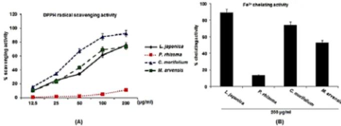
1. Antioxidant activities of herbal teas
Antioxidant activities of herbal teas were evaluated using DPPH radical scavenging assay and Fe
2+chelating assay. Scavenging of DPPH radicals has been used as the basis of a common antioxidant assay. In DPPH radical scavenging activity(Fig. 1A), L. japonica scavenged DPPH radical by 9.8% at 12.5 µg/ml, 24.8%
at 25 µg/ml, 34.3% at 50 µg/ml, 61.1% at 100 µg/ml and 75.8% at 200 µg/ml, respectively. In addition, C.
morifolium and M. arvensis removed DPPH radical by 15.6% and 10.4% at 12.5 µg/ml, 34.8% and 22.8% at 25 µg/ml, 66.9% and 43.3% at 50 µg/ml, 87.4% and 69.1% at 100 µg/ml, and 92.1% and 73.2% at 200 µg/ml, respectively. However, P. rhizoma did not affect DPPH radical scavenging. In Fe
2+chelating activity, the chelating activity was highest in L.
japonica while lowest in P. rhizoma among the herbal teas.
Fig. 1. DPPH radical scavenging activity and Fe2+ chelating activity of the herbal teas. The absorbance values were converted to scavenging activity or chelating activity (%).
2. Protective effect of the herbal teas against oxidative DNA damage
The inhibitory effect of the extracts from the herbal teas was evaluated using invitro fenton reaction between H
2O
2and Fe
2+. Undamaged plasmid DNA was mainly the supercoiled form (SC) in absence of H
2O
2and Fe
2+(Fig. 2, lane 1). When the oxidative damage of plasmid DNA was induced by H
2O
2and Fe
2+, SC was converted into the open-circular form (OC)(Fig. 2, lane 2). The extracts from L. japonica , M. arvensis and C. morifolium attenuated the conversion of SC into OC, which indicates L. japonica , M. arvensis and C. morifolium could protect DNA from oxidative damage.
However, the extracts from P. rhizoma did not affect the protection of oxidative DNA damage.
Fig. 2. Protective effect of the herbal teas against oxidative DNA damage. Oxidative damage of φX-174 RF I plasmid DNA was induced by FeSO4. SC and OC mean supercoiled and open-circular form, respectively.
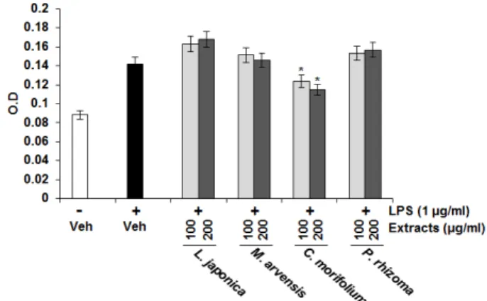
3. Effect of the herbal teas on nitric oxide (NO) production in LPS-stimulated RAW264.7 cells
Since ROS is associated with inflammation, we also evaluated the effect of the herbal teas on NO production in LPS-stimulated RAW264.7 cells. RAW264.7 cells were pretreated with the extracts from the herbal teas for 2 h and then co-treated with LPS (1 µg/ml) for the additional 18 h. As shown in Fig. 3, treatment of LPS without the extracts induced NO overproduction in LPS-stimulated RAW264.7 cells, while pretreatment of the extracts from C. morifolium suppressed LPS-mediated NO overproduction. However, the extracts from L. japonica , M. arvensis and P.
rhizoma did not affect NO production in LPS-stimulated RAW264.7 cells.
Fig. 3. Effect of the extracts from the herbal teas on LPS-induced NO production in RAW264.7 cells. Cells were pre-treated with the extracts from the herbal teas for 2 h and then co-treated with 1 µg/ml of LPS for 15 h. The NO concentration in the medium was measured using Griess reagent. *p < 0.05 compared to LPS-stimulated cells.
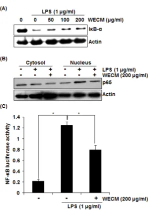
4. Inhibitory effect of the extracts from C.
morifolium on LPS-induced NF-κ B activation in RAW264.7 cells
To elucidate the effect of the extracts from C.
morifolium on NF-κB activation by LPS, we carried out Western blot for Iκ B-α degradation in LPS-stimulated RAW264.7 cells. As shown in Fig. 4A, LPS induced I κB-α degradation at 15 min after the stimulation.
However, pretreatment of the extracts from C. morifolium
dose-dependently attenuated Iκ B-α degradation. NF-κB
p65 nuclear translocation resulted from Iκ B-α degradation
is essential for NF-κ B activation. Thus, we tested if
the extracts from C. morifolium block p65 nuclear
translocation. As shown in Fig. 4B, LPS markedly
increased an amount of p65 in nuclear of RAW264.7
cells, while the extracts from C. morifolium inhibited
LPS-induced p65 nuclear translocation in RAW264.7
cells. Translocated p65 into nucleus directly binds to
the NF-κ B binding site and subsequently induces
transcriptional activation of NF-κ B. So, we
determined whether the extracts from C. morifolium
inhibit transcriptional activity of NF-κ B using pNF-κ
B-Luc-cis-reporter plasmid. In this assay, the
extracts from C. morifolium inhibited LPS-induced
transcriptional activity of NF-κB in RAW264.7 cells
(Fig. 4C). Overall, the results indicated that the
extracts from C. morifolium may inhibit NF-κ B
activation by suppression of p65 translocation into the
nucleus via blocking the IκB-α degradation.
Fig. 4. Effect of the extracts from C. morifolium on NF-κB activation in LPS-stimulated RAW264.7 cells. RAW264.7 cells were pre-treated with indicated concentrations of the extracts from C.
morifolium for 2 h, and then co-treated with LPS (1 µg/ml) for an additional 15 min (A) or 30 min (B). Cell lysate (30 µg) were resolved by 12% SDS–PAGE, transferred to nitrocellulose membranes, and probed with antibodies against IκB-α and p65. The proteins were then visualized using ECL detection. Actin was used as an internal control. For NF-κB luciferase activity (C), pNF-κB-Luc plasmid-transfected cells were pre-treated with the extracts from C. morifolium for 2 h, and then co-treated with LPS (1 µg/ml) for an additional 15 h. The cells were harvested in 1 x luciferase lysis buffer, and luciferase activity was measured using a dual-luciferase assay kit. WECM means the water extracts from C.
morifolium. Values given are the mean ± SD (n = 3). *p < 0.05 compared to LPS-stimulated cells.