Copyright © 2011 Korean Journal of Bronchoesophagology 65
Korean J Bronchoesophagol 2011;17:65-67 ISSN 1226-0916
CASE REPORT
서 론
하인두의 점막구조는 매우 얇고 유연하며, 특히 측방은 경동 맥 초(carotid sheath)와 얇고 작은 하인두 수축근에 의해 경계 되어지는데, 이상와(Pyriform sinus)는 전벽, 외측벽, 내측벽으 로 이루어져 있으며 하방으로 갈수록 좁아져 역 피라미드 모양 이다. 하인두의 천공은 이물, 경부 둔상, 내시경 검사 등 여러 가지 원인에 의한 경우가 보고되었으나, 기관내 삽관 중 발생한 이상와 천공은 매우 드문 합병증으로 국내 문헌에는 1예가 보 고되었다.1) 이상와 천공은 인두 천공에서 두 번째로 흔한 부위 로서2) 이곳의 천공은 이차적인 심경부 감염증을 유발할 수 있고 종격동염, 패혈증 등의 합병증이 병발하는 경우는 생명을 위협 할 수 도 있다.
저자들은 호흡곤란을 주소로 응급실에 내원하여 기관 삽관 술 후 발생한 외상성 이상와 천공 환자를 수술적 치료로 합병 증 없이 치료하였기에 문헌고찰과 함께 보고하고자 한다.
증 례
54세 여자 환자로 수일 전부터 폐염으로 개인의원에서 치료 중 내원 전날부터 점차 심해지는 호흡곤란과 혼미한 의식상태 로 응급실에 내원하였다. 내원 당시 호흡곤란이 심한 상태로 기관 삽관을 여러 차례 시도하여 삽관 하였으나, 수시간 후 환 자가 자가 발관을 하여 다시 여러 차례의 시도 후 재 삽관을 하 였다. 수시간 후 환자의 우측 경부의 경한 종창과 누를 때 피 하기종에 의한 염발음이 발견되어 경부 전산화 단층촬영을 시 행 한 결과 우측 경부 전장에 걸친 피하기종과 상부 종격동의 기종격동(pneumomediatinum)이 보였고 특히 우측 갑상선 부 위에서 기종이 심하였다(Fig. 1). 따라서 인두나 식도의 천공을 의심하였으나 환자의 의식상태와 기도삽관 등으로 확인을 위 한 검사를 보류하고 관찰 하였다. 입원 3일째 의식이 회복되어 기도삽관을 발관 후 GastografinⓇ을 사용한 식도조영술을 시 행 한 결과 우측 이상와 하방으로 적은양의 조영제 누출이 있 어 이상와 천공이 확인되었으나(Fig. 2) 천공부위가 작고 전신 마취시 기존의 폐염이 악화되는 등을 우려하여 금식과 광범위 항생제 사용 등 보존적 치료를 하였다.
식도 조영술 후 7일째 우측 갑상선엽 부위의 통증을 호소하 여 다시 GastografinⓇ을 사용한 식도조영술을 시행 한 결과
기관내 삽관 후 발생한 이상와 천공
조선대학교 의학전문대학원 이비인후-두경부외과학교실
유승우
·박준희
·최지윤
·도남용
Pyriform Sinus Perforation after Intubation
Seung Woo Yu, Jun Hee Park, Ji Yun Choi and Nam Yong Do
Department of Otorhinolaryngology-Head and Neck Surgery, Medical School, Chosun University, Gwangju, Korea
Pyriform sinus perforation is a rare complication of endotracheal intubation. It most commonly occurs at the hands of the less experienced physician in emergency situations. It can occur after traumatic intubation and is potentially lethal. The site most commonly perforated is the pharynx, posterior to the cricopharyngeal muscle; the second most common site is the pyriform sinus. We report a case of pyriform sinus perforation after endotracheal intubation, which was successfully treated with pri-
mary closure. Korean J Bronchoesophagol 2011;17:65-67
KEY WORDSZZ Intubation ㆍintratracheal ㆍPyriform sinus perforation..
논문접수일: 2011년 5월 25일 / 심사완료일: 2011년 6월 24일 교신저자: 도남용, 501-717 광주광역시 동구 서석동 588 조선대학교 의학전문대학원 이비인후-두경부외과학교실 전화: 062-220-3200 ㆍ전송: 062-225-2702 E-mail: nydo@chosun.ac.kr
online©MLComm
66
Korean J Bronchoesophagol
█2011;17:65-67
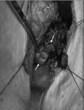
이전 보다 심한 조영제 누출이 있어(Fig. 3) 수술적 치료를 위 해 이비인후과로 전과되었다. 수술소견으로는 우측 이상와 외 측벽에 2 cm정도의 천공이 있고 하인두수축근을 관통하여 갑상선 우엽에 손상을 주어, 동측 갑상선의 괴사와 주위 조직 의 염증소견이 관찰되었다(Fig. 4). 우측 갑상선 엽절제술을 시행하고, 천공 주위의 염증조직을 제거하고 충분히 세척 후 바깥쪽에서 이상와 점막을 4~0 Vicryl을 사용하여 2겹으로 일차봉합하고 하인두수축근을 봉합한 후 견갑설골근과 흉쇄 유돌근을 이용하여 보강하였다.
술 후 10일째 시행한 식도조영촬영상 천공부위의 주영제의 유출은 없으나 경도의 기관내 흡인이 보였으나 유동식의 경구
투여를 시작하고 술 후 14일째 식도조영촬영을 하여 재확인한 결과 조영제 유출이나 흡인이 없고 환자상태 양호하여 퇴원하 였다.
고 찰
기관내 삽관은 안정적 환경에서 시행하는 경우는 비교적 안 전한 술식 이지만 호흡곤란을 호소하는 응급상황에서 시행하
Fig. 1. Computed tomographic scan of the neck shows multiple
subcutaneous emphysema (white arrow).Fig. 2. Gastrografin esophagogram shows fistulous tract and leak-
age of contrast medium from right pyriform sinus apex (white ar- row).Fig. 3. Gastrografin esophagogram at one week after first esoph-
agogram shows more increased leakage of contrast medium from right pyriform sinus to the mediastinum (white arrow).Fig. 4. Intraoperative finding. A perforation 2 cm in diameter
(white arrow) is present in the right pyriform sinus.http://www.korbes.org 67
Pyriform Sinus Perforation after Intubation
█SW Yu et al
는 경우는 후두, 기관지 또는 인두나 식도의 손상 등을 일으킬 수도 있다. 외상성 기관내 삽관시 손상부위는 윤상인두근 뒷 쪽이 가장 흔하며 이상와의 손상은 두 번째로 흔하다.2)
외상성 기관내 삽관으로 인한 하인두 천공을 암시하는 증후 로는 가장 흔한 초기 증후는 경부를 촉진시 염발음을 갖는 피 하기종이 가장 많으며, 출혈, 기종격동, 기흉(pneumothorax), 연하시 인두통 등이며 발열은 초기에는 저명하지 않으나,3,4) 보 다 진행되면 염증에 의한 봉와직염과 경부 부종을 보이며 발 열, 패혈증 증상과 흉부 청진음의 변화 등이 나타난다.5) 본 증 례의 경우는 발열이 없이 경부의 피하기종, 기종격동, 연하시 인두통, 등의 초기 증상과 외상 후부터 항생제 치료를 하여 수 일이 경과한 후 우측 갑상선엽 부위의 경한 통증을 호소하였다.
이상와 천공은 천공의 발견 시간이 중요한데 지연된 경우 불 행 한 결과를 가져올 수 있다. 합병증으로는 후인두부 농양, 종 격동염, 패혈증, 뇌막염 등을 일으켜 사망 한 예도 있다.6) 조기 에 정확한 진단을 위해서는 이학적 검사와 자세한 병력 청취 가 필요하며, 내시경 검사와 방사선학적 검사는 천공의 확인, 천공된 위치와 크기, 주변조직의 손상정도와 치료 도중의 추 가적인 환자 평가에 필요하다. 경부와 흉부의 단순방사선 촬영 은 피하 기종, 기종격동, 기흉 등을 알 수 있으며,7) 경부나 흉부 의 전산화 단층촬영은 경부 농양 및 공기음영으로 손상의 대 략적인 위치 및 정도, 종격동으로의 파급 정도 등을 알 수 있어 수술적 치료 여부의 결정과 수술 접근경로 결정에 필수적이 다.8) 식도조영술은 천공부위를 확인하는 비침습적 방법으로 수용성 조영제인 GastrografinⓇ을 사용하여 시행하는데, 만일 조영제 누출이 없으나 임상적으로 천공이 의심되면 수용성 조 영제 보다 예민한 Barium을 사용하면 작은 천공도 알 수 있 으나 주위조직으로 유출되는 경우 손상이 심하다는 단점이 있 다.9) 본 증례 환자의 경우 초기에 경부 전산화단층술과 Gas- trografinⓇ을 사용한 식도조영술로 천공을 확인하였고, 수술 적 치료의 결정과 술 후 경구식이의 시작 가능여부를 결정할 수 있었다.
치료는 천공 부위가 작은 경우 광범위 항생제 치료와 보존 적 치료가 우선 시도되는데, 천공이 인두에 국한된 2 cm 이하 면 세심한 관찰과 내과적 치료로 치유가 가능하다는 보고와10,11) 보다 작은 0.5 cm 이하인 경우 보존적 치료의 적응이 된다는 보고가 있다.5) 인두나 식도천공의 보존적 치료는 세심한 관찰, 경구를 통한 식이 제한, 비경구 수분공급, 정맥을 통한 광범위 항생제 투여 등과 안전하게 비위관을 삽입 할수 있으면 비위 관을 통한 식이가 필요하다.12) 결손부위가 큰 경우는 조기에 수술적 치료로 결손부위의 봉합 및 재건을 해야 하며 이러한
적극적인 치료로 합병증을 줄이고 치유율을 높일 수 있다고
한다.2,13) 인두-식도 천공의 수술적 치료의 적응증은 0.5~1 cm
이상의 천공, 조영제의 유출이 종격동이나 늑막강까지 있는 경우, 기흉, 늑막액, 패혈증 증상이 있을 때, 그리고 보존적 치 료에 실패한 경우 등이다.5) 수술방법은 작은 결손부는 비위관 위로 일차 봉합만으로 치유될 수 있으나, 큰 결손의 경우는 유 경 흉쇄유돌근 피판의 삽입(pedicled sternocleidomastoid muscle flap interposition) 등으로 봉합부위를 보강해 주어야 한다.14)
따라서 이상와 천공은 빠른 진단과 적극적인 치료가 합병 증을 줄일 수 있을 것으로 생각되는데, 이번 증례에서는 조기 에 우측 이상와 천공이 발생한 것을 알아 금식 및 광범위 항 생제 투여 등으로 보존적 치료를 시행하던 중 갑상선 우엽 쪽 으로 염증이 파급되어 수술적 치료를 통하여 성공적으로 치 유된 증례이다.
