구순구개열 환자에서 상악전방골 신장술
김유진
1, 천강용
1,2, 김수호
1, 박형욱
1, 황순정
1*
서울대학교 치의학대학원 구강악안면외과학교실1, 서울대학교 보라매병원 구강악안면외과2
ABSTRACT
Distraction Osteogenesis of Maxillary Anterior Segment in Cleft Lip and Palate Patients
Eu-Gene Kim
1, Kang-Yong Cheon
1,2, Soo-Ho Kim
1, Hyong-Wook Park
1, Soon-Jung Hwang
1*
Department of Oral and Maxillofacial Surgery, School of Dentistry, Seoul National University,
1Department of Oral and Maxillofacial Surgery, SMG-SNU Boramae Medical center
2Le Fort 1 osteotomy or maxillary advancement with distraction osteogenesis (DO) is main treatment strategy for cleft palate patients with maxillary hypoplasia. Maxillary DO allows greater maxillary advancement within physiological limit than Le Fort 1 osteotomy. Moreover, it is better for velopharyngeal function. However, there is a greater tendency for an increase in nasal sound when maxilla is advanced excessively.
Therefore, the advancement of anterior maxillary segment using DO has been utilized. It offers advantages such as an increase in the length of the palate, a prevention of the change in palatopharyngeal depth, and a preservation of the velopharyngeal function. Moreover, it will obliterate the necessity of bone graft, and it prevents the occurrence of oronasal or oroantral fistula. Finally, it stimulates the regeneration of the soft and hard tissue of alveolus, and subsequently makes possible to place implant.
Key words :
Maxillary anterior segment, Distraction osteogenesis, Velopharyngeal insufficiency, Cleft palate, HypernasalityⅠ. 서론
상악의 열성장은 구순구개열 환자에게서 나타나 는 흔한 발육성 장애 중 하나이다. 이는 흔히 선천 적인 중안면부 성장의 저하 및 이전 구개열 치료에 의한 외과적 반흔의 영향으로 발생한다. 따라서 구
순구개열 환자 대부분은 Class III 부정교합, 후퇴 된 중안면부, 좁은 경구개 등의 증상을 나타낸다.
1970년대 이후로 구순구개열 환자의 이러한 장애 를 악교정술(Orthograthic surgery)로 치료하여
왔고
1,2,3,4, 1990년대 후반부터는 상악골 신장술
(Maxillary distraction osteogenesis)이 새로운
대안으로 선택되어 왔다
5. 이 두 술식의 목적은 두 개골과 교합에 있어 상악을 전진 그리고 하방-이 동하여 정상적인 위치로 회복시키기 위함이다.
하지만 이 두 술식은 비인두 거리의 증가로 구개 범인두 기능부전(velopharyngeal insufficienct, VPI)을 야기하는 문제가 있다. 이러한 문제로 최근 에는 상악 전방골 절단술 후 상악 전방골만의 신장 술(Maxillary anterior segmental distraction osteogenesis)이 수술후 구개범인두 기능부전의 악화를 예방할 수 있는 장점을 가진 새로운 술식으 로 소개되고 있다
12.
이에 저자 구순구개열 환자의 상악 전방골 신장 술과 이를 통한 구개범인두 기능 변화에 대하여 전 반적인 문헌고찰을 통해 살펴보고자 한다.
1. 구개범인두 기능부전
(Velopharyngeal Insufficiency, VPI)
구개범인두는 편도, 혀, 그리고 구개와 인두가 포 함된 구조물들이 튜브모양으로 구성된 구조로서 발 성이나 연하 등의 기능을 하는 동안 구개범인두를 이 루는 근육들이 수축하게 되면 내부 공간을 좁게 하거 나 완전히 폐쇄하여 비강과 구강을 분리할 수 있다.
하지만 발성 시 비강과 구강이 완전히 분리되지 못하 는 경우 구개범인두 기능부전(velopharyngeal insufficiency, VPI)이 발생하게 된다
26. 이러한 구 개범인두 기능부전의 원인은 선천성 구개파열이 있 을 때, 구개파열이나 수술 후 반흔 형성이 심할 때, 연구개나 구개수의 조직량이 부족할 때, 기타 신경 장애 등이 원인으로 나타날 수 있다. 대표적인 증상 으로는 과비음, 비공기 유출로 인한 발음장애와 발 음장애를 보상하기 위해 낮은 목소리와 쉰 목소리 를 내는 경향을 보인다
6.
구순구개열 환자의 치료를 위해 르포씨 I형 골절 단술이나 상악골 신장술을 하여 상악골을 전방이동
함에 따라 구개범인두 부위가 함께 전방이동하게 되면서 이로 인한 합병증으로 구개범인두 기능부전 이 발생하거나 악화되는 현상이 나타난다
4,9. 그 동 안 많은 연구에서 상악의 전방이동으로 발생하는 구개범인두 기능부전에 대하여 보고되었다
7,8. 구개 범인두 기능부전(velopharyngeal insufficiency)과 구개범인두 기능(velopharyngeal function)의 장애 가 수술후 16~23%에 이르는 것으로 보고되었다
9. Cheung 등의 연구에서는 르포씨 I형 골절단술과 상악골 신장술 간에 수술 후 재발, 구개범인두 기 능, 발음 등에서 두 집단간에 큰 차이는 없는 것으 로 나타났다
8. Guyette 등도 상악골 신장술 후에 발생하는 구개범인두 기능부전의 위험성은 르포씨 I형 골절단술과 비슷하다고 하였다
10.
Ⅱ. 연구 방법
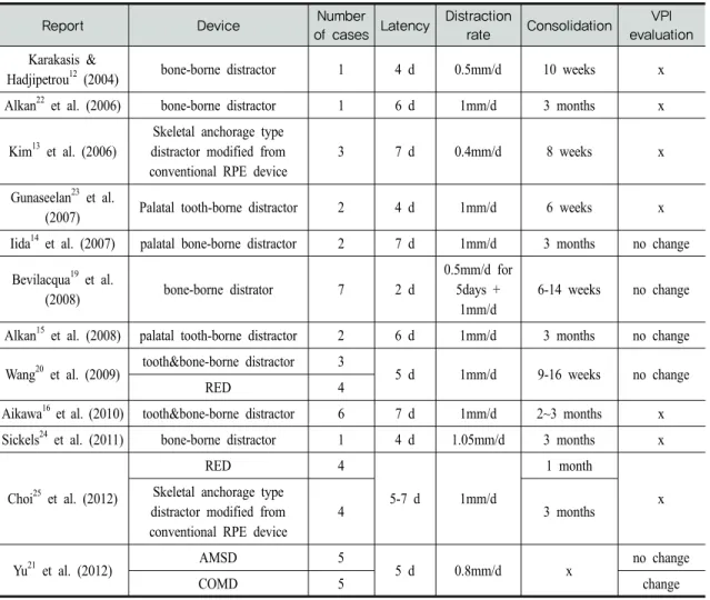
2012년 10월까지 Pubmed에서 ‘maxillary anterior segmental distraction osteogenesis’로 검색한 12 편의 논문을 정리하였다. 이중 상악전방골 신장술에 대한 증례보고가 총 9편이었고, external과 internal type을 비교한 논문이 2편, 상악전방골 신장술과 르 포씨 1형 골절단술 후 골신장술을 비교한 논문이 1편 이 있었다. 또한 구개범인두 기능의 평가는 5편의 논 문에서 이루어졌다(Table 1).
2. 상악 전방골 신장술 (Maxillary anterior segmental distraction osteogenesis)
상악 전방골 절단술은 상악의 치성돌출 환자에서
상악 전방골의 상방 또는 후방이동을 위해 주로 사
용되어 왔다. 현재 시행되는 술식으로는 Wassmund
법
27, Wunderer법
28, Cupar법
29등이 있고 골절단
술은 본질적으로 같으나 상악골에 접근하기 위한
Report Device Number
of cases Latency Distraction
rate Consolidation VPI evaluation Karakasis &
Hadjipetrou12 (2004) bone-borne distractor 1 4 d 0.5mm/d 10 weeks x Alkan22 et al. (2006) bone-borne distractor 1 6 d 1mm/d 3 months x
Kim13 et al. (2006)
Skeletal anchorage type distractor modified from conventional RPE device
3 7 d 0.4mm/d 8 weeks x
Gunaseelan23 et al.
(2007) Palatal tooth-borne distractor 2 4 d 1mm/d 6 weeks x
Iida14 et al. (2007) palatal bone-borne distractor 2 7 d 1mm/d 3 months no change Bevilacqua19 et al.
(2008) bone-borne distrator 7 2 d
0.5mm/d for 5days +
1mm/d
6-14 weeks no change
Alkan15 et al. (2008) palatal tooth-borne distractor 2 6 d 1mm/d 3 months no change Wang20 et al. (2009) tooth&bone-borne distractor 3
5 d 1mm/d 9-16 weeks no change
RED 4
Aikawa16 et al. (2010) tooth&bone-borne distractor 6 7 d 1mm/d 2~3 months x Sickels24 et al. (2011) bone-borne distractor 1 4 d 1.05mm/d 3 months x
Choi25 et al. (2012)
RED 4
5-7 d 1mm/d
1 month Skeletal anchorage type x
distractor modified from conventional RPE device
4 3 months
Yu21 et al. (2012) AMSD 5
5 d 0.8mm/d x no change
COMD 5 change
RED ; rigid external device
AMSD ; anterior maxillary segmental distraction with RED COMD ; conventional maxillary distraction osteogenesis with RED RPE ; rapid palatal expansion
X ; not mentioned
Table 1. The comparison of previous journals about the maxillary anterior segmental distraction osteogenesis 절개방법에서 차이가 있다. 구개부에 절개를 주는
Wassmund법이나 Wunderer법은 구순구개열 환자 에서 시행할 때는 두꺼운 구개점막과 oronasal 또 는 oroantral fistula 등으로 어려움이 있다
23.
2003년 Dolanmaz 등은 상악 열성장을 가진 Class III 환자에서 치조골절단술 후 rapid palatal expansion screw를 가진 tooth-borne palatal
distractor를 이용해 상악 전방분절골을 8mm가량 전 방이동 시켰다
11. 이어 2004년에 karakasis와 Hadijipetrou에 의해 구순구개열 환자의 상악 열성장 치료에 상악 전방골 신장술이 처음으로 보고되었다.
2-stage 수술에서 먼저 구개 누공을 인접 점막피판
으로 닫고 3주 후 2개의 intraoral bone-supported
distractor로 상악 전방골을 전방이동 시켰다. 하지
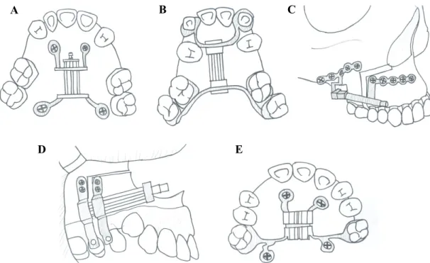
Figure 1. 다양한 구강내 신장기 모식도, A. palatal bone-borne distractor, B. Palatal tooth-borne distractor, C. Bone-borne distractor, D. Tooth&bone-borne distractor, E. Skeletal anchorage type distractor modified from conventional RPE (Rapid palatal expansion) device.
만 이 술식은 술 중에 계획된 방향으로 distractor 를 위치시키는데 어려움이 있었다고 보고되었다
12. 지금까지 구순구개열 환자에서 상악 전방골 신장 술을 이용한 상악골 전방이동의 보고는 Table 1.과 같다.
상악 전방골 신장술의 술식은 먼저 구순구개열 환자의 구치부 교합관계를 확인 뒤 적응증에 해당 되면 미리 환자에 맞게 골신장기를 제작하거나, 적 합한 골신장 장치를 미리 준비한다. 상악 전방부 골절단술을 시행 후 미리 준비된 골신장기를 장착 한다. 통상 일주일간의 휴지기를 거친 후 원하는 이동량만큼 하루에 0.5~1 mm씩 이동시킨다. 이후 8주간의 유지기 후 필요 시 교정치료를 통해 적절 한 교합을 완성한다
13.
상악 전방골 신장술은 장치에 의해 크게 RED
system과 같은 external type과 internal type으로 구분된다. 또한 Internal type의 장치들은 고정원과 장치의 위치에 따라 분류할 수 가 있다. 골을 고정원 으로 하는 장치에는 양쪽 순측에 두개의 장치를 사용 하는 bone-borne distractor
12,22,24와 구개측에 하 나의 장치를 사용하는 palatal bone-borne distractor
14가 있다. 또한 치아를 고정원으로 구개 측에 하나의 장치를 사용하는 palatal tooth-borne distractor
15,21,23가 있고 골과 치아를 동시에 고정원 으로 이용하여 양쪽 순측에 두개의 장치를 사용하는 tooth&bone-borne disctractor
16,20도 있다(Table 1, Fig 1). Internal type은 적용하기 어렵고, 신장 방향을 바꿀 수 없고, 상대적으로 감염이 생길 확률 이 높다. 하지만 환자의 사회생활에는 거의 영향을 미치지 않는다. 이에 반해 RED system은 적용하기
C A B
E
D
쉽고 external hook을 통해 sagittal, vertical plane의 변화를 주기 쉽다. 하지만 external traction hook은 수술에 방해가 되고 cranial frame 은 환자의 사회생활에 영향을 줄 수 밖에 없다
20.
김 등은 RPE를 변형시킨 skeletal anchorage type distractor를 사용하였고
13, Iida 등은 수술 전 에 3D CT 평가를 하여 palatal bone-borne distractor(Dynaform system)의 단점인 수술 중에 위치시키기 어려움을 용이하게 하였다
14. Alkan 등 은 palatal tooth-borne distractor를 시도하였으 나 이는 적절한 신장 방향을 결정하기 어려워 개교합 이 발생하는 단점이 있음을 보고하였다
15. Aikawa 등은 hybrid distractor를 이용해 상악 전방골의 회 전 이동이 필요한 증례에 적용시켰다
16.
Ⅲ. 고찰
구순구개열 환자의 전치부 반대교합을 보다 세 밀히 살펴보면 구치부 관계에 있어 앵글씨 분류상 III급보다는 I급 또는 II급관계가 더 많이 관찰되고
17,18
, 상악 치아의 선천적 결손(측절치나 견치) 또
는 조기 상실에 따른 전방 악궁의 위축으로 납작하 고 부자연스러운 악궁 형태의 모습이 나타난다. 즉 이는 구순구개열 환자의 전치부 반대 교합은 상악 골 자체의 후퇴에 기인한다기 보다는 전상악골의 위축이 주원인일 가능성이 매우 높다는 것을 암시 한다
13.
상악 전방골 신장술은 상악의 전방부를 점진적으 로 움직이게 하여 골이식을 피할 수 있고, oronasal 또는 oroantral fistula가 발생하는 위험을 최소화 할 수 있다. 또 이 술식은 distraction gap에 골을 재생시킬 뿐만 아니라, 구개와 치조골의 연조직 또 한 재생시킬 수 있다. 게다가 재생된 골은 치열의 배 열 또는 임플란트를 위한 골로 사용될 수 있다
10. 상
악 전방분절이 전방 그리고 하방으로 점진적으로 이 동하면서 후방분절의 안정을 유지할 수 있는 것도 큰 장점이다. 따라서 구개범인두 근육은 영향을 받 지 않을 것이며 구개범인두 기능은 최대한으로 보존 될 것이다.
Bevilacqua 등은 7명의 구순구개열을 가진 상악 열 성장 환자에게 bone-borne distractor를 이용한 상악 전방골 신장술을 시행하였다. Video nasoendoscopy 을 이용한 평가에서 구개범인두 폐쇄와 구개범인두 전후방 크기, 측인두벽의 움직임 등은 변하지 않았고 과비음 또한 변화가 없음을 나타내었다. 구내 공간의 증가와 교합의 개선으로 혀의 움직임이 증가해 발음 도 개선이 되었다
19.
Wang 등은 7명의 구순구개열을 가진 상악 열성 장 환자에게(tooth&bone-borne distractor; 4명, RED system; 3명) 상악 전방골 신장술을 시행하였 다. 연구개의 두께와 길이, 연구개-경구개 각도, PNS와 구개수에서 인후벽까지 거리를 측정하였는 데 모두 분명한 변화가 없는 것으로 나타났고 발성 의 평가에서도 변화가 없는 것으로 나타났다
20.
Yu 등은 10명의 구순구개열을 가진 상악 열성장 환자들에게 임의로 상악골 신장술과 상악 전방골 신 장술을 시행하였고 그 차이를 분석하였다. 두 그룹 모두에서 골신장술 후 SNA, NA-FH, overjet, 0-meridian to Sn의 유의한 증가를 가져왔다. 구개 길이(ANS-PNS)에서는 상악 전방골 신장술을 시행 한 그룹이 평균 7.50 mm의 증가로 상악골 신장술을 시행한 그룹에 비교하여 유의한 차이를 나타내었다.
또한 통계적으로 유의한 차이는 얻지 못했지만, 상 악 전방골 신장술을 시행한 그룹이 상악골 신장술을 시행한 그룹에 비해 palatopharyngeal depth와 soft palatal length에서도 변화가 없는 것으로 나 타났다
21.
이렇게 구개범인두 기능부전에 관한 많은 장점
을 가지고 있지만 술식이 더 복잡하고, 상대적으로
높은 치근손상 확률, 전방 분절골 괴사 등의 문제 가 발생할 수 있으므로 큰 집단의 장기간 안정성에 관한 연구가 더 필요하다고 판단된다.
Ⅳ. 결론
상악 전방골 신장술은 골신장술의 장점뿐만 아 니라 구순구개열 환자의 심한 상악 열성장의 치료 에 큰 우월성을 가지고 있다. 하지만 상악 전방골 신장술과 기존의 르포씨 I형골절단술과 상악골 신 장술을 비교하기에는 큰 집단간의 장기간 안정성과 velopharyngeal dynamic evaluation에 관한 연구 가 아직까진 부족한 상황이다. 그럼에도 불구하고 구개길이의 증가, 구개범인두 깊이의 변화 방지, 구개범인두 기능의 보존 능력을 볼 때 상악 전방골 신장술은 먼저 고려되어야 할 대상으로 생각된다.
Reference
1. Bralley RC, Schoeny ZG. Effects of maxillary advancement on the speech of a sub-mucous cleft patient. Cleft Palate J 1977:14:98–101.
2. Fitzpatrick B. Mid-face osteotomy in the adolescent cleft palate patient. Aust Dent J 1977: 22:338–350.
3. Freihofer HPM. Changes in nasal profile after maxillary advancement in cleft and non-cleft patients. J Maxillofac Surg 1977:5:20–27.
4. Witzel MA, Munro IR. Velopharyngeal in- sufficiency after maxillary advancement.
Cleft Palate J 1977:14:176–180.
5. Figueroa AA, Polley JW. Management of se- vere cleft maxillary deficiency with distraction
osteogenesis: Procedure and results. Am J Orthod Dentofacial Orthop 1999:115:1-12.
6. Lee YK, Choi JP, Choi JY. The Diagnosis and Management of Velopharyngeal Insufficiency.
구순구개열 2008:11:12-21.
7. Kuroda S, Araki Y, Oya S, Mishima K, Sugahara T, Takano-Yamamoto T. Maxillary distraction osteogenesis to treat maxillary hypoplasia: Comparison of an internal and an external system. Am J Orthod Dentofacial Orthop 2005:127:493-498.
8. Cheung LK, Chua HDP. A meta-analysis of cleft maxillary osteotomy and distraction osteogenesis. Int J Oral Maxillofac Surg 2006:35:14-24.
9. Watzke I, Turvey TA, Warren DW, Dalston R.
Alterations in velopharyngeal function after maxillary advancement in cleft palate patients. J Oral Maxillofac Surg 1990:48:685-
689.
10. Guyette TW, Polley JW, Figueroa A, Smith BE. Changes in speech following maxillary distraction osteogenesis. Cleft Palate Craniofac J 2001:38:199-205.
11. Dolanmaz D, Karaman AI, Ozyesil AG.
Maxillary anterior segmental advancement by using distraction osteogenesis: a case report. Angle Orthod 2003:73:201-205.
12. Karakasis D, Hadjipetrou L. Advancement of the anterior maxilla by distraction (case report).
J Cranio-maxillofac Surg 2004:32:150-154.
13. Kim KH, Jung YS, Choei JH, Lee SH, Yu HS,
Son BH, Yi CK. Premaxillary Reconstruction
by Distraction Osteogenesis for Cleft
Lip/Palate. 구순구개열 2006:9:63-70.
14. Iida S, Yagi T, Yamashiro T, Okura M, Takada K, Kogo M. Maxillary Anterior Segmental Distraction Osteogenesis with the Dynaform System for Severe Maxillary Retrusion in Cleft Lip and Palate. Plast Reconstr Surg 2007:120:508-516.
15. Alkan A, Bas B, Ozer M, Bayram M, Yuzbasioglu E. Maxillary Anterior Segmental Advancement of Hypoplastic Maxilla in Cleft Patients by Distraction Osteogenesis: Report of 2 Cases. J Oral Maxillofac Surg 2008:66:
126-132.
16. Aikawa T, Haraguchi S, Tanaka S, Uematsu S, Ishibashi M, Kogo M, Iida S. Rotational movement of the anterior maxillary segment by hybrid distractor in patients with cleft lip and palate. Oral Surg Oral Med Oral Pathol Oral Radiol Endod 2010:110:292-300.
17. Schulates G, Gaggl A, Karcher H. A compar- ison of growth impairment and orthodontic results in adult patients with clefts of palate and uilateral clefts of lip, palate and alveolus. Br J of Oral Maxillofac Surg 2000:38:26-32.
18. Gaggl A, Feichtinger M, Schultes G, Santler G, Oichlmaier M, Mossbock R, Karcher H.
Cephalometric and occlusal outcome in adults with unilateral cleft lip, palate, and alveolus after two different surgical techniques. Celft Palate Craniofac J 2003:
40:249-255.
19. Bevilacqua RG, Ritoli EL, Kang C, Mabry K, Castiglione CL. Midmaxillary internal dis- traction osteogenesis: Ideal surgery for the mature cleft patient. Plast Reconstr Surg.
2008:121:1768-1778.
20. Wang XX, Wang X, Li ZL, Yi B, Liang C, Jia YL, Zou BS. Anterior maxillary segmental distraction for correction of maxillary hypo- plasia and dental crowding in cleft palate patients: a preliminary report. Int J Oral Maxillofac Surg 2009:38:1237–1243.
21. Yu H, Wang X, Fang B, Shen SG. Comparative Study of Different Osteotomy Modalities in Maxillary Distraction Osteogenesis for Cleft Lip and Palate. J Oral Maxillofac Surg 2012:70:2641-2647.
22. Alkan A, Bas B, Ozer M, Bayram M. Closure of a Large Palatal Fistula With Maxillary Segmental Distraction Osteogenesis in a Cleft Palate Patient. Cleft Palate-Craniofacial J 2007:44:112-115.
23. Gunaseelan R, Cheung LK, Krishnaswamy R, Veerababu M. Anterior maxillary distraction by tooth-borne palatal distractor. J Oral Maxillofac Surg 2007:65:1044-1049.
24. Sickels JEV, Abadi B, Attisha R. Anterior Segmental distraction for a class III maxil- lary prosthetic defect in a cleft palate patient. J Oral Implantol 2011:37:457-461.
25. Choi HY, Hwang CJ, Kim HJ, Yu HS, Cha JY.
Maxillary anterior segmental distraction os- teogenesis with 2 different types of distractors. J Craniofac Surg 2012:23:706-711.
26. Korean Associations of Oral and Maxillofacial Surgery, editors. Textbook of oral and maxillofacial surgery. 2nd ed.
Seoul: Dental & Medical Publishing Co.;
2005.
27. Wassmund M: Lehrbuch der praktischen
교신 저자