Korean Circulation Journal
Introduction
Advances in percutaneous transcatheter device closure of se- cundum atrial septal defect (ASD) have resulted in high success rates.
1)2)Nowadays, it is becoming a primary option for treating se- cundum ASD because transcatheter closures may avoid the com-
plications associated with open-heart surgery and can reduce the complication rates and the length of the hospital stay, compared with surgical repairs.
3)4)However, certain patients still require sur- gical repairs, even when the defect size indicates that transcathe- ter closures are feasible. It has been reported that the risk factors for the failure of transcatheter closure included the patient being a small child, having a large-sized single defect, and having a defi- cient surrounding the rims.
5)For such difficult cases, modified transcatheter techniques have been recently introduced. For ex- ample, balloon assisted (BA) method and methods using pulmo- nary vein (PV) have improved the success rates and overcame the anatomical disadvantages, but those methods still have limitations with varying success rates.
3)4)6)To find the risk factors for the complicated conventional method of the transcatheter device closure, we investigated the echocar- diographic parameters for the anatomy of ASD and for the size of the left atrium (LA).
Print ISSN 1738-5520 • On-line ISSN 1738-5555
Small Left Atrial Size Complicating Percutaneous Transcatheter Device Closure of Secundum Atrial Septal Defect with Conventional Approach
Hong Ki Ko, MD
1, So Yeon Kang, MD
2, Jeong Jin Yu, MD
2, Jae-Kon, Ko, MD
2, and Young-Hwue Kim, MD
21Department of Pediatrics, Ajou University Hospital, Ajou University School of Medicine, Suwon,
2Division of Pediatric Cardiology, Department of Pediatrics, Asan Medical Center, University of Ulsan College of Medicine, Seoul, Korea
Background and Objectives: Transcatheter device closure becomes the first option for treating secundum atrial septal defect (ASD), but the conventional method is sometimes unsuccessful even when the defect size indicates the closure to be feasible. To increase the suc- cess rate, modified methods have been introduced and used. This study aimed to find predictors for using the modified methods in the device closure of secundum ASDs.
Subjects and Methods: Between October 2010 and December 2012, 92 patients with ASDs underwent the transcatheter device closure.
We analyzed the sizes of the defect, the surrounding rims, and the ratios of the left atrium (LA) dimensions to the device size in the patients who underwent the procedure either using the conventional or modified methods.
Results: Among the 88 successful cases (95.7%), 22 patients (25%) required modified methods (12 using pulmonary vein and 10 using balloon). The modified method group had the larger size of ASDs and smaller posterosuperior rim. The mean ratios of the LA anteroposte- rior diameter, width, and length to the device size were all significantly smaller in the modified methods group than in the conventional group (1.20 vs. 1.56, 1.32 vs. 1.71, and 1.61 vs. 2.07, respectively). We found that the risk factors for the modified methods were smaller retroaortic rim, larger ASD, and smaller LA dimension/device size.
Conclusion: In addition to larger defects and smaller retroaortic rim, the smaller ratios of the LA dimensions to the device size influenced the need for the application of modified methods in the transcatheter device closure of ASDs. (Korean Circ J 2015;45(3):216-224) KEY WORDS: Heart defect, congenital; Heart septal defects, atrial; Cardiac catheterization; Septal occluder device.
Received: October 24, 2014 Revision Received: January 7, 2015 Accepted: February 9, 2015
Correspondence: Young-Hwue Kim, MD, Division of Pediatric Cardiology, Department of Pediatrics, Asan Medical Center, University of Ulsan College of Medicine, 88 Olympic-ro 43-gil, Songpa-gu, Seoul 138-736, Korea Tel: 82-2-3010-3378, Fax: 82-2-473-3725
E-mail: [email protected]
• The authors have no financial conflicts of interest.
This is an Open Access article distributed under the terms of the Creative Commons Attribution Non-Commercial License (http://creativecommons.
org/licenses/ by-nc/3.0) which permits unrestricted non-commercial use,
distribution, and reproduction in any medium, provided the original work
is properly cited.
Subjects and Methods
Patient selection and collecting data
This study analyzed 92 consecutive patients who had undergone percutaneous transcatheter closure of the secundum ASD at the congenital heart diseases center at the Asan Medical Center, Seoul, Korea between October 2010 and December 2012. The median age of the study group was 3.5 years (range, 0.9-57.4 years).We ex- cluded cases of combined cardiac anomalies, except for mild valvar pulmonary stenosis, multiple ASDs except cribriform ASD, or asso- ciated syndromes. The medical records of the patients were retro- spectively reviewed. Age at intervention, sex, weight, height, and 2D images of the pre-interventional transthoracic echocardiogra- phy (TTE) were analyzed with a post-processing program by Image Arena (Tomtech Imaging Systems, Munich, Germany). The defect
sizes were measured by intraoperative transesophageal echocar- diography (TEE) and the sizes of device used were also collected from the catheterization reports. The institutional review board at Asan Medical Center approved this study. Informed consents were waived due to the retrospective study design.
Pre-interventional echocardiographic parameters
Prior to each intervention, each patient had been admitted and examined by a single physician through a TTE using an iE33 with a S5-1 transducer (Philips Medical Systems, Andover, MA, USA) after resting at least 15 minutes in the supine position. Children under 3 years old were sedated with chloral hydrate (0.5 cc/kg) while mon- itoring the vital signs and oxygen saturation, at room air.
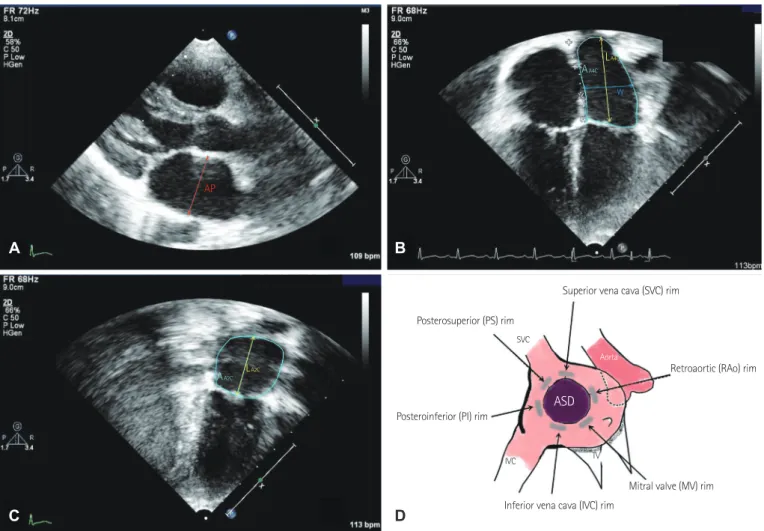
The rims around the secundum ASD were measured in 3 planes and were defined as follows: superior vena cava and inferior vena
A
C
B
AP
A A4C
A A2C
L A4C
w
D
Fig. 1. Measurements of the LA diameters (A, B, C) and schematic diagram of the surrounding rims from the pre-interventional TTE (D). A: AP, antero-
posterior diameter measured at the midline of the LA from a PLAX view during an end-systolic phase (red). B: parameters measured from an A4C view at an end systolic phase; W, width (blue); LA4C, length (yellow); AA4C, area measured from an A4C view (sky blue). C: parameters measured from an A2C at an end systolic phase; LA2C, length (yellow); AA2C, area measured from an A2C (sky blue). D: gray regions represent the surrounding rims measured from a subcoastal, an A4C, and a PSAX view. LA: left atrium, TTE: transthoracic echocardiography, PLAX: parasternal long axis, A4C: apical four chambers, A2C: apical two chambers, PSAX: parasternal short axis, FR: frame rate, C: compression rate, P: persistent grade, HGen: general harmony, BPM: beat per minute, ASD: secundum atrial septal defect, TV: tricuspid valve.
Superior vena cava (SVC) rim
Retroaortic (RAo) rim
Mitral valve (MV) rim ASD
Inferior vena cava (IVC) rim Posteroinferior (PI) rim
Posterosuperior (PS) rim SVC
Aorta
IVC TV
L A2C
cava rim from a subcostal view, posterosuperior (PS) and mitral valve rim from an apical four-chamber (A4C) view, and posteroinferior and retroaortic (RAo) rim from a parasternal short axis view (Fig. 1). The LA dimensions were measured at the end of the systolic phase; the anteroposterior diameter (AP) was measured in a parasternal long- axis view. The width was measured in an A4C view. The mean value of the length was calculated from the lengths measured in A4C and apical two chamber (A2C) views. The LA volume was calculated by the length and areas of the LA in the A4C and A2C views {biplane area-length method: 0.85×(Area
A2C)×(Area
A4C)/D
L}.
Percutaneous transcatheter intervention
The transcatheter device closure was performed under general anesthesia, and the secundum ASD size was repeatedly measured by an intraoperative TEE by a single physician who was different from the TTE-examiner with an iE33 machine (Philips Medical Sys- tems, Andover, MA, USA). Prior to the cardiac catheterization, anti- coagulation was initiated with heparin (100 units/kg, intravenously) and a standard right cardiac catheterization was performed for a hemodynamic study. We did not measure the balloon that routinely stretched the defect size except when a large aneurysmal interatrial septum was present. All the patients underwent transcatheter
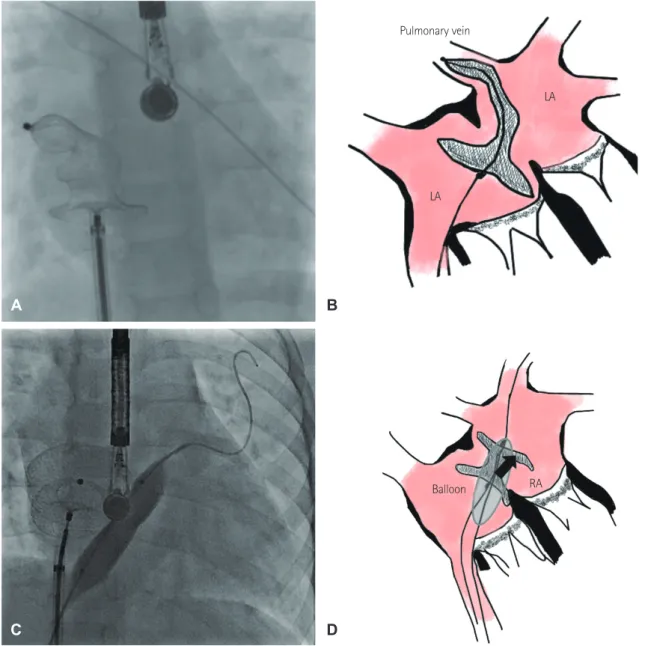
Fig. 2. Pulmonary vein method and schematic diagram. Angiographic film showed the approximation of the RA disc by pushing
the cable while the LA disc was in the orifice of the right upper PV (A). Schematic diagram of the PV method (B). Modified bal- loon-assisted method and schematic diagram. Angiographic film showed both wings of the device were deployed before implan- tation by supporting the peripheral balloon (C). Schematic diagram of BA method (D). RA: right atrium, LA disc of the device was prevented from prolapsed into the RA by balloon (arrow head). LA: left atrium, PV: pulmonary vein, BA: balloon assisted.
A
C
B
D
Balloon RA Pulmonary vein
LA
LA
closure with the Amplatzer septal occluder (AGA Medical Corpora- tion, Golden Valley, MN, USA) and device implantation was initially tried by the conventional method that had already been extensive- ly described.
3)4)Devices measuring1-2 mm larger than the defect sizes were measured by an intraoperative TEE and were employed in the transcatheter closure. If the device implantation was not successful after multiple attempts using the conventional method, the so-called ‘PV method’ was attempted as Papa et al.
4)reported.
The PV method is described as follows: after the sheath was locat- ed in the right upper PV, the LA disc was deployed with the re- maining unspread and stretched from the orifice of PV through the LA cavity until the right atrial disc was delivered and contacted to the vicinity of interatrial septum from the right atrial side. By pushing the cable with the device, the LA disc fell out of the PV and spread automatically and fully with an approximation to the interatrial septum
4)(Fig. 2A and B).
If neither the conventional nor the PV method was successful, we tried the BA method next, as Kammache et al.
6)reported through another transvenous approach (usually in the opposite side femoral vein) for peripheral balloon insertion. The supporting wire for the balloon was located into either the right or left PV depending upon the locations of the deficient rims. The sheath for the device was lo- cated on the other side. We preferred to use peripheral balloons in- stead of the bigger test-occlusion ones. When a balloon was intro- duced through the wire and positioned at the interatrial septum, the LA disc that was ‘assisted’ or supported by the inflating balloon was delivered first. This procedure prevented the prolapse of the LA disc by keeping the plane of the LA disc parallel to the interatrial septum.
The right atrial disc was deployed in this position, leaving the device in a dumbbell shape. The subsequent deflation of the balloon ap- proximated the disc towards the interatrial septum. By carefully steadying the device position, the balloon and wire were extracted.
After the successful removal of the balloon and the wire, we con- firmed the device position and the lack of the atrial shunt using the TEE, prior to the final release of the device (Fig. 2C and D).
If the device implantation was unsuccessful after all three methods, we aborted further attempts at the percutaneous trans- catheter closure. Devices were not deployed and extracted in cas- es of unstable positioning with either a Minnesota wiggling, a compression of the mitral valve or aortic wall, or significant re- sidual leakage during the TEE evaluation. Patients, whose percuta- neous transcatheter closure was unsuccessful, underwent surgical closure at a later time.
Post-interventional follow-up protocols
An antiplatelet agent (aspirin 3.5-5.0 mg/kg once daily) was prescribed for six months following all successful closures. Device
stability, residual shunt, or compression of the cardiac valve and aortic wall were evaluated at post-interventional day 1, 6 months, and 12 months, using the TTE. We also evaluated any clinical symptoms and signs of the patients in the outpatient clinic.
Statistical analysis
We used the Statistical Package for Social Science (SPSS) for Windows, version 18 (SPSS Inc., Chicago, IL, USA) for the statistical analysis. Student ttest and Mann-Whitney test for continuous variables and chi-square test for categorical variables were used for comparing the groups, as appropriate. The logistic regression test was used and receiver-operating curves were plotted for the cut-off values. The p value <0.05 was considered significant.
Results
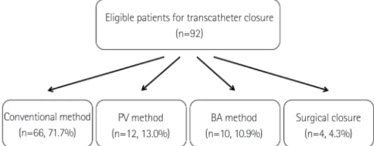
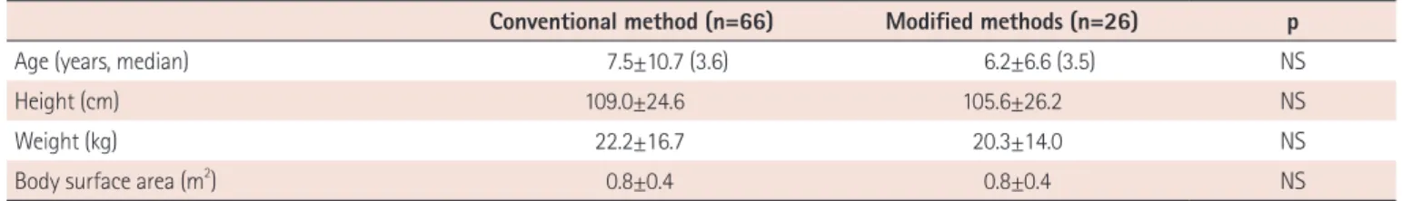
A percutaneous transcatheter closure had been attempted in a total of 92 patients during this study’s period. Successful closures at each stage are summarized as a flow chart (Fig. 3). Sixty-six pa- tients (71.7%) underwent successful closures by the conventional method, and 22 patients (23.9%) underwent successful closures by the PV (n=12, 13.0%) and BA method (n=10, 10.9%). A percutane- ous closure was unsuccessful in four patients (4.3%), who subse- quently underwent surgical repair. We divided the successful pa- tients into the conventional method group and the modified methods group, including the PV and BA method. The demograph- ic data of the conventional and modified methods groups are characterized in Table 1. There were no significant differences in age, height, weight, and body surface area between the groups.
The mean values of the two groups’ anatomical parameters of ASDs by echocardiography are shown in Table 2. We found that the modified methods group had the smaller PS rim (6.1 mm vs.
7.7 mm) and larger ASD (18.1 mm vs. 13.8 mm). The mean sizes of the RAo rim of both groups were deficient (<5 mm) and were not
Fig. 3. Flow chart of the strategy with treating ASD patients. All the pa-
tients started with the conventional method and then the modified meth- ods were used; the BA method was the last approach after the PV method failed. If all three methods failed, surgical closure was indicated. ASD: se- cundum atrial septal defect, BA: balloon assisted; PV: pulmonary vein.
Eligible patients for transcatheter closure (n=92)
Conventional method (n=66, 71.7%)
Surgical closure (n=4, 4.3%) BA method
(n=10, 10.9%) PV method
(n=12, 13.0%)
significantly different. The LA diameters and LA volumes indexed by body surface area were also similar in the two groups. Table 3 shows the anatomical risk factors for using the modified risk fac- tors from the result of the logistic regression test. Larger ASD and smaller RAo rim were found to be significant factors for the modi- fied methods group.
We examined the LA dimensions/device size because we pos- tulated that the relatively large size of the LA disc might techni- cally hinder the device implantation with conventional method.
The ratios of the LA dimensions (AP, width, and length) to the
device sizes were significantly smaller in the modified group com- pared to the conventional method group (p=0.001). Fig. 4 illus- trates the statistical differences, in the ratios of the LA dimensions to the employed device size, between the conventional and modi- fied methods groups. The mean values of the LA AP diameter/de- vice size were 1.56 in the conventional method group and 1.20 in the modified methods group, which was significantly smaller (p=0.001). The mean LA width/device size and LA length/device size were also smaller in the modified methods group (1.32 vs.
1.71 and 1.61 vs. 2.07, respectively). Among the three ratios, the LA
Table 1. Demographic data of the study groupsConventional method (n=66) Modified methods (n=26) p
Age (years, median) 7.5±10.7 (3.6) 6.2±6.6 (3.5) NS
Height (cm) 109.0±24.6 105.6±26.2 NS
Weight (kg) 22.2±16.7 20.3±14.0 NS
Body surface area (m
2) 0.8±0.4 0.8±0.4 NS
Values are mean±SD. NS: not significant
Table 2. Comparison of the echocardiography parameters between the conventional method and modified methods group
Conventional method
(n=66) Modified methods
(n=22) p
Surrounding rims (mm)
SVC rim 8.3±3.3 7.8±4.1 0.585
IVC rim 11.3±4.3 11.0±3.7 0.805
PS rim 7.7±3.3 6.1±2.3 0.013*
MV rim 10.0±3.5 10.2±3.0 0.731
PI rim 7.2±2.9 6.6±3.0 0.365
RAo rim 3.8±1.4 3.3±1.2 0.102
ASD size measured by TEE (mm) 13.8±4.2 18.2±5.3 0.001*
Employed device size (mm) 12.0±2.2 15.6±5.1 0.001*
Indexed LA dimensions (mm/m
2)
D
AP(at PLAX) 31.5±7.1 34.9±10.3 0.156
D
W(at A4C) 34.9±9.3 36.9±7.9 0.364
D
L(at A4C) 41.9±9.6 45.8±10.4 0.131
Indexed LA volume (mL/m
2) 21.1±8.6 21.5±7.7 0.846
Values are mean±SD. *Significant. SVC: superior vena cava, IVC: inferior vena cava, PS: posterosuperior, MV: mitral valve, PI: posteroinferior, RAo: retroaor- tic rim, ASD: secundum atrial septal defect, TEE: transesophageal echocardiography, LA: left atrium, D
AP(at PLAX): anteroposterior diameter measured at a parasternal long axis, D
W(at A4C): left atrium width measured at an apical four chamber, D
L(at A4C): left atrium length measured at an apical four chamber
Table 3. Parameters associated with using the modified methodsParameters Univariate analysis Multivariate analysis
p Crude OR (95% CI) p Adjusted OR (95% CI)
ASD size (mm) <0.001 1.33 (1.138-1.553) 0.001 1.26 (1.114-1.423)
RAo rim (mm) 0.013 0.51 (0.304-0.869) 0.022 0.56 (0.341-0.922)
PS rim (mm) 0.031 0.72 (0.535-0.971) 0.080 0.82 (0.658-1.024)
OR: odds ratio, CI: confidence interval, ASD: secundum atrial septal defect, RAo: retroaortic rim, PS: posterosuperior
AP diameter/device size were the most important parameter based on the result of the logistic regression test (odds ratio 0.059, 95%
confidence interval 0.008-0.448, p=0.006). Comparing the two modified methods (PV and BA) groups, the BA group showed signifi- cantly larger sizes of ASD and smaller LA diameters/Amplatzer septal occluder sizes (p=0.031 and p=0.027, respectively). The receiver-op- erating characteristic curve showed the cut-off values for the three ratios (Fig. 5). For the cut-off values for using the conventional methods with the LA AP diameter/device size, the LA width/device size and LA length/device size might be 1.69, 1.64 and 1.78, respec- tively (95.0% of specificity and 46.6% of sensitivity).
When we reviewed the four patients who failed to have the per- cutaneous closure, Fig. 6 illustrates the only LA AP diameter/device that was significantly smaller than those of the successful cases (p=0.031) while the LA width/device size and LA length/device size were not significantly smaller than those of the successful cases (p=0.066 and 0.443, respectively). The 3 of 4 failed cases showed that the LA AP diameters/device sizes were below 1 and that the LA lengths were smaller than the LA disc of the device.
Complications
There was no peri-procedural complication, such as device dis- placement, sustained arrhythmia, or vascular complications, within the initial 24-hours follow-up during the study period. No early
Fig. 5. ROC curve for the ratios of the LA AP diameter (red), LA width (blue),and LA length (yellow) to the ASO size. Areas under the curve were 0.719 (p=0.004), 0.716 (p=0.04), and 0.716 (p=0.004), respectively. ROC: receiver- operating characteristic, LA: left atrium, AP: anteroposterior, ASO: Am- platzer septal occluder, W: width, L: length
ROC curve
0.0 0.2 0.4 0.6 0.8 1.0 1-specificity
LA AP/ASO size LA W/ASO size LA L/ASO size 1.0
0.8
0.6
0.4
0.2
0.0
Sensitivity
Fig. 4. Comparisons of the two groups in the ratios of the LA diameters
(AP, width, and length) to the ASO size. A: ratio of LA AP diameter to ASO size. B: ratio of LA width to ASO size. C: ratio of LA length to ASO size. Pink box represented conventional group and blue box represented modified group. *Significant with p=0.001. LA: left atrium, AP: anteroposterior, ASO:
Amplatzer septal occluder.
Conventional method
Conventional method
Conventional method
Modified method
Modified method
Modified method 3.5
3.0
2.5
2.0
1.5
1.0
0.5
3.5
3.0
2.5
2.0
1.5
1.0
0.5
5.0
4.0
3.0
2.0
1.0
Applied methods
Applied methods
Used methods
LA AP diamter/ASO sizeLA width/ASO sizeLA length/ASO size
A
B
C
complication, such as aortic erosion or thrombosis, (within the first 6 months) after the hospital discharge was observed in any of the patients during the outpatient follow-up period.
Discussion
This study showed an overall success rate of 95.7% for the transcatheter device closure of the secundum ASD and these re- sults were comparable to other reports.
2)3)5)Among the successful closures, the conventional method accounted for 85%, with the remaining 15% being closed with the modified methods, such as the PV or BA method. Because the latter group represented a con- siderable portion of patients, regarded as technically challenging cases, we tried to identify the predictors for these technical chal- lenges prior to an intervention, possibly helping to prepare and to shorten the procedure time.
7)This study found two main predictors. One was the anatomical characteristic of a larger defect and smaller retroaortic rim, agree- ing with previous reports.
4)8-10)Large ASDs and the locations and the extent of deficient surrounding the rims were previously known to be risk factors of the modified methods.
8-10)Papa et al.
4)reported that the method using PV was helpful in patients with a deficient PS rim. This study showed that a larger defect and small- er RAo rim were the most powerful risk factors, although the modified methods group had a smaller PS rim. The RAo rim size, which was known to be unrelated to the success rate,
8)was one of the risk factors for using the modified methods. These anatomical parameters should be precisely measured; however, we sometimes had difficulty in applying them to the pediatric population because the pre-interventional TEE is not a routine procedure in every cen- ter. The clinical cut-off values of the defect and surrounding rim size were not reported in children whose body surface area widely varied according to age and hemodynamic status.
The second predictor was the ratio of the LA dimensions to the employed device sizes. At times, we experienced a deformation of the left atrial disc into an abnormal shape in a small LA and it was frequently prolapsed into the right atrium. This resulted not from the LA size but from the relationship with the LA size and device size; this conclusion was supported by the fact that the indexed LA dimensions and LA volumes were not significantly different be- tween the two groups. Because we postulated that the instability of the LA disc in a relatively small LA might be a risk factor for us- ing the modified methods, resulting in the right atrium disc to be settled down first, we found that the LA dimensions/device size were significantly smaller in patients who failed with the con- ventional method. Those ratios that were not influenced by the patient’s body surface area could be easily applicable to the pediatric
Fig. 6. Comparisons between the failed and successful cases in the modifiedmethods group. A: ratio of LA AP diameter to ASO size. B: ratio of LA width to ASO size. C: ratio of LA length to ASO size. Black circle represented each of the failed cases and the blue circle represented each of the successful cases in the ratios of the LA diameters (AP, W and L) to the ASO size. LA: left atrium, AP:
anteroposterior, ASO: Amplatzer septal occluder, W: width, L: length
LA AP diameter to ASO size of failed and successful cases in modified methods group
LA width to ASO size of failed and successful cases in modified methods group
LA length to ASO size of failed and successful cases in modified methods group Failed
Failed
Failed
Successful
Successful
Successful LA AP/ASO sizeLA W/ASO sizeLA L/ASO size
1.6
1.4
1.2
1.0
0.8
2.0
1.8
1.6
1.4
1.2
1.0
0.8
2.4 2.2 2.0 1.8 1.6 1.4 1.2 1.0