• 검색 결과가 없습니다.

A Case of Acute Carbon Monoxide Poisoning Resulting in an ST Elevation Myocardial Infarction

N/A
N/A
Protected

Academic year: 2022

Share "A Case of Acute Carbon Monoxide Poisoning Resulting in an ST Elevation Myocardial Infarction"

Copied!
3
0
0

로드 중.... (전체 텍스트 보기)

전체 글

(1)

133 Copyright © 2012 The Korean Society of Cardiology

Korean Circulation Journal

Introduction

Carbon monoxide (CO) is known as a silent killer, because it is an odorless, colorless, and non-irritating gas. Myocardial infarction related to CO poisoning has been frequently reported in the litera- ture; however, an ST elevation myocardial infarction (STEMI) due to coronary occlusion is a very rare presentation.

1)

Although an increased tendency for thrombogenesis during CO poisoning has been reported,

2)

the precise mechanism and treat- ment of STEMI related to CO poisoning remain uncertain. Here, we report a rare case of acute STEMI complicated by increased throm- bogenicity secondary to acute CO poisoning and complete revas- cularization after anti-thrombotic treatment.
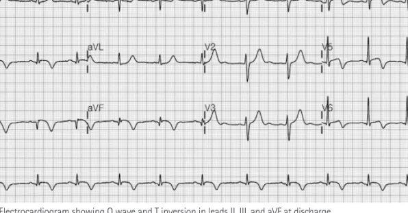
Case Report

http://dx.doi.org/10.4070/kcj.2012.42.2.133 Print ISSN 1738-5520 • On-line ISSN 1738-5555

A Case of Acute Carbon Monoxide Poisoning Resulting in an ST Elevation Myocardial Infarction

Soohyun Kim, MD 1 , Joo Han Lim, MD 2 , Youngjoong Kim, MD 1 , Sewon Oh, MD 1 , and Woong Gil Choi, MD 1

1

Division of Cardiology, Internal Medicine, Konkuk University Chungju Hospital, Chungju,

2

Division of Hemato-Oncology, Internal Medicine, Inha University Hospital, Incheon, Korea

Carbon monoxide (CO) is a well-known chemical asphyxiant, which causes tissue hypoxia with prominent neurological and cardiovascular injury. After exposure to CO, several cardiac manifestations have been reported, including arrhythmias, acute myocardial infarction, and pul- monary edema. However, an ST elevation myocardial infarction (STEMI) due to CO poisoning is a very rare presentation, and the treatment for STEMI due to CO poisoning is not well established. Here, we report a rare case of STEMI complicated by increased thrombogenicity sec- ondary to acute CO poisoning and complete revascularization after antithrombotic treatment. (Korean Circ J 2012;42:133-135) KEY WORDS: Carbon monoxide; Myocardial infarction.

Received: July 6, 2011

Revision Received: August 2, 2011 Accepted: August 3, 2011

Correspondence: Woong Gil Choi, MD, Division of Cardiology, Internal Me- dicine, Konkuk University Chungju Hospital, 82 Gugwon-daero, Chungju 380- 704, Korea

Tel: 82-43-840-8213, Fax: 82-43-843-6655 E-mail: gilll@paran.com

• The authors have no financial conflicts of interest.

This is an Open Access article distributed under the terms of the Creative Commons Attribution Non-Commercial License (http://creativecommons.

org/licenses/by-nc/3.0) which permits unrestricted non-commercial use, distribution, and reproduction in any medium, provided the original work is properly cited.

Case

A 47-year-old male presented to the emergency department with prolonged chest pain, which developed during underground min- ing. Upon arrival at the emergency department, his vital signs were blood pressure, 158/119 mm Hg; heart beat, 68/min; respiratory rate, 12/min; body temperature, 36.5°C; and O

2

saturation, 100%. Initial laboratory data revealed the following: carboxyhemoglobin (COHb), 2.6%; troponin-I, 3.06 ng/mL; myoglobin, 321 ng/mL; and creatine kinase (CK)-MB, 22.9 ng/mL. Electrocardiography (ECG) revealed an ST elevation in leads II, III, and aVF (Fig. 1). Transthoracic ECG, which was performed when the patient arrived at hospital, demonstrated hypokinesia of the inferior wall and preserved systolic function (ejec- tion fraction=50%). The patient was diagnosed with STEMI and un- derwent emergency coronary angiography.

Coronary angiography revealed total occlusion of the posterior de-

scending (PD) branch of the right coronary artery (RCA) with a

large occlusive thrombus burden (Fig. 2). Because the thrombotic

occlusive lesion was far from the distal site of the RCA, we decided

to closely observe the patient in the coronary care unit and adminis-

ter anti-thrombotic medications including heparin, aspirin, and clo-

pidogrel. An electrocardiogram obtained 8 hours after admission

showed normalization of the ST segment elevation, Q wave, and T

waves in leads II, III, and aVF (Fig. 3). Cardiac enzymes were elevat-

ed, with a CK-MB of 153.9 ng/mL and troponin I of 30.3 ng/mL on

the first admission day. Three days after admission, a follow-up co-

ronary angiography was normal (Fig. 4). The patient was discharged

(2)

134 Carbon Monoxide Poisoning Resulting in Acute Myocardial Infarction

http://dx.doi.org/10.4070/kcj.2012.42.2.133 www.e-kcj.org

later in a stable condition.

Discussion

Carbon monoxide is one of the leading causes of poisoning-relat- ed deaths in the United States. CO binds to hemoglobin with greater affinity than oxygen and forms COHb, which leads to impaired oxy- gen transport and subsequent tissue hypoxia.

3)4)

After exposure to CO, several cardiac manifestations have been reported, including arrhythmias and electrocardiographic altera- tions,

5)

acute myocardial infarction, pulmonary edema, and cardio- genic shock.

6)

Patients with coronary artery disease are more suscep-

tible to CO-induced cardiotoxicity.

7)

Inhaling even relatively small amounts of CO can lead to hypoxic injury, neurological damage, and possibly death. Additionally, even if the initial COHb level is not very high, acute CO poisoning can still lead to severe cardiovascular complications in high cardiovascular risk cases.

8)

Toxicity also increases due to several factors, including increased activity and ventilation rate, preexisting cerebral or cardio- vascular disease, reduced cardiac output, anemia or other hemato- logical disorders, decreased barometric pressure, and high metabolic rate.

9)

Myocardial ischemic changes often reveal T-wave inversion or ST depression in patients with CO poisoning. However, an ST segment elevation is a rare presentation during CO poisoning. Some cases re- ported with an ST elevation had normal coronary arteries while un- dergoing coronary angiography.

1)

However, Hsu et al.

8)

reported a case of CO poisoning complicated with STEMI, which had total oc- clusion of the left anterior descending artery and underwent prima- ry percutaneous coronary intervention.

The proposed mechanisms of myocardial damage are myocardial stunning as a result of CO poisoning or unmasking of underlying co- ronary arterial disease by creating a myocardial demand/supply mis- match.

10)

Furthermore, hematocrit, blood viscosity, and platelet func- tion have been implicated as very important pathophysiological me- chanisms in patients with acute myocardial infarction but normal coronary arteries. An increasing thrombotic tendency secondary to platelet stickiness and polycythemia has been reported in patients with CO poisoning.

11)

A COHb level >3% in nonsmokers or >10% in smokers confirms exposure to CO.

12)

In our case, even if the initial COHb level was not very high, emergency coronary angiography revealed total occlu- sion of the PD branch of the RCA. However, the follow-up coronary Fig. 1. Electrocardiogram showing ST elevation in leads II, III, and aVF at admission.

Fig. 2. Coronary angiogram showed total occlusion of the posterior de-

scending branch of the right coronary artery with a large occlusive throm-

bus burden.

(3)

135 Soohyun Kim, et al.

http://dx.doi.org/10.4070/kcj.2012.42.2.133 www.e-kcj.org

Fig. 3. Electrocardiogram showing Q wave and T inversion in leads II, III, and aVF at discharge.

angiography revealed a normal coronary angiogram after anti-th- rombotic treatment, unlike other cases. The present case was uni- que in that the mechanism leading to the STEMI in the CO poisoned state was illustrated by coronary angiography, and complete revas- cularization was performed after anti-thrombotic treatment without percutaneous coronary intervention.

In conclusion, this case highlights that anti-thrombotic treat- ment is the most essential therapy for CO poisoning complicated by STEMI in some specific cases.

References

1. McCabe MJ, Weston CF, Fraser AG. Acute myocardial infarction related

to smoke inhalation and myocardial bridging. Postgrad Med J 1992;68:

758-61.

2. Aronow WS. Effect of carbon monoxide on cardiovascular disease.

Prev Med 1979;8:271-8.

3. Weaver LK. Clinical practice: carbon monoxide poisoning. N Engl J Med 2009;360:1217-25.

4. Chong DY, Lee Y, Cho SH, Song KY. Electron Microscopic Study on mi- tochondria and cytochrome oxidase activity following acute carbon monoxide exposure in rat myocardium. Korean Circ J 1988;18:69-83.

5. Gandini C, Castoldi AF, Candura SM, et al. Carbon monoxide cardio- toxicity. J Toxicol Clin Toxicol 2001;39:35-44.

6. McMeekin JD, Finegan BA. Reversible myocardial dysfunction follow- ing carbon monoxide poisoning. Can J Cardiol 1987;3:118-21.

7. Allred EN, Bleecker ER, Chaitman BR, et al. Short term effects of car- bon monoxide exposure on the exercise performance of subjects with coronary artery disease. N Engl J Med 1989;321:1426-32.

8. Hsu PC, Lin TH, Su HM, et al. Acute carbon monoxide poisoning result- ing in ST elevation myocardial infarction: a rare case report. Kaohsiung J Med Sci 2010;26:271-5.

9. Raub JA, Mathieu-Nolf M, Hampson NB, Thom SR. Carbon monoxide poisoning: a public health perspective. Toxicology 2000;145:1-14.

10. Satran D, Henry CR, Adkinson C, Nicholson CI, Bracha Y, Henry TD. Car- diovascular manifestations of moderate to severe carbon monoxide poisoning. J Am Coll Cardiol 2005;45:1513-6.

11. Marius-Nunez AL. Myocardial infarction with normal coronary arteries after acute exposure to carbon monoxide. Chest 1990;97:491-4.

12. Hampson NB, Hauff NM. Carboxyhemoglobin levels in carbon mon- oxide poisoning: do they correlate with the clinical picture? Am J Emerg Med 2008;26:665-73.

Fig. 4. Follow-up coronary angiogram showed a normal right coronary artery.

수치

Fig. 2. Coronary angiogram showed total occlusion of the posterior de- de-scending branch of the right coronary artery with a large occlusive  throm-bus burden.
Fig. 3. Electrocardiogram showing Q wave and T inversion in leads II, III, and aVF at discharge.

참조

관련 문서

수출과 수입으로 나누어 볼 때,수출과 수입의 경우 모두 ODCY를 경유하는 물동.. 도로운송을 통해 부산항에서 처리된 수출입컨테이너 가운데 경북권을 기․종점 으로

(Background) Gallbladder wall thickening(GWT) and gallbladder contraction are often observed in patients with acute hepatitis.. The incidence of acute hepatitis A

- Basic research on a compact, high b ST (Spherical Torus) with elongated chamber in partial solenoid configuration - Study on advanced tokamak scenario

- For gas-phase reactions, the volumetric flow rate most often changes during the course of the reaction due to a often changes during the course of the reaction due to a change

하였다.이호성 연구에서는 남북한 화학 교과서 용어의 차이점만 분석이 되 었고,이러한 차이점을 나타내는 이유에 대한 원인은 분석이 되어있지 않았 다.따라서

Therefore, cTnT is a recommended biomarker for use in the detection of myocardial infarction (MI) and in acute coronary syndromes.[8] Indeed, several authors

ECG is a valuable noninvasive diagnostic method for detection of NSTE-ACS. In addition, it has a important role in the management of NSTE-ACS such as

Approved clinical use of bone marrow stem cells for myocardial infarction treatment... Cardiac