Introduction
In early December 2019, the first case of pneumonia of un- known origin was identified in Wuhan. As of May 17, 2020, approximately 4,710,614 cases have been reported from over a hundred countries worldwide. More than 315,023 patients have died from severe infection with this novel virus, known as severe acute respiratory syndrome coronavirus 2 (SARS-CoV-2). This enveloped RNA, beta coronavirus was first identified by high-throughput sequencing. SARS-CoV-2 has a receptor-binding domain structure similar to that of SARS-CoV, as shown with the help of homology modeling [1].
Zhou et al. [2] found that 96% of SARS-CoV-2 and bat coro- naviruses are similar at the whole-genome level. The clini- cal disease course of this highly contagious illness, known coronavirus disease 2019 (COVID-19), has not yet been well
described in the medical literature. During this ongoing pan- demic, the center-specific clinical experience of COVID-19 is being shared by researchers worldwide. To date, there is no
A systematic review of clinical and laboratory parameters of 3,000 COVID-19 cases
Harsh Goel, M.Tech
1, Ishan Gupta, MBBS-Student
2, Meenakshi Mourya, MBBS
3, Sukhdeep Gill, MS, DABR
4, Anita Chopra, MD, DM
1, Amar Ranjan, MD
1, Goura Kishor Rath, MD
5, Pranay Tanwar, MD
11Laboratory Oncology Unit, Dr.B.R.A.-Institute Rotary Cancer Hospital, 2All India Institute of Medical Sciences, New Delhi; 3Department of Anaesthesia and Intensive Care, Vardhaman Mahavir Medical College & Safdarjung Hospital, New Delhi, India, 4UPMC Hillman Cancer Center, Erie, PA, USA,
5Department of Radiotherapy, Dr. B.R.A.- Institute Rotary Cancer Hospital, All India Institute of Medical Sciences, New Delhi, India
The coronavirus disease 2019 (COVID-19) has spread worldwide. It is still a pandemic and poses major health problem across the globe. In our review, clinical characteristics and laboratory parameters of COVID-19 patients were compiled systematically, with special reference to pregnant women in order to understand the disease course. An extensive literature search on various scientific databases for relevant manuscripts was conducted, which yielded 7 manuscripts for final analysis. The most common symptoms were fever (85%), cough (70.63%), chest tightness (37.36%), expectoration (33.27%), fatigue (32%), dyspnea (31.95%), and shortness of breath (31.19%), while hemoptysis (1.0%) was the least common. The associated comorbidities were hypertension (21.6%) and diabetes (10.0%). In terms of hematological parameters, lower total leukocyte counts were observed in 65% of cases and biochemical parameters, patients demonstrated elevated levels of albumin (53.72%), lactate dehydrogenase (45.71%), and natriuretic peptide (34.84%); however, total bilirubin was elevated in only 8% of cases. In the acute inflammatory cytokine profile, C-reactive protein (59.0%), tumor necrosis factor (58.0%), erythrocyte sedimentation rate (57.0%), interleukin-2 (IL- 2, 54.0%), and IL-6 (52.0%) levels were increased, while prolactin levels (6.5%) were minimally elevated. The recovery rate was approximately 41%, and mortality was about 6.5%. The study also concluded that the clinical symptoms of COVID-19 were similar among pregnant and non-pregnant women. There was no evidence of vertical transmission of COVID-19 infection. This review critically analyzed COVID-19 as a public health hazard in order to help policy makers, health care givers, and primary physicians to promote early diagnosis and prevention.
Keywords: COVID-19; SARS-CoV-2; 2019-nCoV; Coronavirus
Received: 2020.06.23. Revised: 2020.09.17. Accepted: 2021.01.11.
Corresponding author: Pranay Tanwar, MD
Laboratory Oncology Unit, Dr.B.R.A.-Institute Rotary Cancer Hospital, All India Institute of Medical Sciences, Dr.B.R.A.- Institute Rotary Cancer Hospital, Sri Aurobindo Marg, Ansari Nagar, New Delhi 110029, India
E-mail: [email protected] https://orcid.org/0000-0002-2357-976X
Articles published in Obstet Gynecol Sci are open-access, distributed under the terms of the Creative Commons Attribution Non-Commercial License (http://creativecommons.
org/licenses/by-nc/3.0/) which permits unrestricted non-commercial use, distribution, and reproduction in any medium, provided the original work is properly cited.
Copyright © 2021 Korean Society of Obstetrics and Gynecology https://doi.org/10.5468/ogs.20174
eISSN 2287-8580
effective vaccine or targeted therapy available. Therefore, supportive treatment alone is the mainstay of clinical man- agement [3]. Subsequently, it’s proven that the virus causing COVID-19 is similar to SARS-CoV and was designated as SARS-CoV-2 [4,5]. The COVID-19 outbreak was declared a
“Public Health Emergency of International Concern” by the World Health Organization (WHO) on January 30, 2020 [6].
SARS-CoV-2 is capable of human-to-human transmission, and has spread worldwide by SARS-CoV-2-infected interna- tional travelers. In this study, we explored the clinical features and related laboratory parameters of more than 3,000 cases of COVID-19 and evaluated critical factors that may affect the disease course and prognosis. This review also focuses on the COVID-19 disease course and symptoms among preg- nant women, with the intent to analyze the effect of COV- ID-19 on the progress of pregnancy and neonatal outcomes.
We also evaluated the possibilities of vertical transmission of COVID-19 infection. This analysis is critically important and discusses COVID-19 as a public health hazard to “sensitize”
the policymakers, health care givers, and primary physicians.
The human coronavirus is one of the primary pathogens of respiratory infections worldwide. The 2 highly pathogenic coronaviruses, SARS-CoV and the Middle East respiratory syndrome-related coronavirus (MERS-CoV), cause severe respiratory syndrome in humans and have led to global epidemics in the past decades. Severe acute respiratory syn- drome coronavirus 2 (SARS-CoV-2) is the coronavirus strain that causes a COVID-19 spread worldwide, posing a critical threat to global health. MERS-CoV is a species of coronavirus was first reported in Saudi Arabia in 2012, which is respon- sible for a viral respiratory disease [7,8]. In addition, 4 differ- ent human coronaviruses (HCoV) have been shown to induce mild upper respiratory disease, including HCoV-OC43, HCoV- 229E, HCoV-NL63, and HCoV-HKU1 [9]. Despite the high number of infections linked with COVID-19, it seems to have a lower death rate than either SARS-CoV or MERS- CoV [10-12].
The similarity of the phylogenetic profiles between SARS- CoV-2 and SARS-CoV also lead to some overlapping clinical symptoms. Certain organs are more predisposed to COV- ID-19 infection when compared with SARS-CoV, MERS-CoV, and seasonal influenza infections. For example, seasonal influenza is more common in respiratory outpatient clinics and wards [13-15]. There are 6 coronavirus species currently known to cause infections in humans. SARS-CoV-2 is most closely linked to SARS-CoV as they share a similar recep-
tor, that is, angiotensin-converting enzyme receptor-2. This evidence indicates that COVID-19 may partially mimic SARS [2]. The SARS-CoV mortality has been reported as more than 10%, while MERS- CoV has been reported to be higher than 35% [16].
1. Definition of COVID-19 cases
The WHO recently updated and grouped the COVID-19 case definition into 3 categories: suspected, probable, and con- firmed [17]. A suspected case is one that meets the clinical and epidemiological characteristics. Clinical features include acute onset of fever and cough with or without an acute onset of any three of the following, fever, cough, fatigue, headache, myalgia, sore throat, coryza, dyspnea, vomiting, and diarrhea. Epidemiological features include residing in a high transmission area, having traveled to a pandemic zone in the last 14 days, or working in a healthcare setting. A pa- tient with severe acute respiratory illness (acute respiratory infection with a history of fever or measured fever of ≥38°C;
and cough; with onset within the last ten days; and requires hospitalization) is also considered as a suspected case. The criteria for a probable case are as follows: a) a patient who meets the clinical criteria mentioned above and is a contact of a probable or confirmed case, or epidemiologically linked to a cluster with at least one confirmed case; b) a suspected case with chest imaging showing findings suggestive of CO- VID-19 disease; c) a person with a recent onset of anosmia (loss of smell) or ageusia (loss of taste) in the absence of any other identified cause; d) death, not otherwise explained, in an adult with respiratory distress preceding death, and the person was a contact of a probable or confirmed case or epi- demiologically linked to a cluster with at least 1 confirmed case. A confirmed case is a person with laboratory confirma- tion of COVID-19 infection, irrespective of clinical signs and symptoms.
Methods
1. Literature search and selection
We performed a systematic literature search of online data-
bases, including PubMed, Google Scholar, Embase, and Web
of Science, from December 2019 to May 2020, using the fol-
lowing search terms: COVID-19, 2019-nCoV, clinical charac-
teristics, 2019 novel coronavirus, and SARS-CoV-2. All search
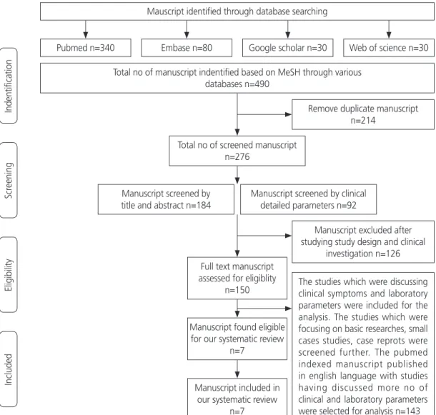
results were evaluated according to the algorithm (Fig. 1), as mentioned. A total of 490 manuscripts were identified across various databases using the initial keyword search, as mentioned above. After eliminating 214 manuscripts as du- plicates due to common MeSH terms, a total of 276 manu- scripts were identified, which included 184 by abstracts and initial headings and 92 by detailed clinical parameters. Out of the 276 screened manuscripts, 126 were excluded after evaluating their scientific study design and clinical investiga- tion. The remaining 150 were retrieved for full-text assess- ment. The studies that discussed clinical symptoms and labo- ratory parameters were included in the analysis. The studies that focused on basic research, small case studies, and case reports were screened further. After qualitative synthesis and
application of our inclusion criteria, we further excluded 143 articles. Finally, we identified 7 manuscripts that were eligible for our systematic review.
2. Study definitions, inclusion and exclusion criteria Studies were included if they satisfied the following criteria:
identified target population; included cases with diagnosed COVID-19; and provided detailed accounts of clinical features and related laboratory parameters. Studies related to mater- nal and neonatal outcomes were compiled and evaluated separately using the same selection criteria. Studies mention- ing follow-up and detailed assessment were only utilized for analysis. This review has a selection bias towards patients from the Asia Pacific region based on the geographic loca-
Fig. 1. Schematic representation of searching eligible manuscripts for analysis.
Mauscript identified through database searching
Total no of manuscript indentified based on MeSH through various databases n=490
Remove duplicate manuscript n=214
Manuscript excluded after studying study design and clinical
investigation n=126 The studies which were discussing clinical symptoms and laboratory parameters were included for the analysis. The studies which were focusing on basic researches, small cases studies, case reprots were screened further. The pubmed indexed manuscript published in english language with studies having discussed more no of clinical and laboratory parameters were selected for analysis n=143 Total no of screened manuscript
n=276
Full text manuscript assessed for eligiblity
n=150
Manuscript found eligible for our systematic review
n=7
Manuscript included in our systematic review
n=7 Manuscript screened by title and abstract n=184
Manuscript screened by clinical detailed parameters n=92 Pubmed n=340
IndentificationScreeningEligibilityIncluded
Embase n=80 Google scholar n=30 Web of science n=30
tion of the initial COVID-19 outbreak. Of the seven studies, six were retrospective [4,18-22], and 1 by Li et al. [23] was an ambispective cohort study. All studies [24-28] describing COVID-19 symptoms in pregnant women were retrospective studies.
3. Data extraction and quality assessment
Both authors independently assessed the quality of the man- uscript based on scientific methodology and case selection.
Studies containing detailed clinical features and investiga- tions were selected for analysis. Variables related to demo- graphic characteristics, clinical features, associated comorbid conditions and related blood and biochemical investigations, treatment-related interventions, complication records, and duration of hospital stay were collected. None of the studies reported any variables; hence, the denominator was based on the availability of specific variable-related information in the manuscript. The entire process of data compilation was individually performed by two reviewers independently. Dis- agreements were resolved by mutual consensus.
4. Bias risk assessment
All manuscripts were selected based on strict inclusion cri- teria containing sufficient information of maximum possible variables. Scientific methodology as well as consecutive case selection were also ensured. However, stringent criteria for risk bias assessment related to the geographical region could not be applied, as selection bias favored the Asia-Pacific region. The identification of the bias coincided with the in- cidence of the initial COVID-19 epidemic in the Asia-Pacific region. Therefore, a majority of the recent pioneering studies evaluating clinical and laboratory parameters of COVID-19 have emerged from this region.
5. Statistical analysis
The relevant variables were analyzed from the individual manuscripts and were tabulated in Microsoft Excel sheets and expressed as event numbers, percentages, total events, and absolute frequency.
Results
1. Observations of COVID-19 infection
The overall analysis was categorized into two different
groups: the general population (Tables 1-4), and pregnant women (Table 5).
2. Age and sex distribution
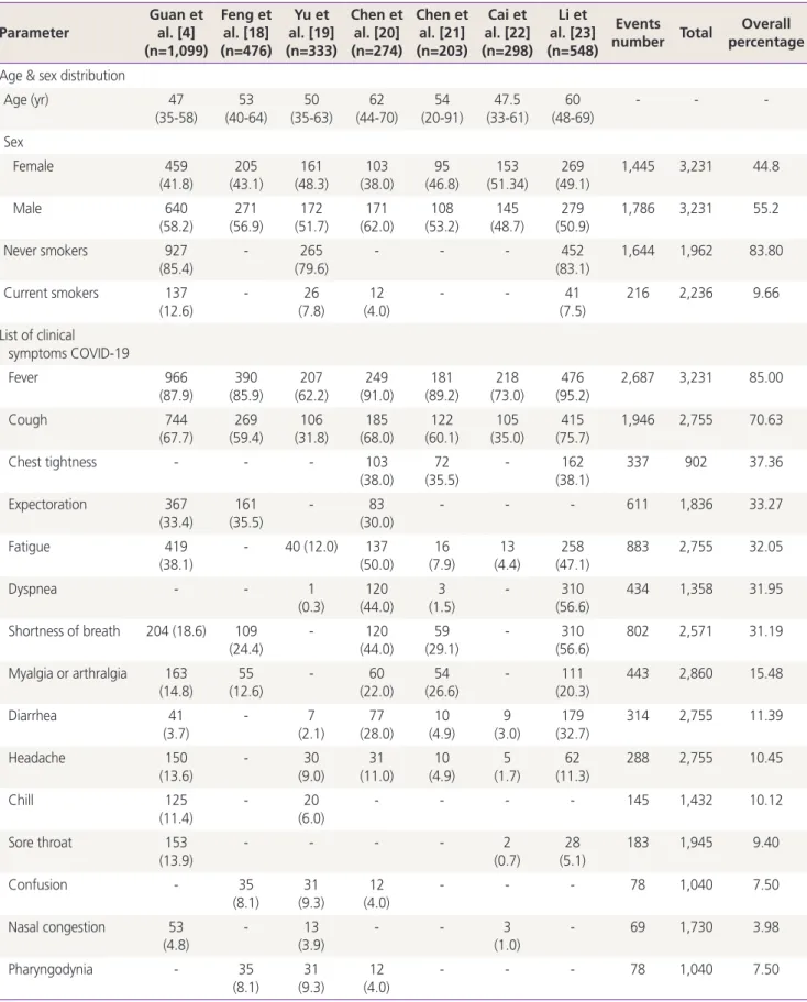
Seven studies from China, with a total of 3,231 subjects (1,786 male and 1,445 female), were analyzed. The median age was 53 years (range, 47-62 years) (Table 1), indicating that middle-aged adults were more prone to COVID-19.
The minimum age of the patients was 20 years and the maximum was 91 years. The proportion of males (55.2%) infected was higher than that of females (44.8%). A history of smoking was not mentioned in any of the included stud- ies; however, infection among current smokers was not as prominent as among non-smokers (83.80%).
3. Clinical symptoms
A detailed account of the patients’ clinical history and symp- toms was collected from different studies and tabulated in order to determine the frequency list. Information on a total of 23 symptoms was collected from all the published studies.
Although information on many symptoms was lacking, a de- tailed analysis was carried out from the available data. Fever (85.0%) was the most common clinical manifestation, fol- lowed by cough (70.63%) (Table 1). The data regarding fever and cough can be considered homogeneous as it was ana- lyzed by all the authors. Other common symptoms included, in decreasing frequency, chest tightness (37.36%), expecto- ration (33.27%), fatigue (32.05%), dyspnea (31.95%), and shortness of breath (31.19%). Minor symptoms included myalgia (15.48%), diarrhea (11.39%), headache (10.45%), chills (10.12%), sore throat (9.40%), nausea, and vomiting (5.97%). Abdominal pain and hemoptysis were considered rare symptoms as they reported very low frequencies (less than 5%).
4. Association among comorbid conditions
Most infectious diseases affect immunocompromised indi- viduals with associated comorbidities. The reports of associ- ated comorbid conditions was also analyzed in detail (Table 2).
However, there was little uniformity of data as associated
morbidity-related cardinal information was not available in
some of the analyzed studies. The final frequency percent-
age was calculated after reducing the number of total cases
from the denominators if the data of a particular illness was
not available in a specific study. The associated comorbid
Table 1. Demographic details and list of clinical symptoms COVID-19 Parameter
Guan et al. [4]
(n=1,099)
Feng et al. [18]
(n=476)
Yu et al. [19]
(n=333)
Chen et al. [20]
(n=274)
Chen et al. [21]
(n=203)
Cai et al. [22]
(n=298)
Li et al. [23]
(n=548)
Events
number Total Overall percentage Age & sex distribution
Age (yr) 47
(35-58) 53
(40-64) 50
(35-63) 62
(44-70) 54
(20-91) 47.5
(33-61) 60
(48-69) - - -
Sex
Female 459
(41.8)
205 (43.1)
161 (48.3)
103 (38.0)
95 (46.8)
153 (51.34)
269 (49.1)
1,445 3,231 44.8
Male 640
(58.2) 271
(56.9) 172
(51.7) 171
(62.0) 108
(53.2) 145
(48.7) 279
(50.9) 1,786 3,231 55.2
Never smokers 927
(85.4)
- 265
(79.6)
- - - 452
(83.1)
1,644 1,962 83.80
Current smokers 137
(12.6) - 26
(7.8) 12
(4.0) - - 41
(7.5) 216 2,236 9.66
List of clinical symptoms COVID-19
Fever 966
(87.9) 390
(85.9) 207
(62.2) 249
(91.0) 181
(89.2) 218
(73.0) 476
(95.2) 2,687 3,231 85.00
Cough 744
(67.7) 269
(59.4) 106
(31.8) 185
(68.0) 122
(60.1) 105
(35.0) 415
(75.7) 1,946 2,755 70.63
Chest tightness - - - 103
(38.0)
72 (35.5)
- 162
(38.1)
337 902 37.36
Expectoration 367
(33.4) 161
(35.5) - 83
(30.0) - - - 611 1,836 33.27
Fatigue 419
(38.1)
- 40 (12.0) 137 (50.0)
16 (7.9)
13 (4.4)
258 (47.1)
883 2,755 32.05
Dyspnea - - 1
(0.3) 120
(44.0) 3
(1.5) - 310
(56.6) 434 1,358 31.95
Shortness of breath 204 (18.6) 109 (24.4)
- 120
(44.0)
59 (29.1)
- 310
(56.6)
802 2,571 31.19
Myalgia or arthralgia 163
(14.8) 55
(12.6) - 60
(22.0) 54
(26.6) - 111
(20.3) 443 2,860 15.48
Diarrhea 41
(3.7)
- 7
(2.1)
77 (28.0)
10 (4.9)
9 (3.0)
179 (32.7)
314 2,755 11.39
Headache 150
(13.6) - 30
(9.0) 31
(11.0) 10
(4.9) 5
(1.7) 62
(11.3) 288 2,755 10.45
Chill 125
(11.4)
- 20
(6.0)
- - - - 145 1,432 10.12
Sore throat 153
(13.9) - - - - 2
(0.7) 28
(5.1) 183 1,945 9.40
Confusion - 35
(8.1)
31 (9.3)
12 (4.0)
- - - 78 1,040 7.50
Nasal congestion 53
(4.8) - 13
(3.9) - - 3
(1.0) - 69 1,730 3.98
Pharyngodynia - 35
(8.1)
31 (9.3)
12 (4.0)
- - - 78 1,040 7.50
conditions analyzed were hypertension (21.60%), diabetes (9.96%), cardiovascular disease (5.78%), chronic pulmonary obstructive disease (2.96%), malignancy (2.23%), cerebro- vascular disease (2.19%), and chronic nephropathy (1.30%) were investigated in this study. Although the number of
subjects was higher in the study by Guan et al. [4], the asso- ciated comorbid conditions were not so prevalent; this could be because the subjects were relatively younger, with the lowest median age of 47 years, compared with those in the other studies [18-23].
Parameter Guan et
al. [4]
(n=1,099)
Feng et al. [18]
(n=476)
Yu et al. [19]
(n=333)
Chen et al. [20]
(n=274)
Chen et al. [21]
(n=203)
Cai et al. [22]
(n=298)
Li et al. [23]
(n=548)
Events
number Total Overall percentage
Dizziness - - 8
(2.4)
21 (8.0)
4 (2.0)
- 56
(10.2)
89 1,358 6.55
Shivering - 24
(6.4) 19
(5.7) - - - - 43 707 6.08
Anorexia - 24
(6.4)
19 (5.7)
- - - - 43 707 6.08
Nausea or vomiting 55 (5.0) - - 24
(9.0) 3
(1.5) - 45
(8.2) 127 2,124 5.97
Chest pain - 21
(4.4)
- - 4
(2.0)
- 41
(7.5)
66 1,191 5.54
Abdominal pain - - - 19
(7.0) 4
(2.0) - 16
(2.9) 39 1,025 3.80
Hemoptysis 10
(0.9)
5 (1.1)
- 7
(3.0)
- - - 22 1,808 1.21
Data are presented as number of patients (%) or median (range) unless otherwise indicated.
COVID-19, coronavirus disease 2019.
Table 1. Continued
Table 2. Co-morbidities of patients with COVID-19 Co-morbid
conditions
Guan et al. [4]
(n=1,099)
Feng et al. [18]
(n=476)
Yu et al.
[19]
(n=333)
Chen et al. [20]
(n=274)
Chen et al. [21]
(n=203)
Cai et al. [22]
(n=298)
Li et al.
[23]
(n=548)
Events
number Total
number Overall percent
Hypertension 164
(14.9) 113
(23.7) - 93
(34.0) 43
(21.2) 47
(15.8) 166
(30.3) 626 2,898 21.60
Diabetes 81
(7.4)
49 (10.0)
28 (8.4)
47 (17.0)
16 (7.9)
18 (6.0)
83 (15.0)
322 3,231 9.96
Cardiovascular disease 27
(2.5) 38 (8.0) 24
(7.2) 23
(8.0) 16
(7.9) 25
(8.4) 34
(6.2) 187 3,231 5.78
Chronic obstructive pulmonary disease
12 (1.1)
22 (4.6)
- 18
(7.0)
8 (3.9)
- 17
(3.1)
77 2,600 2.96
Cancer 10
(0.9) 12
(2.5) - 7
(3.0) 7
(3.4) 4
(1.3) 24
(4.7) 64 2,863 2.23
Cerebrovascular diseases 15 (1.4)
17 (3.6)
- 4
(1.0)
9 (4.4)
- - 45 2,052 2.19
Chronic renal diseases 8
(0.7) 4
(0.8) - 4
(1.0) 8
(3.9) - 10
(1.8) 34 2,599 1.30
Data are presented as number of patients (%) unless otherwise indicated.
COVID-19, coronavirus disease 2019.
Table 3. Hematological biochemical and inflammatory parameters profile Guan et al.
[4]
(n=1,099)
Feng et al. [18]
(n=476)
Yu et al.
[19]
(n=333)
Chen et al.
[20]
(n=274)
Chen et al. [21]
(n=203)
Cai et al. [22]
(n=298)
Li et al.
[23]
(n=548)
Events
number Total
number Overall percent Hematological
parameters Haemoglobin
(g/L) 134.0
(119.0-148.0) 132
(121-144) - 128.0
(116.0-140.0) - - - -
Platelets (decreased)
315/869 (36.2)
- - - 22/203
(10.8)
- 157/539
(29.1)
494 1,611 28.80
TLC
(decreased) 330/978
(33.7) 91/475
(19.2) 56/333
(18.6) 58/274
(21.0) 79/203
(38.9) 114/298
(38.9) 130/542
(24.0) 858 3,098 27.69
TLC (increased)
58/978 (5.9)
49/475 (10.3)
1/333 (0.3)
62/274 (23.0)
14/203 (6.9)
63/542 (11.6)
247 2,805 8.80
Lymphocytes
(decreased) 731/890
(82.1) 225/476
(47.3) 28/333
(10.8) 179/274
(65.32) 117/203
(57.6) - 489/542
(90.2) 1,769 2,718 65.08 Neutrophils
(increased)
- - 1/333
(0.4)
93/274 (34.0)
- - 118/542
(21.8)
212 1,149 18.45
Biochemical parameters (increased) Albumin
(increased) - - - 96/274
(35.0) 131/203
(64.5) - 320/541
(59.1) 547 1,018 53.72
LDH (increased)
277/675 (41.0)
- - 116/274
(42.0)
87/203 (42.9)
23/274 (8.4)
393/534 (73.6)
896 1,960 45.71
Natriureti peptide (increased)
- - - 85/173 (49.0) - - 92/33
(27.5) 177 508 34.84
Globulin
(increased) - - - - 68/203
(33.5) - 218/540
(40.4) 68 203 33.5
AST (increased)
168/757 (22.2)
- - 84/274
(31.0)
69/203 (34.0)
25/298 (8.4)
179/540 (33.1)
525 2,072 25.33
ALT
(increased) 158/741
(21.3) - - 60/274 (22.0) 30/203
(14.8) 39/298
(13.1) 125/541
(23.1) 412 1,884 21.86
BUN
(increased) - - - 85/539
(15.8) 85 539 15.8
Creatinine (increased)
12/752 (1.6)
- - - 25/203
(12.3)
- 146/539
(27.1)
183 1,494 12.24
Total bilirubing
(increased) 76/722 (10.5) - - - - 24/298
(8.1) 24/541
(4.4) 124 1,561 7.94
Inflammatory cytokines (increased) C-reactive
protein level
481/793 (60.7)
266/41 (64.1)
84/333 (42.9)
80/243 (33.0) 110/203 (54.2)
196/291 (67.4)
460/540 (85.2)
1,677 2,818 59.51 Tumor necrosis
factor - - - 93/163 (57.0) - - 182/309
(58.9) 275 472 58.26
Erythrocyte sedimentation rate
- - - - 82/203
(40.4)
117/287 (40.8)
377/518 (72.8)
576 1,008 57.14
Guan et al.
[4]
(n=1,099)
Feng et al. [18]
(n=476)
Yu et al.
[19]
(n=333)
Chen et al.
[20]
(n=274)
Chen et al. [21]
(n=203)
Cai et al. [22]
(n=298)
Li et al.
[23]
(n=548)
Events
number Total
number Overall percent
Interleukin-6 - - - 119/163
(73.0)
44/203 (21.7)
146/294 (49.7)
221/312 (70.8)
530 972 54.52
Interleukin-2 - - - 84/163
(52.0) - - 164/309
(53.1) 248 472 52.54
Interleukin-10 - - - 58/163
(36.0)
- - 83/307
(27.0)
141 470 30.00
Interleukin-8 - - - 24/163
(15.0) - - 24/309
(7.8) 48 472 10.16
Procalcitonin level
- - - 8/236
(3.0)
7/203 (3.4)
- 46/486
(9.5)
61 925 6.59
Data are presented as number of patients (%) or mean (range) unless otherwise indicated.
TLC, total leukocytes count; LDH, lactate dehyadrogenase, AST, aspartate transaminase; ALT, alanine transaminase; BUN, blood urea nitrogen.
Table 3. Continued
Table 4. Complications, treatment and outcomes of patients with COVID-19 Characteristic
Guan et al.
[4]
(n=1,099)
Feng et al. [18]
(n=476)
Chen et al. [20]
(n=274)
Chen et al. [21]
(n=203)
Cai et al.
[22]
(n=298)
Li et al. [23]
(n=548)
Total events number
Total number
Overall percent Oxygen therapy 418 (38.0) 36 (77.0) 251 (92.0) 123 (60.6) - 355 (64.8) 1,515 2,600 58.26 Antiviral therapy 393 (35.8) 286 (60.1) 236 (86.0) 131 (64.8) 236 (79.2) 398 (72.6) 1,680 2,898 57.97 Intravenous antibiotics 632 (57.5) 319 (67.0) 249 (91.0) - - 7 (1.3) 1,207 2,397 50.35 Systemic corticosteroids 204 (18.6) 127 (26.0) - 107 (52.7) 91 (30.5) 341 (62.2) 870 2,624 33.15 Intravenous immunoglobin 143 (13.0) - 54 (20.0) - 94 (31.5) 213 (38.9) 504 2,219 22.71
Mechanical ventilation 67 (6.1) - 119 (43.0) 39 (19.2) 30 (10.1) - 255 1,874 13.60
Invasive 24 (2.2) 39 (8.2) 102 (37.0) - - 25 (4.6) 190 2,397 7.92
Non-invasive 56 (5.1) 34 (7.1) 17 (6.0) - - 78 (14.2) 185 2,397 7.71
Concurrent chemoradiation 9 (0.8) - 3 (1.0) - - 2 (0.4) 14 1,921 0.72
Extracorporealmembrane
oxygenation 5 (0.5) 4 (0.8) 1 (<1) - 3 (1.0) - 13 1,873 0.69
Cardiac complications - - 89 (44.0) - - 119 (21.7) 208 751 27.69
ARDS 37 (3.4) - 196 (72.0) - - 210 (38.3) 443 1,921 23.06
Liver dysfunction - - 13 (5.0) - - 106 (19.3) 119 822 14.47
Acute kidney injury 6 (0.5) - 29 (11.0) - - 95/ (17.3) 130 1,921 6.76
Septic shock 11 (1.0) - 46 (17.0) - - - 57 1,373 4.15
DIC 1 (0.1) - 21 (8.0) - - - 22 1,373 1.60
Staying in hospital 1,029 (93.6) 23 (4.8) - - - 168 (30.8) 1,220 2,120 57.54
Discharge from hospital 55 (5.0) 403 (84.7) - - 268 (89.9) 287 (52.7) 1,013 2,418 41.89
Death 15 (1.4) 38 (8.0) - 26 (12.8) 3 (1.0) 90 (16.5) 172 2,621 6.56
Data are presented as number of patients (%) unless otherwise indicated.
COVID-19, coronavirus disease 2019; ARDS, acute respiratory distress syndrome; DIC, disseminated intravascular coagulopathy.
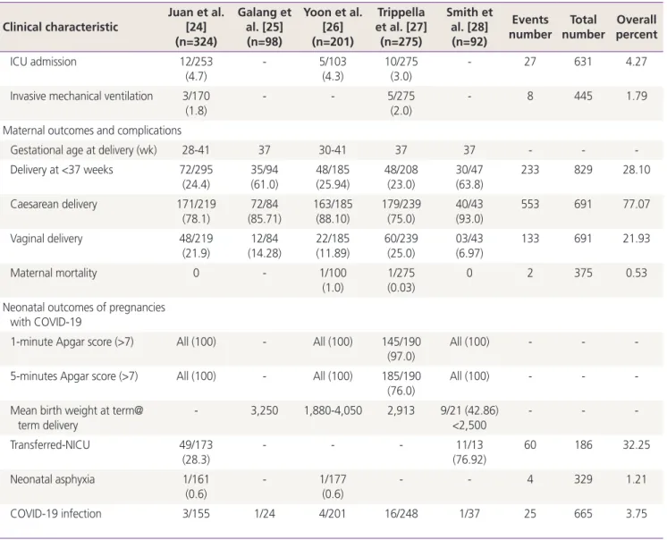
Table 5. Maternal & neonatal outcome of COVID-19 in pregnant women Clinical characteristic
Juan et al.
[24]
(n=324)
Galang et al. [25]
(n=98)
Yoon et al.
[26]
(n=201)
Trippella et al. [27]
(n=275)
Smith et al. [28]
(n=92)
Events
number Total
number Overall percent Symptoms of COVID-19
in pregnant women
Fever 138/295
(46.8) 76/92
(83.0) 85/201
(42.3) 155/275
(58.0) 57/92
(61.96) 511 955 53.50
Cough 101/295
(34.2)
34/66 (52.0)
64/201 (31.8)
95/275 (36.0)
35/92 (38.04)
329 929 35.41
Malaise 0 14/31
(45.0) 18/95
(18.9) 37/275
(14.0) 25/82
(30.49) 94 483 19.46
Shortness of breath 39/295 (13.2)
12/47 (26.0)
16/142 (11.3)
- 10/83
(12.05)
77 567 13.58
Myalgia 27/295
(9.2) 5/21
(24.0) 15/56
(21.4) 37/275
(14.0) 6/28
(21.4) 90 675 13.33
Diarrhea/GI symptoms 11/295 (3.7)
76/92 (83.0)
10/134 (7.5)
9/275 (3.0)
4/38 (10.43)
110 834 13.18
Fatigue 28/295
(9.5) 7/29
(24.0) - 28/275
(10.0) 25/82
(30.49) 88 681 12.92
Sore throat 10/295
(3.4)
1/24 (4.0)
8/84 (9.5)
9/275 (3.0)
6/50 (12.0)
34 728 4.67
Hemogram findings of
pregnant women with COVID-19
TLC (decreased) 146/182
(80.2)
- - - - 146 182 80.2
Lymphocytes (decreased) 85/197
(43.1) 27/50
(54.0) 52/120
(43.3) 31/108
(29.0) 46/69
(66.67) 241 544 44.30
Thrombocytopenia - 8/18
(44.0)
7/31 (22.6)
- - 15 49 30.61
TLC (increased) 4/19
(21.05) 12/47
(26.0) 28/89
(31.5) - - 40 155 28.38
Biochemical of pregnant women with COVID-19
Elevated CRP 90/197
(45.7)
- 65/103
(63.1)
52/108 (48.0)
- 207 408 50.73
Elevated AST 5/42
(11.9) 7/28
(25.0) - 9/108
(8.0) - 21 178 11.79
Elevated ALT 5/42
(11.9)
6/28 (21.0)
- 9/108
(8.0)
- 20 178 11.23
Treatments of pregnant women with COVID-19
Antibiotics 111/157
(70.7)
46/49 (94.0)
- - - 157 206 76.21
Antivirals 82/217
(37.8) 43/57
(75.0) - - - 125 274 45.62
Non-invasive mechanical ventilation
21/170 (12.4)
- 5/50
(10.0)
- - 26 220 11.81
5. Laboratory parameters in COVID-19 patients
Routine blood test reports of 3,231 COVID-19 cases were available from different sets of studies. The mean hemo- globin values were in the normal range in 3 analyzed stud- ies (Table 3), which was probably due to the acute course of the illness. Predominantly, the cases had reduced total leukocyte count (TLC, 28.80%) along with reduced platelet counts (27.69%). Hemoglobin levels were not as acutely affected; however, thrombocytopenia was observed in ap- proximately one-third of the patients. A minority of cases also presented with increased TLC (8.80%). About 65% of cases had lymphocytopenia, as determined by a differential
leukocyte count (DLC), which would be considered an ini- tial response in viral illnesses. A few cases presented with increased neutrophil count (18.45%), probably reflecting a component of superadded non-viral bacterial infection. The laboratory biochemical parameters (Table 3), such as albu- min (53.72%) and lactate dehydrogenase (LDH, 45.71%), were affected in all cases. The least affected parameter in our analysis was bilirubin. One third (34.84%) of the cases presented with the raised natriuretic peptide responsible for natriuresis. There were no symptoms related to hypovolemic shock or increased frequency of urination. The impact of the raised natriuretic peptide could have been worse in cases
Clinical characteristic Juan et al.[24]
(n=324)
Galang et al. [25]
(n=98)
Yoon et al.
[26]
(n=201)
Trippella et al. [27]
(n=275)
Smith et al. [28]
(n=92)
Events
number Total
number Overall percent
ICU admission 12/253
(4.7)
- 5/103
(4.3)
10/275 (3.0)
- 27 631 4.27
Invasive mechanical ventilation 3/170
(1.8) - - 5/275
(2.0) - 8 445 1.79
Maternal outcomes and complications
Gestational age at delivery (wk) 28-41 37 30-41 37 37 - - -
Delivery at <37 weeks 72/295 (24.4)
35/94 (61.0)
48/185 (25.94)
48/208 (23.0)
30/47 (63.8)
233 829 28.10
Caesarean delivery 171/219
(78.1) 72/84
(85.71) 163/185
(88.10) 179/239
(75.0) 40/43
(93.0) 553 691 77.07
Vaginal delivery 48/219
(21.9)
12/84 (14.28)
22/185 (11.89)
60/239 (25.0)
03/43 (6.97)
133 691 21.93
Maternal mortality 0 - 1/100
(1.0) 1/275
(0.03) 0 2 375 0.53
Neonatal outcomes of pregnancies with COVID-19
1-minute Apgar score (>7) All (100) - All (100) 145/190
(97.0) All (100) - - -
5-minutes Apgar score (>7) All (100) - All (100) 185/190 (76.0)
All (100) - - -
Mean birth weight at term@
term delivery - 3,250 1,880-4,050 2,913 9/21 (42.86)
<2,500 - - -
Transferred-NICU 49/173
(28.3)
- - - 11/13
(76.92)
60 186 32.25
Neonatal asphyxia 1/161
(0.6) - 1/177
(0.6) - - 4 329 1.21
COVID-19 infection 3/155 1/24 4/201 16/248 1/37 25 665 3.75
Data are presented as number of patients (%) unless otherwise indicated.
COVID-19, coronavirus disease 2019; GI, gastro-intestinal; TLC, total leukocytes count; CRP, C-reactive protein; AST, aspartate transaminase;
ALT, alanine transaminase; ICU, Intensive Care Unit; NICU, Neonatal Intensive Care Unit.
Table 5. Continued
![Table 2. Co-morbidities of patients with COVID-19 Co-morbid conditions Guan et al. [4] (n=1,099) Feng et al](https://thumb-ap.123doks.com/thumbv2/123dokinfo/5460870.657009/6.892.80.820.586.911/table-morbidities-patients-covid-morbid-conditions-guan-feng.webp)
![Table 3. Hematological biochemical and inflammatory parameters profile Guan et al. [4] (n=1,099) Feng et al](https://thumb-ap.123doks.com/thumbv2/123dokinfo/5460870.657009/7.892.75.827.155.1096/table-hematological-biochemical-inflammatory-parameters-profile-guan-feng.webp)