INTRODUCTION
Since severe acute respiratory syndrome coronavirus 2 (SARS- COV-2) was first identified in Wuhan, China, it has spread rap- idly and the World Health Organization declared a global pan- demic on March 11, 2020 [1]. As of June 30, 2020, more than 10 million people have been infected and more than 500000 have died [2]. Although the main clinical manifestations of novel coro- navirus disease 2019 (COVID-19) are pneumonia and respira- tory symptoms, serious cardiovascular complications have also been reported [3-5]. Cardiovascular involvement in COVID-19 is associated with increased mortality risk [6]. Cardiac manifes- tations of COVID-19 include myocarditis, heart failure, stress- induced cardiomyopathy, arrhythmia, and thromboembolism [3,7-9]. Patients with underlying cardiovascular disease are par- ticularly vulnerable to cardiovascular complications caused by COVID-19 and have a poor prognosis [10,11]. Therefore, car- diovascular radiologists and clinicians must be aware of the car- diovascular complications of COVID-19.
UNDERLYING CARDIOVASCULAR DISEASE IN COVID-19 PATIENTS
A considerable proportion of patients have preexisting car-
diovascular diseases, such as coronary artery disease, heart fail- ure, arrhythmias, which are related to poor prognosis after CO- VID-19 infection. A recent meta-analysis reported that the pooled prevalence of cardiovascular disease in COVID-19 pa- tients was 12.1% [12]. Pre-existing cardiovascular disease sig- nificantly increases the risk of in-hospital death and fatal out- comes [10]. Recent meta-analysis also reported that preexisting cardiovascular disease increased the risk of severe COVID-19 [odds ratio (OR), 3.14; 95% confidence interval (CI), 2.32–4.24]
and of all-cause mortality (OR, 11.08; 95% CI, 2.59–47.32) [13].
Severe COVID-19 in this meta-analysis was defined as a com- posite of: 1) respiratory distress, defined as a respiratory rate
≥30 per minute; 2) oxygen saturation on room air at rest ≤93%;
3) partial pressure of oxygen in arterial blood/fraction of inspired oxygen ≤300 mm Hg; 4) requirement for mechanical ventilation vital life support, or intensive care unit (ICU) admission; 5) and death [13]. This is thought to be because patients with underly- ing cardiovascular disease tend to be older and are more likely to have other comorbidities and an impaired immune system than the younger patients [14].
CARDIOVASCULAR INVOLVEMENT OF COVID-19
Pathophysiology
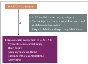
Several mechanisms that explain the pathophysiology of car- diac injury in COVID-19 have been proposed (Fig. 1) [14]. The
cc This is an Open Access article distributed under the terms of the Creative Commons Attribution Non-Commercial License (https://creativecommons.org/licenses/by- nc/4.0) which permits unrestricted non-commercial use, distribution, and reproduc- tion in any medium, provided the original work is properly cited.
CVIA Cardiovascular Manifestations of COVID-19
Jin Young Kim
Department of Radiology, Dongsan Hospital, Keimyung University College of Medicine, Daegu, Korea
Received: July 20, 2020 Revised: July 29, 2020 Accepted: July 29, 2020 Corresponding author Jin Young Kim, MD Department of Radiology, Dongsan Hospital,
Keimyung University College of Medicine, 1035 Dalgubeol-daero, Dalseo-gu, Daegu 42601, Korea
Tel: 82-53-258-4151 Fax: 82-53-258-4153 E-mail: [email protected]
Although novel coronavirus disease 2019 (COVID-19) mainly affects lung, it also affects the cardiovascular system. Cardiac manifestations of COVID-19 include myocardial injury, myo- carditis, heart failure, acute coronary syndrome, thromboembolism, and arrhythmias. Cardio- vascular involvement in COVID-19 is related to poor prognosis and increased mortality. There- fore, the recognition of cardiac complications and prompt treatment of cardiac injury in patients with suspected COVID-19 is essential. In this review article, we discuss the cardiovas- cular manifestations of COVID-19 and their related imaging findings.
Key words COVID-19 · Cardiovascular abnormalities · CT · MRI.
pISSN 2508-707X / eISSN 2508-7088 Cardiovasc Imaging Asia 2020;4(3):74-80 https://doi.org/10.22468/cvia.2020.00066
REVIEW ARTICLE
Jin Young Kim
CVIA
first is direct cardiac damage caused by the virus. SARS-CoV-2 is a single-stranded RNA virus that enters cells after binding to the angiotensin-converting enzyme 2 (ACE2) protein [15,16].
ACE2 is highly expressed in type 2 lung alveolar cells, as well as in the heart and blood vessels [17]. ACE2 mediates direct en- try of the virus into cells which subsequently causes cardiotox- icity. Another mechanism is myocardial injury secondary to viral driven inflammation and cytokine storm syndrome. CO- VID-19 has been demonstrated to cause severe inflammation by increasing the expression of plasma cytokines and chemo- kines such as interleukin (IL)-6, IL-2, IL-7, IL-8, granulocyte colony-stimulating factor, and tumor necrosis factor [18]. This systemic cytokine storm can lead to multi-organ dysfunction and cardiac overload that progresses to cardiac damage due to a mismatch between myocardial oxygen supply and demand.
Moreover, the severe inflammation caused by SARS-CoV-2 leads to a hypercoagulable state and increased plaque vulnerability which predisposes patients acute coronary syndrome (ACS) and venous thromboembolism. Further research is required to im- prove our understanding of the multifactorial etiology of car- diovascular complications in COVID-19 patients.
Myocardial injury
Myocardial injury is defined as an increase of the cardiac bio- markers such as troponin I and troponin T above the 99th per- centile of the upper normal reference [19,20]. As cardiac tropo- nin level cannot distinguish the cause of myocardial injury, considering them together with electrocardiogram changes, or global or regional left ventricular (LV) wall motion abnormali- ties can help to determine the extent and potential cause of myo- cardial injury. The reported prevalence of myocardial injury in COVID-19 patients ranges from 7–28% [6,21]. Patients with se- vere COVID-19 requiring mechanical ventilation, ICU admis-
sion, and those who ultimately die have more myocardial inju- ry than those with milder forms [22]. Furthermore, patients with underlying cardiovascular disease are more likely to have myocardial injury than those without [23]. Myocardial injury significantly increases the risk of poor outcome and death com- pared to patients without myocardial injury [23-25]. A recent study reported that even mildly increased troponin I (troponin I 0.03–0.09 ng/mL) was significantly associated with death [ad- justed hazard ratio (HR) 1.75, 95% CI 1.37–2.24; p<0.001], and this association was even stronger in patients with troponin I
>0.09 ng/dL (adjusted HR 3.03, 95% CI 2.42–3.80; p<0.001) [23].
Myocarditis
COVID-19 related myocarditis has been reported in various countries, but the exact incidence is unclear [3,26-30]. It com- monly affects children and young adults [3,30,31]. The clinical presentation of myocarditis is variable and nonspecific and can include fatigue, chest pain, or dyspnea [32]. Myocarditis should be suspected in patients with a history of exposure to SARS- CoV-2 or those with a diagnosis of COVID-19 that have symp- toms of myocarditis and elevated levels of cardiac enzymes and inflammatory markers [32]. Distinguishing COVID-19-related myocarditis from other etiologies requires a positive SARS-CoV-2 polymerase chain reaction result from cardiac tissue. However, performing such an invasive procedure with the accompany- ing risk of contamination should be carefully considered. Al- though, several reported cases of COVID-19-related myocar- ditis exhibited SARS-CoV-2 in the cardiac tissue [30,33], most were diagnosed based on the clinical and radiological findings [27,29,34,35]. Cardiac imaging study may help to diagnose CO- VID-19 related myocarditis in these patients. The European So- ciety of Cardiology (ESC) recommends the use of cardiac mag- netic resonance (CMR) imaging for suspected acute myocarditis [36]. CMR is the best imaging modality to detect myocardial edema and myocardial injury in acute myocarditis. Regional and globally increased T2 signal intensity on short-tau inversion recovery sequence indicates myocardial edema, and regional late gadolinium enhancement (LGE) indicates myocardial in- jury. Emerging CMR techniques, T1 and T2 mapping, and ECV were added to the Revised 2018 Lake Louise Criteria [37].
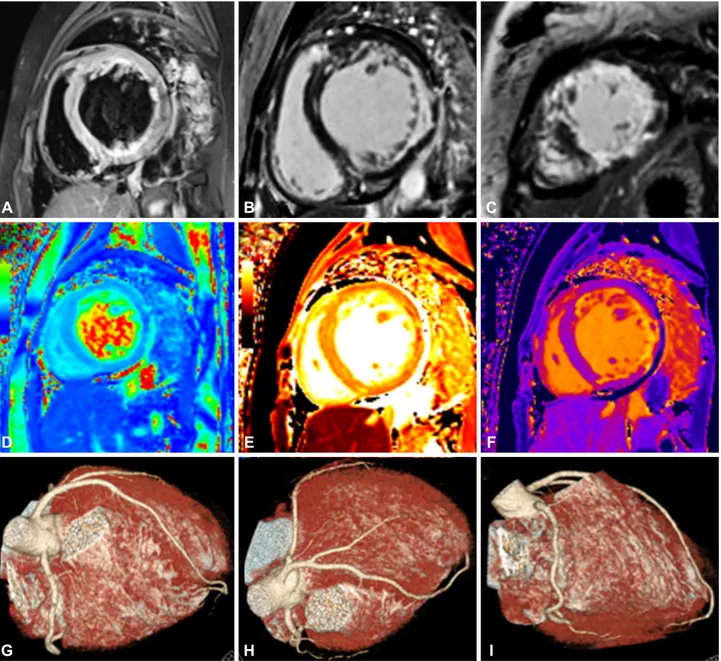
Myocardial edema prolongs T1 and T2 relaxation time of the myocardium. Extracellular volume fraction can also detect ex- panded extracellular space caused by myocardial necrosis or fibrosis. Cardiac CT angiography is an alternative non-invasive imaging modality to distinguish myocarditis from coronary artery disease [36]. Fig. 2 illustrates a case of COVID-19 relat- ed myocarditis in a 21-year-old female patient. Extensive LGE with myocardial edema was noted on CMR and coronary ar- tery disease was excluded on coronary CT angiography.
SARS-CoV-2 infection
- ACE2 mediated direct myocyte injury - Cardiac injury secondary to cytokine storm and
viral driven inflammation
- Plaque instability and hypercoagulability state
Cardiovascular involvement of COVID-19 - Myocarditis, myocardial injury - Heart failure
- Acute coronary syndrome - Thromboembolic complications - Arrhythmia
Fig. 1. Pathophysiology of cardiovascular involvement in COVID-19.
SARS-CoV-2: severe acute respiratory syndrome-coronavirus 2, ACE:
angiotensin converting enzyme, COVID-19: novel coronavirus disease 2019.
Cardiovascular Manifestations of COVID-19
CVIA
Pericardial effusion
Few cases of pericardial effusion complicated by COVID-19 have been reported [38,39]. Pericardial effusion might develop as a result of pericarditis or myo-pericarditis [40]. Farina et al.
[39] reported the detection of SARS-CoV-2 in pericardial fluid.
Another paper reported that pericardial fluid was exudative in COVID-19 patient with elevated lactate dehydrogenase and al- bumin levels [41]. At our institution, there was one case of peri- cardial effusion complicated by COVID-19 in an 86-year-old fe-
male patient (Fig. 3). A large pericardial effusion was noted on CT at the time of diagnosis of COVID-19 that completely re- solved after treatment of COVID-19. This was presumed to be caused by myo-pericarditis. There is still little known about clini- cal significance of pericardial effusion in COVID-19 patients.
As in our case, pericardial effusion in COVID-19 can demon- strate a self-limiting course, but there are also reported cases of life-threatening cardiac tamponade. Further research on the mechanism of development of pericardial effusion and its clin-
Fig. 2. COVID-19-related myocarditis in a 21-year-old female patient. Myocarditis was suspected during hospitalization because of an in- crease in the cardiac biomarkers Troponin I (1.26 ng/mL, reference value <0.3 ng/mL) and NT-proBNP (1929 pg/mL, reference <125 pg/mL) with worsening of shortness of breath. Cardiac MRI (3T system, Vida, Siemens) showed diffuse high signal intensity of the LV myocardium on T2 short tau inversion recovery image (A, signal intensity ratio of myocardium over skeletal muscle=2.16), reflecting myocardial edema. Late gadolinium enhanced images (B and C) showed extensive transmural enhancement of the LV myocardium. T2 mapping (D, septal wall 45 ms, reference value ~37 ms), native T1 mapping (E, septal wall 1431 ms, lateral wall 1453 ms, reference value ~1200 ms) values and extracellular volume fraction (F, septal wall 30%, lateral wall 60%, reference ~25%) were also increased. Coronary CT angiography (G, H, and I) excluded coronary artery disease. Finally, an endomyocardial biopsy revealed myocarditis. COVID-19: novel coronavirus disease 2019, NT-proBNP: N- terminal pro brain natriuretic peptide, LV: left ventricular.
A
D
G
E
H
F
I
B C
Jin Young Kim
CVIA
ical significance in COVID-19 patient is needed.
Heart failure
Several studies have reported that heart failure is a complica- tion of COVID-19, but its exact prevalence is unknown [42]. A cohort study from Wuhan, China reported that 23% (44/191) of COVID-19 patients had heart failure [42]. The prevalence of heart failure was significantly higher in non-survivors than sur- vivors (52% vs. 12%, p<0.001, respectively) [42]. Although ex- act mechanism of acute heart failure in COVID-19 has not been identified some possible cause have been proposed [43]. Older patients with underlying cardiovascular disease, such as coro- nary artery disease, hypertension, diabetes, or subclinical heart disease, may progress to heart failure while being in the inflam- matory state caused by COVID-19. Myocardial injury, myocar- ditis, stress-induced cardiomyopathy, acute illness due to sys- temic inflammation, cytokine storm induced by COVID-19, and sepsis may lead acute cardiac dysfunction [43]. An increased natriuretic peptide level may help to diagnose heart failure in COVID-19 patients. Additionally, transthoracic echocardiog- raphy with appropriate personal protective equipment could be considered to evaluate LV dysfunction in COVID-19 patients with symptoms of heart failure [3]. Heart failure has been ob- served after extensive COVID-19 related myocarditis (Fig. 2) at our institution. Globally decreased LV wall motion (LV ejec- tion fraction 28%) and dilated LV have been seen on transtho- racic echocardiography (Supplemental Video 1 in the online- only Data Supplement).
Thromboembolism
The severe inflammatory response and hypoxia caused by COVID-19 may lead to endothelial dysfunction and an increase
in circulating thrombotic factors [9,44,45]. One study demon- strated that the levels of D-dimer and fibrin degradation prod- ucts, fibrin, and fibrinogen were significantly higher in COV- ID-19 patients than in normal subjects [45]. In another study, venous thromboembolism was confirmed in 58% (7/12) of pa- tients at autopsy and pulmonary embolism was the direct cause of death in 4 patients [46]. Among 100 COVID-19 patients who underwent contrast-enhanced CT, pulmonary thromboembo- lism was found in 23%, and the incidence of pulmonary embo- lism was significantly higher in severely unwell patients requir- ing mechanical ventilation and critical care admission [47]. In another study of 184 patients with severe COVID-19, the cu- mulative incidence of thrombotic events including pulmonary embolism, deep vein thrombosis, ischemic stroke, and myocar- dial infarction was 49% [48]. Pulmonary thromboembolism was the most common thrombotic event in COVID-19 patients (87%). Pulmonary embolism should be suspected in COVID-19 patients with worsening respiratory symptoms and increased D-dimer levels. Pulmonary CT angiography is the diagnostic modality of choice in these patients; however, physicians must weigh the potential risks of contamination by intravenous injec- tion of contrast media and the expected benefits of performing CT in this population. The World Health Organization recom- mends that contrast-enhanced CT is considered when throm- boembolic complications are suspected [49]. Fig. 4 illustrates the imaging findings of an 87-year old female with COVID-19 and pulmonary thromboembolism treated in our institution.
Contrast-enhanced chest CT showed pulmonary embolism and venous thrombosis in the left internal jugular vein.
Acute coronary syndrome
The pathophysiology of ACS in COVID-19 involves plaque
Fig. 3. Pericardial effusion in an 86-year-old female COVID-19 patient. A large pericardial effusion was noted on chest CT at the time of ad- mission (A). Two months after discharge, pericardial effusion was completely resolved (B). COVID-19: novel coronavirus disease 2019.A B
Cardiovascular Manifestations of COVID-19
CVIA
instability, hypercoagulable state, microvascular dysfunction, hemodynamic change, and hypoxemia caused by severe in- flammation [50,51]. The majority of ACS cases caused by CO- VID-19 are type 2 which result from a mismatch between oxy- gen supply and demand [20]. Type 1 myocardial infarction caused by plaque disruption may also be precipitated by COV- ID-19. Since chest pain is common in COVID-19 patients, this can lead to a delay in the diagnosis of ACS. Therefore, clinicians should maintain a high index of suspicion and rule out ACS by assessing cardiac biomarker levels and electrocardiogram find- ings. In case of ST-elevation myocardial infarction, primary per- cutaneous coronary intervention remains the therapy of choice, to be performed in a dedicated COVID-19 catheterization lab- oratory. In patients with non-ST-segment elevation ACS with low to intermediate risk, coronary CT angiography is the pre- ferred non-invasive diagnostic imaging modality because it minimizes the exposure time of patients [36,52].
ARRHYTHMIA
Although the incidence and the exact mechanism of arrhyth- mia in COVID-19 are uncertain, various types of arrhythmias have been reported in recent studies of COVID-19 patients [53].
Myocardial injury and antiviral therapies are possible drivers of arrhythmias in COVID-19 patients [54]. Among a cohort of 137 patients in Hubei, China, 10 patients (7.3%) had palpitations [55]. In another study of 187 Chinese patients, 11 (5.9%) had ma- lignant arrhythmias such as ventricular tachycardia and ven- tricular fibrillation [6]. In this study, patients with elevated tro- ponin I level were at a significantly higher risk of developing malignant arrhythmias [6]. In a cohort of 393 patients in New York, Goyal et al. [56] found that the risk of atrial arrhythmia was significantly higher in patients who received invasive me- chanical ventilation compared to those who did not (17.7% vs.
1.9%, respectively). Cardiac CT with delayed phase scanning (1 minute after contrast administration) is preferred to trans-
esophageal echocardiography in the evaluation of thrombi in the left atrial appendage and assessment of complications of cardiac implantable devices inserted for the management of ar- rhythmias [36]. Moreover, since arrhythmia in COVID-19 pa- tients may be caused by structural heart disease such as myo- cardial injury or myocarditis, imaging studies such as CMR can be considered to identify the cause of the arrhythmia, while weighing the risks and benefits to each patient [57,58].
ROLE OF CARDIOVASCULAR IMAGING IN COVID-19
Imaging COVID-19 patients is difficult because of the risk of transmission of the virus. Therefore, routine cardiac imaging is not recommended for COVID-19 patients and should be re- served for patients with suspected cardiovascular injury. The Society of Cardiovascular Computed Tomography and the ESC have published updated guidance on the use of cardiac CT and MRI in COVID-19 patients [36,59]. The risk of con- tamination during CT and CMR is considered lower than that during echocardiography. Cardiac CT angiography is the pre- ferred noninvasive imaging tool for patients with suspected coronary artery disease, acute symptomatic valvular heart dis- ease, pulmonary embolism, intracardiac thrombus, and for those requiring urgent structural interventions and urgent management of complications of intracardiac devices. CMR is recommended for patients with suspected acute myocarditis.
Prevention of virus transmission from COVID-19 patients to healthcare workers and other patients is the foremost priority.
To prevent disease dissemination during CT or MR scans, ra- diographers are required to wear personal protective equip- ment [60,61], which consists of goggles, face masks, fluid-resis- tant gowns, and gloves. Patients should wear a surgical mask in the scanning room, and in the case of MR scanning, the metal strip in the mask should be removed prior to arrival at the im- aging suite or MR-safe masks should be used [62]. Non-contrast
Fig. 4. Thromboembolic complications in an 87-year-old female COVID-19 patient. Contrast enhanced CT showed a thrombus in the left in- ternal jugular vein (A, arrow) and pulmonary thromboembolism at the right main and lower lobar pulmonary arteries (B and C, arrows). COV- ID-19: novel coronavirus disease 2019.A B C
Jin Young Kim
CVIA
enhanced CT is usually recommended in COVID-19 patient because of the increased risk of virus transmission through in- travenous contrast injection. After the imaging study is com- pleted, the CT or MR scanner and the rooms are sanitized [60].
High frequency contact surfaces should be wiped with an alco- hol-based disinfectant or other environmental protection agen- cy-approved disinfectants [61,63]. Depending on the air exchange rate, the imaging suite may need to be closed for 60 minutes to ventilate and exchange the room after sanitation [60,61]. Some hospitals use negative pressure individual isolation systems of sizes suitable for a CT scanner’s gantry [64]. Given the risk of transmission, the appropriate use of diagnostic imaging in CO- VID-19 patients with suspected cardiovascular complications is essential.
CONCLUSION
Cardiovascular injury is a serious complication of COVID-19 that is related to fatal outcomes. Current studies are ongoing to try and elucidate the exact incidence and mechanism of cardio- vascular complications in COVID-19. To improve outcomes, it is essential that clinicians are aware of the various cardiovascu- lar manifestations COVID-19 and that they are familiar with the latest guidance on the appropriate use of cardiac biomarkers and cardiovascular imaging in suspected patients.
Supplementary Video Legends
Video 1. Transthoracic echocardiography shows globally decreased left ventricular wall motion (ejection fraction 28%) because of COVID-19 relat- ed myocarditis.
Supplementary Materials
The online-only Data Supplement is available with this article at https://
doi.org/10.22468/cvia.2020.00066.
Conflicts of Interest
The author has no potential conflicts of interest to disclose.
Acknowledgments
None.ORCID iD
Jin Young Kim https://orcid.org/0000-0001-6714-8358
REFERENCES
1. WHO Director-General’s opening remarks at the media briefing on CO- VID-19-11 March 2020. World Health Organization Web site. https://
www.who.int/dg/speeches/detail/who-director-general-s-opening-re- marks-at-the-media-briefing-on-covid-19---11-march-2020. Published March 11, 2020. Accessed June 30, 2020.
2. WHO coronavirus disease (COVID-19) dashboard. World Health Orga- nization Web site. https://covid19.who.int/. Accessed June 30, 2020.
3. Kim IC, Kim JY, Kim HA, Han S. COVID-19-related myocarditis in a 21- year-old female patient. Eur Heart J 2020;41:1859.
4. Inciardi RM, Lupi L, Zaccone G, Italia L, Raffo M, Tomasoni D, et al. Car-
diac involvement in a patient with coronavirus disease 2019 (COVID-19).
JAMA Cardiol 2020;5:819-824.
5. Buja LM, Wolf DA, Zhao B, Akkanti B, McDonald M, Lelenwa L, et al. The emerging spectrum of cardiopulmonary pathology of the coronavirus disease 2019 (COVID-19): report of 3 autopsies from Houston, Texas, and review of autopsy findings from other United States cities. Cardiovasc Pathol 2020;48:107233.
6. Guo T, Fan Y, Chen M, Wu X, Zhang L, He T, et al. Cardiovascular impli- cations of fatal outcomes of patients with coronavirus disease 2019 (CO- VID-19). JAMA Cardiol 2020;5:811-818.
7. Fried JA, Ramasubbu K, Bhatt R, Topkara VK, Clerkin KJ, Horn E, et al.
The variety of cardiovascular presentations of COVID-19. Circulation 2020;141:1930-1936.
8. Kochi AN, Tagliari AP, Forleo GB, Fassini GM, Tondo C. Cardiac and ar- rhythmic complications in patients with COVID-19. J Cardiovasc Electro- physiol 2020;31:1003-1008.
9. Bikdeli B, Madhavan MV, Jimenez D, Chuich T, Dreyfus I, Driggin E, et al. COVID-19 and thrombotic or thromboembolic disease: implications for prevention, antithrombotic therapy, and follow-up: JACC state-of-the- art review. J Am Coll Cardiol 2020;75:2950-2973.
10. Wu Z, McGoogan JM. Characteristics of and important lessons from the coronavirus disease 2019 (COVID-19) outbreak in China: summary of a report of 72 314 cases from the Chinese Center for Disease Control and Prevention. JAMA 2020;323:1239-1242.
11. Bansal M. Cardiovascular disease and COVID-19. Diabetes Metab Syndr 2020;14:247-250.
12. Emami A, Javanmardi F, Pirbonyeh N, Akbari A. Prevalence of underly- ing diseases in hospitalized patients with COVID-19: a systematic review and meta-analysis. Arch Acad Emerg Med 2020;8:e35.
13. Aggarwal G, Cheruiyot I, Aggarwal S, Wong J, Lippi G, Lavie CJ, et al. As- sociation of cardiovascular disease with coronavirus disease 2019 (COV- ID-19) severity: a meta-analysis. Curr Probl Cardiol 2020;45:100617.
14. Clerkin KJ, Fried JA, Raikhelkar J, Sayer G, Griffin JM, Masoumi A, et al.
COVID-19 and cardiovascular disease. Circulation 2020;141:1648-1655.
15. Shang J, Ye G, Shi K, Wan Y, Luo C, Aihara H, et al. Structural basis of re- ceptor recognition by SARS-CoV-2. Nature 2020;581:221-224.
16. Hoffmann M, Kleine-Weber H, Schroeder S, Krüger N, Herrler T, Erich- sen S, et al. SARS-CoV-2 cell entry depends on ACE2 and TMPRSS2 and is blocked by a clinically proven protease inhibitor. Cell 2020;181:271-280.
17. Chen L, Li X, Chen M, Feng Y, Xiong C. The ACE2 expression in human heart indicates new potential mechanism of heart injury among patients infected with SARS-CoV-2. Cardiovasc Res 2020;116:1097-1100.
18. Huang C, Wang Y, Li X, Ren L, Zhao J, Hu Y, et al. Clinical features of pa- tients infected with 2019 novel coronavirus in Wuhan, China. Lancet 2020;
395:497-506.
19. Li B, Yang J, Zhao F, Zhi L, Wang X, Liu L, et al. Prevalence and impact of cardiovascular metabolic diseases on COVID-19 in China. Clin Res Car- diol 2020;109:531-538.
20. Thygesen K, Alpert JS, Jaffe AS, Chaitman BR, Bax JJ, Morrow DA, et al.
Fourth universal definition of myocardial infarction (2018). J Am Coll Cardiol 2018;72:2231-2264.
21. Wang D, Hu B, Hu C, Zhu F, Liu X, Zhang J, et al. Clinical characteristics of 138 hospitalized patients with 2019 novel coronavirus-infected pneu- monia in Wuhan, China. JAMA 2020;323:1061-1069.
22. Lippi G, Lavie CJ, Sanchis-Gomar F. Cardiac troponin I in patients with coronavirus disease 2019 (COVID-19): evidence from a meta-analysis.
Prog Cardiovasc Dis 2020 Mar 10 [Epub]. http://doi.org/10.1016/j.
pcad.2020.03.001.
23. Lala A, Johnson KW, Januzzi JL, Russak AJ, Paranjpe I, Richter F, et al.
Prevalence and impact of myocardial injury in patients hospitalized with COVID-19 infection. J Am Coll Cardiol 2020 Jun 8 [Epub]. https://doi.
org/10.1016/j.jacc.2020.06.007.
24. Lim GB. Myocardial injury in patients with COVID-19. Nat Rev Cardiol 2020 Jun 22 [Epub]. http://doi.org/10.1038/s41569-020-0408-6.
25. Shi S, Qin M, Shen B, Cai Y, Liu T, Yang F, et al. Association of cardiac in-
Cardiovascular Manifestations of COVID-19
CVIA
jury with mortality in hospitalized patients with COVID-19 in Wuhan, China. JAMA Cardiol. 2020;5:802-810.
26. Pavon AG, Meier D, Samim D, Rotzinger DC, Fournier S, Marquis P, et al. First documentation of persistent SARS-Cov-2 infection presenting with late acute severe myocarditis. Can J Cardiol 2020 Jun 6 [Epub].
https://doi.org/10.1016/j.cjca.2020.06.005.
27. Zeng JH, Liu YX, Yuan J, Wang FX, Wu WB, Li JX, et al. First case of CO- VID-19 complicated with fulminant myocarditis: a case report and in- sights. Infection 2020 Apr 10 [Epub]. http://doi.org/10.1007/s15010-020- 01424-5.
28. Trogen B, Gonzalez FJ, Shust GF. COVID-19-associated myocarditis in an adolescent. Pediatr Infect Dis J 2020;39:e204-e205.
29. Paul JF, Charles P, Richaud C, Caussin C, Diakov C. Myocarditis revealing COVID-19 infection in a young patient. Eur Heart J Cardiovasc Imaging 2020 Apr 27 [Epub]. http://doi.org/10.1093/ehjci/jeaa107.
30. Kesici S, Aykan HH, Orhan D, Bayrakci B. Fulminant COVID-19-related myocarditis in an infant. Eur Heart J 2020 Jun 12 [Epub]. http://doi.
org/10.1093/eurheartj/ehaa515.
31. Grimaud M, Starck J, Levy M, Marais C, Chareyre J, Khraiche D, et al.
Acute myocarditis and multisystem inflammatory emerging disease fol- lowing SARS-CoV-2 infection in critically ill children. Ann Intensive Care 2020;10:69.
32. Siripanthong B, Nazarian S, Muser D, Deo R, Santangeli P, Khanji MY, et al. Recognizing COVID-19-related myocarditis: the possible pathophysi- ology and proposed guideline for diagnosis and management. Heart Rhythm 2020 May 5 [Epub]. http://doi.org/10.1016/j.hrthm.2020.05.001.
33. Wenzel P, Kopp S, Göbel S, Jansen T, Geyer M, Hahn F, et al. Evidence of SARS-CoV-2 mRNA in endomyocardial biopsies of patients with clini- cally suspected myocarditis tested negative for COVID-19 in nasopha- ryngeal swab. Cardiovasc Res 2020;116:1661-1663.
34. Irabien-Ortiz Á, Carreras-Mora J, Sionis A, Pàmies J, Montiel J, Tauron M.
Fulminant myocarditis due to COVID-19. Rev Esp Cardiol (Engl Ed) 2020;
73:503-504.
35. Beşler MS, Arslan H. Acute myocarditis associated with COVID-19 in- fection. Am J Emerg Med 2020 Jun 2 [Epub]. http://doi.org/10.1016/
j.ajem.2020.05.100.
36. ESC guidance for the diagnosis and management of CV disease during the COVID-19 pandemic. European Society of Cardiology Web site.
https://www.escardio.org/Education/COVID-19-and-Cardiology/ESC- COVID-19-Guidance. Published June 10, 2020. Accessed July 26, 2020.
37. Ferreira VM, Schulz-Menger J, Holmvang G, Kramer CM, Carbone I, Sechtem U, et al. Cardiovascular magnetic resonance in nonischemic myocardial inflammation: expert recommendations. J Am Coll Cardiol 2018;72:3158-3176.
38. Dabbagh MF, Aurora L, D’Souza P, Weinmann AJ, Bhargava P, Basir MB.
Cardiac tamponade secondary to COVID-19. JACC Case Rep 2020;2:
1326-1330.
39. Farina A, Uccello G, Spreafico M, Bassanelli G, Savonitto S. SARS-CoV-2 detection in the pericardial fluid of a patient with cardiac tamponade. Eur J Intern Med 2020;76:100-101.
40. Oakley CM. Myocarditis, pericarditis and other pericardial diseases.
Heart 2000;84:449-454.
41. Allam HH, Kinsara AJ, Tuaima T, Alfakih S. Pericardial fluid in a COV- ID-19 patient: is it exudate or transudate? Eur J Case Rep Intern Med 2020;
7:001703.
42. Zhou F, Yu T, Du R, Fan G, Liu Y, Liu Z, et al. Clinical course and risk fac- tors for mortality of adult inpatients with COVID-19 in Wuhan, China: a retrospective cohort study. Lancet 2020;395:1054-1062.
43. Mehra MR, Ruschitzka F. COVID-19 illness and heart failure: a missing link? JACC Heart Fail 2020;8:512-514.
44. Maier CL, Truong AD, Auld SC, Polly DM, Tanksley CL, Duncan A. CO- VID-19-associated hyperviscosity: a link between inflammation and throm- bophilia? Lancet 2020;395:1758-1759.
45. Han H, Yang L, Liu R, Liu F, Wu KL, Li J, et al. Prominent changes in blood coagulation of patients with SARS-CoV-2 infection. Clin Chem Lab Med
2020;58:1116-1120.
46. Wichmann D, Sperhake JP, Lütgehetmann M, Steurer S, Edler C, Heine- mann A, et al. Autopsy findings and venous thromboembolism in pa- tients with COVID-19. Ann Intern Med 2020 May 6 [Epub]. http://doi.
org/ 10.7326/M20-2003.
47. Grillet F, Behr J, Calame P, Aubry S, Delabrousse E. Acute pulmonary em- bolism associated with COVID-19 pneumonia detected by pulmonary CT angiography. Radiology 2020 Apr 23 [Epub]. http://doi.org/ 10.1148/
radiol.2020201544.
48. Klok FA, Kruip MJHA, van der Meer NJM, Arbous MS, Gommers D, Kant KM, et al. Confirmation of the high cumulative incidence of throm- botic complications in critically ill ICU patients with COVID-19: an up- dated analysis. Thromb Res 2020;191:148-150.
49. World Health Organization. Use of chest imaging in COVID-19: a rapid advice guide. Geneva: World Health Organization;2020.
50. Bangalore S, Sharma A, Slotwiner A, Yatskar L, Harari R, Shah B, et al. ST- segment elevation in patients with covid-19 - a case series. N Engl J Med 2020;382:2478-2480.
51. Kim HN, Lee JH, Park HS, Yang DH, Jang SY, Bae MH, et al. A case of COVID-19 with acute myocardial infarction and cardiogenic shock. J Korean Med Sci 2020;35:e258.
52. Mahmud E, Dauerman HL, Welt FG, Messenger JC, Rao SV, Grines C, et al. Management of acute myocardial infarction during the COVID-19 pan- demic. J Am Coll Cardiol 2020 Apr 21 [Epub]. http://doi.org/10.1016/
j.jacc.2020.04.039.
53. Kochav SM, Coromilas E, Nalbandian A, Ranard LS, Gupta A, Chung MK, et al. Cardiac arrhythmias in COVID-19 infection. Circ Arrhythm Electrophysiol 2020;13:e008719.
54. Lazzerini PE, Boutjdir M, Capecchi PL. COVID-19, arrhythmic risk, and inflammation: mind the gap! Circulation 2020;142:7-9.
55. Liu K, Fang YY, Deng Y, Liu W, Wang MF, Ma JP, et al. Clinical character- istics of novel coronavirus cases in tertiary hospitals in Hubei province.
Chin Med J (Engl) 2020;133:1025-1031.
56. Goyal P, Choi JJ, Pinheiro LC, Schenck EJ, Chen R, Jabri A, et al. Clinical characteristics of covid-19 in New York city. N Engl J Med 2020;382:2372- 2374.
57. Nelson T, Garg P, Clayton RH, Lee J. The role of cardiac MRI in the man- agement of ventricular arrhythmias in ischaemic and non-ischaemic di- lated cardiomyopathy. Arrhythm Electrophysiol Rev 2019;8:191-201.
58. Leong DP, Delgado V, Bax JJ. Imaging for atrial fibrillation. Curr Probl Cardiol 2012;37:7-33.
59. Choi AD, Abbara S, Branch KR, Feuchtner GM, Ghoshhajra B, Nieman K, et al. Society of Cardiovascular Computed Tomography guidance for use of cardiac computed tomography amidst the COVID-19 pandemic endorsed by the American College of Cardiology. J Cardiovasc Comput Tomogr 2020;14:101-104.
60. Kooraki S, Hosseiny M, Myers L, Gholamrezanezhad A. Coronavirus (COVID-19) outbreak: what the department of radiology should know. J Am Coll Radiol 2020;17:447-451.
61. Mossa-Basha M, Azadi J, Ko K, Meltzer C, COVID-19 Task Force. RSNA COVID-19 Task Force: best practices for radiology departments during covid-19. Radiological Society of North America Web site. https://www.
rsna.org/-/media/Files/RSNA/Covid-19/RSNA-COVID-19-bestpractic- es.ashx?la=en&hash=58700DDDEDB3E5A9C8EDE80BE534B4ABB102 91B7. Published April 27, 2020. Accessed July 26, 2020.
62. ACR guidance on COVID-19 and MR use. American College of Radiol- ogy Web site. https://www.acr.org/Clinical-Resources/Radiology-Safety/
MR-Safety/COVID-19-and-MR-Use. Accessed Jul 26, 2020.
63. List N: disinfectants for use against SARS-CoV-2 (COVID-19). United States Environmental Protection Agency Web site. https://www.epa.gov/
pesticide-registration/list-n-disinfectants-use-against-sars-cov-2-cov- id-19. Published July 23, 2020. Accessed July 25, 2020.
64. Cheah PK, Krisnan T, Abdul Kadir MH, Steven EM. Use of negative pres- sure individual isolation system for CT scan of patients with suspected COVID-19. Emerg Med J 2020;37:467.