A case of COVID-19 immediately after liver transplantation:
Not only bad news
Mikel Prieto1,2, Mikel Gastaca1,2, Patricia Ruiz1, Alberto Ventoso1, Ibone Palomares1,
Regino José Rodríguez-Álvarez3, Patricia Salvador2,4, Javier Bustamante2,4, and Andrés Valdivieso1,2
1Hepatobiliary Surgery and Liver Transplant Unit, Cruces University Hospital,
2Medicine Faculty, University of the Basque Country, 3Infectious Disease Unit, Cruces University Hospital,
4Hepatology Unit, Cruces University Hospital, Bilbao, Spain
COVID-19, the illness caused by the SARS-CoV-2 virus originated in December 2019 in Wuhan, China and has caused more 3,3 million cases and more than 230,000 deaths throughout the world, with 25,000 of them only in Spain, where the first case was diagnosed on January 31st, 2020. As COVID-19 is a “new” disease, we still do not have data on prognosis or treatment in transplant patients or on how to manage immunosuppression in this complex scenario.
We present a case of COVID-19 diagnosed during the early postoperative period in a recipient whose liver trans- plantation was performed on late March during the lockdown in Spain, with donor and recipient previously negative rRT-PCR to SARS-CoV-2. In the first post-operative week the patient suffered COVID-19 pneumonia that was treated with immunosuppression minimization, oral Hydroxycloroquine and Azithromycin with favorable outcome. The patient was discharged on POD 21 without complications. To date, few early post-liver transplantation SARS-CoV-2 infected recipients have been published, but only one was an early postoperative infection. In our case the outcome was favor- able, even though it was an early post -liver transplantation COVID-19 in a frail patient. (Ann Hepatobiliary Pancreat Surg 2020;24:314-318)
Key Words: Coronavirus; SARS-CoV-2; COVID-19; Transplantation; Liver
Received: May 6, 2020; Revised: June 2, 2020; Accepted: June 7, 2020 Corresponding author: Mikel Gastaca
Hepatobiliary Surgery and Liver Transplant Unit, Cruces University Hospital, Plaza de Cruces S/n, CP 48903, Bilbao, Spain Tel: +34-946002264, Fax: +34-946006372, E-mail: [email protected]
Copyright Ⓒ 2020 by The Korean Association of Hepato-Biliary-Pancreatic Surgery
This is an Open Access article distributed under the terms of the Creative Commons Attribution Non-Commercial License (http://creativecommons.org/
licenses/by-nc/4.0) which permits unrestricted non-commercial use, distribution, and reproduction in any medium, provided the original work is properly cited.
Annals of Hepato-Biliary-Pancreatic Surgery ∙ pISSN: 2508-5778ㆍeISSN: 2508-5859
INTRODUCTION
The coronavirus disease-19 (COVID-19) originated in December 2019, in the city of Wuhan (Hubei, China) as an outbreak caused by a virus named “severe acute respi- ratory syndrome coronavirus 2” (SARS-CoV-2). The World Health Organization (WHO) declared COVID-19 a pandemic on March 11, 2020. As of May 3rd, 2020, the disease has been reported in 215 countries all over the world with 3,349,786 confirmed cases and over 238,628 deaths.1 In Spain, the first case was diagnosed on January 31st, 2020, and to date, up to 217,466 cases have been diagnosed with 25,264 deaths reported.2 To date, Spain is the country with the second-highest number of confirmed COVID-19 cases.1
In general population, 20% of COVID-19 patients de- velop severe illness requiring hospital admission. Moreover,
5% of patients need intensive care support, with a re- ported case fatality rate of 1-6% (11.6% in Spain).2,3 Patients under chronic immunosuppression such as liver transplantation recipients (LT) may present atypical respi- ratory infections, difficult to distinguish from other post- operative infections.4 To date, few clinical experiences have been published on how SARS-CoV-2 affects im- munosuppressed or transplant patients.5-7 On the other hand, there are some data among surgical patients with postoperative COVID-19 infection, 44.1% requiring Intensive Care Unit (ICU) with a mortality rate approaching 50%,8 reflecting a much more higher ICU admission and mortal- ity rates than non-surgical COVID19 patients.3 Lei et al.8 suggest that surgery may accelerate and exacerbate dis- ease progression of COVID-19.
To date, only one case of early postoperative COVID-19 disease has been described in LT.9 We present a case of
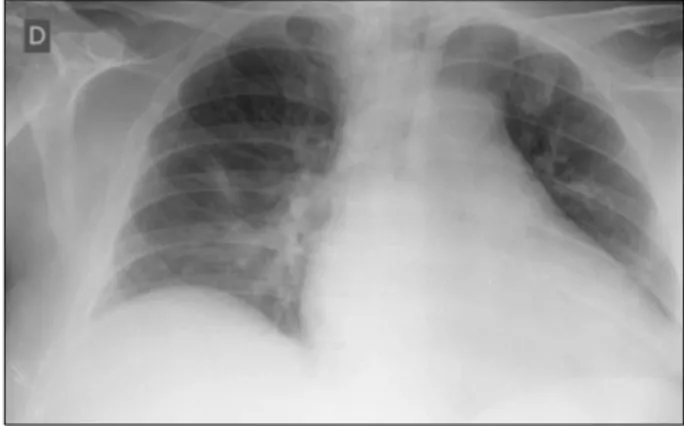
Fig. 1. Chest radiography showing diffuse bilateral infiltrates.
COVID-19 diagnosed during the early postoperative peri- od in a recipient whose LT was performed at the end of March during the lockdown in Spain.
CASE
The liver recipient was a blood group A positive 52- year-old male, with decompensated alcoholic cirrhosis and severe portal hypertension (MELD 20, encephalopathy, ascites, hepatorenal syndrome type II with an preoperative estimated glomerular filtration rate [eGFR] of 52 ml/min), atrial fibrillation and a gastric by-pass surgery due to mor- bid obesity performed 7 years ago (actual body mass in- dex was 35). He had been hospitalized until 2 days before the transplant due to recurrent liver complications and, de- spite not having fever or clinically evident infection, sur- veillance nasopharyngeal and oropharyngeal swabs for SARS-CoV-2 RT-PCT were obtained but resulted negative.
The donor was a blood group 0 positive, 47-year-old man without any previous disease who died from intra- cranial hemorrhage after a work-related accident. Following the current recommendations of the Spanish National Transplant Organization,10 the donor and next of kin were investigated for epidemiological risks or presence of clin- ical symptoms compatible with COVID-19. Even though they showed no specific risks or symptoms, a nasophar- yngeal and oropharyngeal swab SARS-CoV-2 real-time reverse-transcriptase–polymerase-chain-reaction (rRT-PCR) was performed, which was negative. The liver was procured by standard methods and appeared healthy upon retrieval.
The cold ischemia time was 283 minutes, the warm is- chemia time was 25 minutes and the total surgery time was 225 minutes. All anastomoses were done in a stand- ard way following a piggyback technique. The patient re- quired 1 unit of packed red blood cells administered intraoperatively.
The patient received Basiliximab for induction therapy (day 0 and day 4) combined with Mycophenolate Mofetil (MMF), 1 g/12 hours and glucocorticoids (Prednisone 20 mg per day), according to our protocol for patients with pretransplant renal dysfunction, with good postoperative graft function.11 On postoperative day (POD) 3, MMF was reduced to 500 mg/12 hours due to pancytopenia. At that point, pancytopenia was thought to be to be related with surgery and his previous cirrhosis. The patient showed an
uneventful initial course, so he was discharged from the ICU on the following day, without any infectious or respi- ratory symptoms. On POD 5 he presented with fever (38°C), dyspnea, hypoxia (89% on room air), and tachyp- nea without respiratory distress. Supplemental oxygen via nasal cannula (2 liters/minute), antipyretic (Paracetamol 1 g) and intravenous broad-spectrum antibiotic therapy (Mero- penem 1 g/8 hours and Linezolid 600 mg/12 hours) were started. Blood cultures were negative, and chest radiog- raphy showed diffuse bilateral infiltrates (Fig. 1). White blood cell count on peripheral blood was 4.9×10³/ml with lymphopenia (0.4×10³/ml) and with a platelet count of 48×103/ml, with a high ferritin (783 ng/ml) and high D-dimers (16.200 ng/ml) levels. The lactate dehydrogen- ase was normal (243 UI/L). Kidney (eGFR>90 ml/min) and liver graft function were normal. Considering the pa- tient’s immunosuppression and symptoms, and the pan- demic state at the end of March, (at that time 335 patients with confirmed COVID-19 infection were hospitalized at Cruces University Hospital),12 a committee of transplant surgeons, hepatologist and infectious diseases specialists, decided to develop a secure pathway for all the contacts, and to repeat a nasopharyngeal/oropharyngeal swab SARS- CoV-2 rRT-PCR to the patient, which was again negative.
Two days after the chest radiography, we performed a CT on POD 7, but it did not show any characteristic images.
In spite of the patient’s clinical improvement, and due to the high clinical suspicion, the secure pathway was maintained and a new nasopharyngeal/oropharyngeal swab for SARS-CoV-2 rRT-PCR was performed 48 hours later, that turned out positive. We contacted the donor center in order to retrieve new samples from the donor to per-
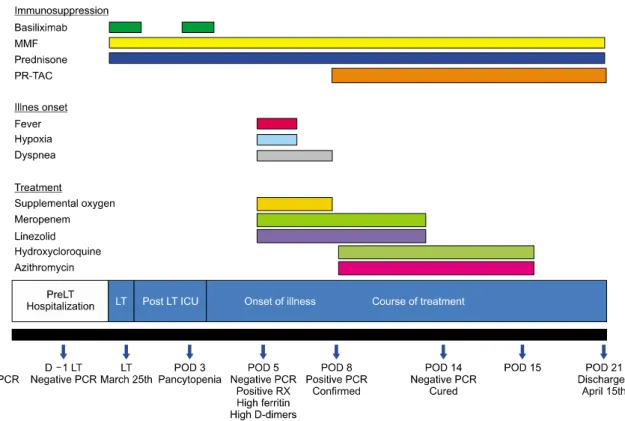
Fig. 2. Case report evolution. PR-TAC, prolonged released tacrolimus; D, day; LT, liver transplantation; POD, postoperative day.
form a SARS-CoV-2 serology, which were confirmed nega- tive. Previous to start the treatment with oral Hydroxyclor- oquine and Azithromycin our patient was evaluated by the cardiologist, without contraindication. That day we de- cided to start treatment with oral Hydroxycloroquine (400 mg/12 hours for the first 24 hours and then 200 mg/12 hours) and Azithromycin (500 mg/24 hours the first 24 hours and then 250 mg/24 hours).13 Graft showed pro- gressive cholestatic dysfunction (gGT 484 U/L, ALP 634 U/L and bilirubin 7.2 mg/dl) which was considered related to under immunosuppression so 0.05 mg/kg/day prolonged release Tacrolimus was introduced, in order to keep im- munosuppression close to the lower range of our standard target levels for the initial posttransplant period (between 4-6 ng/ml).11 The patient was moved to a COVID-19 spe- cific ward. Meropenem and Linezolid were discontinued after 8 days. Patient’s general condition continued to improve. Nasopharyngeal/oropharyngeal swab for SARS- CoV-2 rRT-PCR performed on POD 14 turned out nega- tive. Lymphopenia (2×10³/ml), platelet count (252×103/ml), ferritin (469 ng/ml) and D-Dimers (4.240 ng/ml) levels progressively normalized. Graft function quickly returned to normal after several adjustments of triple immuno-
suppressive therapy. Hydroxycloroquine and Azithromycin were prescribed for 8 days and discontinued after the neg- ative rRT-PCR. At the time the patient was diagnosed and treated only one negative SARS-CoV-2 rRT-PCR after treatment was the policy of the Spanish health ministry.
The patient was discharged on POD 21 without further complications (Fig. 2).
DISCUSSION
Severe acute respiratory syndrome coronavirus (SARS- CoV) and Middle East respiratory syndrome coronavirus (MERS-CoV) were reported in transplant recipients dur- ing prior outbreaks of these viruses.14 Although it is not yet known if immunosuppression is a risk factor for more severe disease, the transplant community eagerly awaits more studies in this patient population.6 The first two re- ported cases of solid organ transplantation (SOT) with COVID-19 were two heart transplant recipients from the city of Wuhan (Hubei, China).15 To date, few clinical cas- es of LT with COVID-198,9,16,17 and three series from Italy and USA with small number7,18,19 have been published and only one was an early infection just after LT.8 Recently,
the first COVID-19 related death after LT has been reported.17 In our case, even though it was an early post-LT COVID- 19 in a frail patient, a favorable outcome was observed.
As SARS-CoV-2 rt-PCR was negative for both donor and recipient, we decided to initiate intravenous broad-spec- trum antibiotic therapy. The MMF dose had already been reduced due to pancytopenia. Due to the pandemic state at our country and center, and the high suspicion derived from the clinical status compatible with a stage IIa (moderate) pulmonary COVID-19 involvement,20 a new nasopharyngeal/oropharyngeal swab SARS-CoV-2 rRT-PCR was performed 48 hours later and COVID-19 was confirmed. Hydroxycloroquine and Azithromycin were im- mediately added while previous antibiotic therapy was progressively withdrawn.
Our patient showed symptoms on POD 5, in accord- ance with the findings by Lei et al.8 who suggest that pa- tients may develop COVID-19 symptoms after general surgery procedures within a shorter period of time (between 2 and 6 days) than the conventional COVID- 19 patients. The secure pathway was maintained during all the postoperative period. The was no clinical evidence of SARS-CoV-2 infection in the transplantion group which includes surgeons, anesthetists, hepatologists, nurses and all manpower in direct contact with the donor or the re- cipient’s family. Rt-PCR for SARS-CoV-2 has high sensi- tivity and specificity but these tests can show false negatives. Usually these are due to the sample being in- sufficient or unrepresentative, taken too early or too late in the course of the disease, or degraded during transport or handling. In negative cases in which suspicion or symptoms persist, it is recommended to repeat the Rt-PCR within a few days.20 The theory supporting a shorter in- cubation period may be difficult to apply here, because it is unknown whether the patient was in incubation peri- od upon admission or if this was acquired during his hos- pital stay and showed two false negative Rt-PCR tests. It seems more clear that it is not a donor to recipient trans- mission of COVID-19 as the donor and next of kin were investigated for epidemiological risks or presence of clin- ical symptoms compatible without suspicion, the Rt-PCR and the serology were also negative.
It is known that in these initial stages, symptoms can be mild and non-specific; however, chest imaging may re- veal bilateral infiltrates or ground glass opacities as, in
our case.21 Currently, there is no strong evidence from controlled clinical trials to recommend a specific treat- ment for the SARS-CoV-2 coronavirus or the manage- ment of immunosuppression in LT in patients with con- firmed COVID-19. The Spanish Society of Liver Trans- plantation guidelines recommend a reduction in MMF dose and maintenance of calcineurin inhibitor levels to the desired range in recipients with Stage I or IIa COVID-19.13 It is known that immunosuppressed patients have higher risk of developing infections; however, it is also possible that immunosuppressants could downregulate the delete- rious inflammatory cascade characteristic of Stage III of this viral infection.6 Therefore, it may be possible that maintaining immunosuppressive treatment could help overcome the immunoreactive phase of the disease. In our case, we followed our standard immunosuppression policy for recipients with renal dysfunction: induction therapy, prednisone, MMF and delayed/reduced once-daily Tacro- limus.11 MMF was already reduced when COVID-19 was suspected, so we initially maintained Tacrolimus levels in the lower range; later Tacrolimus was progressively in- creased as needed, without clear impact on COVID19 evolution. Our local policy is to consider liver biopsy only when cholestatic dysfunction continues, after achieving ta- crolimus levels within therapeutic range and other poten- tial causes have been rule out. Regarding specific thera- pies for COVID-19 we followed the recommendations of the Spanish Society of Liver Transplantation and decided not to use Lopinavir/Ritonavir (very weak evidence of ef- ficacy and significant drug interactions) but added oral Hydroxycloroquine and Azithromycin, which the risk/ben- efit ratio was considered acceptable despite the absence of clear evidence.13 Clinical trials, mostly for Stage III COVID-19 infection, are currently evaluating potential therapies including Remdesivir, which has been pre- viously administered to Ebola virus patients, and others such as Tocilizumab.22
In Spain, universal screening (through nasopharyngeal and oropharyngeal rRT-PCR) is now mandatory for all donors across the country. In the Basque Country, with 193.04 COVID-19 cases per million population,2 we should balance the risks of postponing a livesaving trans- plant with the rationing of healthcare resources and the high risk of postoperative infection even when secure pathways are organized. In this pandemic scenario, a
phased approach to decrease transplant activity has been recommended.23 In Spain a dramatic decrease in organ donation has been observed: from a mean of 15-18.6 do- nors per day in the first months of 2020 to a mean of 0.3-1.4 donors per day during the lockdown period.10
In conclusion, although liver recipients could be consid- ered as potentially high-risk patients, favorable outcomes could be achieved even in case of postoperative COVID- 19 infection, provided that a quick diagnosis is made.
The fact that immunosuppressive state could be pro- tective in severe COVID-19, as proposed by some au- thors, needs to be proved.
ORCID
Mikel Prieto: https://orcid.org/0000-0001-6662-4252 Mikel Gastaca: https://orcid.org/0000-0003-2771-9640 Patricia Ruiz: https://orcid.org/0000-0002-2598-0370 Alberto Ventoso: https://orcid.org/0000-0003-4635-8545 Ibone Palomares: https://orcid.org/0000-0002-0002-7436 Regino José Rodríguez-Álvarez:
https://orcid.org/0000-0001-8779-412X
Patricia Salvador: https://orcid.org/0000-0001-7741-9465 Javier Bustamante: https://orcid.org/0000-0002-5280-3038 Andrés Valdivieso: https://orcid.org/0000-0002-2614-3670
REFERENCES
1. World Health Organization. Coronavirus disease (COVID-19) outbreak [Internet]. Geneva: World Health Organization; 2020 [cited 2020 May 3]. Available from: https://www.who.int/emergencies/
diseases/novel-coronavirus-2019.
2. Spanish Ministry of Health. COVID-19 disease [Internet]. Paseo del Prado: Ministerio de Sanidad; 2020 [cited 2020 May 3]. Avail- able from: https://www.mscbs.gob.es/en/profesionales/saludPublica/
ccayes/alertasActual/nCov-China/documentos/Actualizacion_
67_COVID-19.pdf.
3. Wu Z, McGoogan JM. Characteristics of and important lessons from the coronavirus disease 2019 (COVID-19) outbreak in China:
summary of a report of 72 314 cases from the Chinese Center for Disease Control and Prevention. JAMA 2020;323:1239-1242.
4. Shelhamer JH, Toews GB, Masur H, Suffredini AF, Pizzo PA, Walsh TJ, et al. NIH conference. Respiratory disease in the im- munosuppressed patient. Ann Intern Med 1992;117:415-431.
5. Michaels MG, La Hoz RM, Danziger-Isakov L, Blumberg EA, Kumar D, Green M, et al. Coronavirus disease 2019: implications of emerging infections for transplantation. Am J Transplant 2020;20:1768-1772.
6. D'Antiga L. Coronaviruses and immunosuppressed patients: the facts during the third epidemic. Liver Transpl 2020;26:832-834.
7. Pereira MR, Mohan S, Cohen DJ, Husain SA, Dube GK, Ratner
LE, et al. COVID-19 in solid organ transplant recipients: Initial report from the US epicenter. Am J Transplant 2020;20:1800- 1808.
8. Lei S, Jiang F, Su W, Chen C, Chen J, Mei W, et al. Clinical characteristics and outcomes of patients undergoing surgeries during the incubation period of COVID-19 infection. Version 2. EClinical- Medicine 2020. doi: 10.1016/j.eclinm.2020.100331. [in press]
9. Qin J, Wang H, Qin X, Zhang P, Zhu L, Cai J, et al. Perioper- ative presentation of COVID-19 disease in a liver transplant recipient. Hepatology 2020. doi: 10.1002/hep.31257. [in press]
10. National Transplant Organization. Spanish recommendations to manage organ donation and transplantation regarding the infec- tion associated with the new coronavirus (SARS-CoV-2) produ- cer of COVID-19 [Internet]. Paseo del Prado: Ministerio de Sani- dad; 2020 [cited 2020 Apr 16]. Available from: http://www.ont.
es/infesp/RecomendacionesParaProfesionales/Spanish%20Recom mendations%20on%20Organ%20Donation%20and%20Transplan tation%20COVID-19%20%20ONT.pdf.
11. Gastaca M, Prieto M, Palomares I, Bustamante J, Fernandez JR, Ruiz P, et al. Long-term outcomes of liver transplantation in pa- tients with pretransplant renal dysfunction treated with induction therapy and delayed reduced de novo once-daily tacrolimus.
Transplant Proc 2020;52:1489-1492.
12. Osakidetza. Basque Health Department [Internet]. San Juan: De- partamento de Salud; 2020 [cited 2020 Mar 30]. Available from:
http://www.euskadi.eus/contenidos/informacion/boletin_corona virus/es_def/adjuntos/30_marzo_Boletin.pdf.
13. Spanish Society of Liver Transplant. Recommendations of the SSLT against COVID-19 in liver transplantation [Internet]. [cited 2020 Apr 13]. Available from: https://www.sethepatico.org/docs/2020/
Reco mend_IMS_COVID_TH.pdf.
14. AlGhamdi M, Mushtaq F, Awn N, Shalhoub S. MERS CoV in- fection in two renal transplant recipients: case report. Am J Transplant 2015;15:1101-1104.
15. Li F, Cai J, Dong N. First cases of COVID-19 in heart trans- plantation from China. J Heart Lung Transplant 2020;39:496-497.
16. Liu B, Wang Y, Zhao Y, Shi H, Zeng F, Chen Z. Successful treatment of severe COVID-19 pneumonia in a liver transplant recipient. Am J Transplant 2020;20:1891-1895.
17. Huang JF, Zheng KI, George J, Gao HN, Wei RN, Yan HD, et al. Fatal outcome in a liver transplant recipient with COVID-19.
Am J Transplant 2020;20:1907-1910.
18. Bhoori S, Rossi RE, Citterio D, Mazzaferro V. COVID-19 in long-term liver transplant patients: preliminary experience from an Italian transplant centre in Lombardy. Lancet Gastroenterol Hepatol 2020;5:532-533.
19. Webb GJ, Moon AM, Barnes E, Barritt AS, Marjot T.
Determining risk factors for mortality in liver transplant patients with COVID-19. Lancet Gastroenterol Hepatol 2020;5:643-644.
20. Fang Y, Zhang H, Xie J, Lin M, Ying L, Pang P, et al.
Sensitivity of chest CT for COVID-19: comparison to RT-PCR.
Radiology 2020;296:E115-E117.
21. Siddiqi HK, Mehra MR. COVID-19 illness in native and im- munosuppressed states: a clinical-therapeutic staging proposal. J Heart Lung Transplant 2020;39:405-407.
22. Costanzo M, De Giglio MAR, Roviello GN. SARS-CoV-2: re- cent reports on antiviral therapies based on lopinavir/ritonavir, darunavir/umifenovir, hydroxychloroquine, remdesivir, Favipiravir and other drugs for the treatment of the new coronavirus. Curr Med Chem 2020;27:4536-4541.
23. Kumar D, Manuel O, Natori Y, Egawa H, Grossi P, Han SH, et al. COVID-19: a global transplant perspective on successfully navigating a pandemic. Am J Transplant 2020;20:1773-1779.