359
http://dx.doi.org/10.4046/trd.2011.71.5.359 ISSN: 1738-3536(Print)/2005-6184(Online) Tuberc Respir Dis 2011;71:359-362
CopyrightⒸ2011. The Korean Academy of Tuberculosis and Respiratory Diseases. All rights reserved.
Streptococcus parasanguinis
에 의한 폐렴과 부폐렴성 흉수 1예충남대학교 의학전문대학원 내과학교실
박명린, 박동일, 유수진, 정선영, 은혁수, 김민정, 박지원, 박희선, 정성수, 김주옥, 김선영, 이정은
A Case of Pneumonia and Parapneumonic Effusion Caused by Streptococcus parasanguinis
Myoung Rin Park, M.D., Dong Il Park, M.D., Su Jin Yoo, M.D., Sun Young Jung, M.D., Hyuk Soo Eun, M.D., Min Jung Kim, M.D., Ji Won Park, M.D., Hee Sun Park, M.D., Sung Soo Jung, M.D., Ju Ock Kim, M.D., Sun Young Kim, M.D., Jeong Eun Lee, M.D.
Department of Internal medicine, Chungnam National University School of Medicine, Daejeon, Korea
Streptococcus parasanguinis is a Viridans group bacteria that is most often discovered in the oral cavity and causes dental plaque and endocarditis in a rat model. It has low virulence but an unknown relationship to human respiratory infections. We report on a 61-year-old woman who developed hemoptysis followed by pleuritic chest pain after conscious sedation during a gastroscopic polypectomy and was diagnosed with pneumonia and parapneumonic effusion from Streptococcus parasanguinis isolated in pleural fluid. Microaspiration during the procedure was presumed to play a role in the pathogenesis.
Key Words: Streptococcal Infections; Pneumonia; Pleural Effusion
Address for correspondence: Jeong Eun Lee, M.D.
Department of Internal Medicine, Chungnam National University College of Medicine, 640, Daesa-dong, Jung-gu, Daejeon 301-721, Korea
Phone: 82-42-280-8035, Fax: 82-42-257-5753 E-mail: [email protected]
Received: Jun. 2, 2011 Accepted: Jun. 30, 2011
서 론
Viridans group streptococci는 사람의 구강 내에 상재 하는 세균으로 Streptococcus mutans군, Streptococcus salivarius군, Streptococcus mitis군, Streptococcus san- guinis군, Streptococcus anginosus군 이렇게 크게 다섯 가지로 분류된다1. 위장관계 및 요로계에서도 발견되며1, 인간에서는 병원체로서 심내막염, 항암화학치료 후 호중 구 감소상태에서의 균혈증 및 뇌농양 등을 일으킬 수 있 다2-4. Streptococcus sanguinis 군에 속하는 Streptococcus parasanguinis는 다른 Viridans group streptococci와 마찬 가지로 치석을 형성하고5, in vitro에서 심내막염을 일으킬
수 있다는 보고만 있을 뿐 인간에서 병원체로서의 역할은 아직 알려진 바가 없다6. 저자들은 특별한 기저질환이 없 는 61세 여자환자가 객혈을 주소로 내원하여 흉수에서 균 이 동정되어 Streptococcus parasanguinis에 의한 폐렴과 부폐렴성 흉수로 진단된 증례를 경험하였으므로 보고하 고자 한다.
증 례
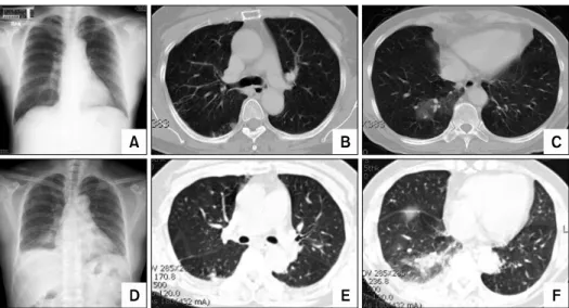
61세 여자가 4일 전부터 발생한 객혈과 3일 전부터 열 감, 오한, 전신근육통과 함께 우측의 흉막성 흉통이 발생 하여 개인의원에 방문하여 흉부 단순촬영 및 흉부 전산화 단층촬영 후 외래로 내원하였다(Figure 1A∼C). 평소 특 이 질환을 진단받은 적은 없었으나 내원 6개월 전 민물게 장을 먹었고, 10일 전 개인의원에서 의식하 진정내시경으 로 위 용종절제술을 시행 받은 과거력이 있었다. 활력징 후는 혈압 130/90 mm Hg 심박수는 분당 88회, 호흡수는 분당 20회였고, 38.3oC의 발열이 있었다. 의식은 명료하 였고, 급성 병색을 띄고 있었으며 흉부청진에서 심잡음은
Case Report
MR Park et al: Pneumonia and parapneumonic effusion caused by Streptococcus parasanguinis
360
Figure 1. Chest PA view and chest CT. (A) Chest PA at a local clinic shows no active lung lesion. (B, C) Chest CT at a local clinic shows ill defined nodules in the posterior segment of the right upper lobe with patchy ground glass opacity in the posterobasal segment of the right lower lobe. (D) Chest PA on admission. Focal consolidation is seen in the right lower lung field. (E, F) Chest CT on admission shows increased extent of consolidation with ground glass opacity in the right lower lobe and a small volume of pleural effusion in a right-sided hemithorax. PA: posterior anterior;
CT: computed tomography.
없었으나 양측 하폐야에 수포음을 동반한 거친 호흡음이 있었다.
흉부 단순촬영에서 우측 하폐야에 초점성의 폐경화 소 견이 관찰되었다(Figure 1D). 말초혈액 검사에서 백혈구 10,620/μL (호중구 81.6%, 림프구 10.9%, 단핵구 7.1%, 호산구 0.3%, 호염구 0.1%), 혈색소 13 g/dL, 혈소판 193,000/μL이었고, C반응단백은 9.41 mg/dL로 증가되어 있었다. 생화학 검사에서 aspartate aminotransferasae 35 IU/L, alanine aminotransferase 36 IU/L, 총빌리루빈 1.16 mg/dL, lactate dehydrogenase 438 IU/L, 총단백 7.7 g/dL, 알부민 4.3 g/dL, BUN 9 mg/dL, 크레아티닌 0.5 mg/dL이었다. 동맥혈 가스 검사는 room air에서 pH 7.47, 이산화탄소 분압 35 mm Hg, 산소 분압 86 mm Hg, 중탄산염 25.5 mmol/L, 산소포화도 97%였다. 흉부 전산 화 단층촬영에서 우하엽에 이전보다 확장되고 음영이 증 가한 폐경화 소견과 함께 소량의 흉수가 있었다(Figure 1E, F). 폐결핵이나 폐경색에 동반된 폐렴 또는 기생충감 염이나 혈관염에 의한 폐포출혈로 의심되어 폐기관지 내 시경과 객담 항산성 염색 검사, 자가면역질환에 대한 혈청 검사를 시행하였다. 폐기관지 내시경에서 우하엽의 후기 저분절에 출혈이 지속되어 기관지 폐포세척 검사를 시행 하였고 적혈구가 다수, 대식세포 88%, 호중구 10%, 림프 구 2%로 관찰되었다. 혈액 및 객담 배양 검사에서 동정된
세균은 없었으며, 객담의 그람염색 및 항산성 염색도 음성 소견이었다. FANA는 speckled pattern (1:40), C-ANCA 94 AAU, P-ANCA 92 AAU, Anti GBM Ab <5 units, Anti dsDNA antibody 1.95 IU/mL, SCL 70 antibody 및 Anti mitochondrial antibody는 음성 소견이었다. 입원 시 경험 적으로 Ceftriaxone과 Azithromycin을 사용하였고 발열은 입원 2일째 소실되었다. 입원 4일째 더 이상의 객혈은 없 었으나 우측 흉막성 흉통을 여전히 호소하는 상태로 흉부 단순촬영에서 우하엽의 폐경화는 호전되었고, 우측와위 흉부사진에서 흉수가 지속되어 무균기술로 진단적 흉수 천자를 실시하였다. 흉수는 맑은 담황색이었다. 흉수에 대한 생화학 검사에서 pH 7.6, 적혈구 50,000, 백혈구 6,600, 다형 백혈구 80%, 단핵구 20%, 총단백 4.8 g/dL, 알부민 3.0 g/dL, lactate dehydrogenase 675 IU/L, ad- enosine deaminase 21 IU/L였으며 기생충 검사에서 Cysti- cercus, Paragonimus, Sparganum, Clonorchis 모두 음성 이었다. 이때 시행한 흉수 세포면역 검사에서 결핵균 중 합효소 연쇄반응 검사는 음성이었다. 흉수 그람염색 검사 에서는 그람양성 구균이 검출되었다. 말초혈액 검사에서 백혈구 4,640/μL (호중구 56%, 림프구 24.8%, 단핵구 6.3%, 호산구 12.3.0%, 호염구 0.6%), 혈색소 12.3 g/dL, 혈소판 273,000/μL이었고 입원 9일째 환자의 호흡기적 증상은 모두 호전되었다. 말초혈액 검사에서 백혈구
Tuberculosis and Respiratory Diseases Vol. 71. No. 5, Nov. 2011
361 Figure 2. Chest PA view and chest CT. (A) Chest PA on day 9 of admission. Improved consolidation and an increased volume of pleural effusion is shown in the right lung. (B∼D) Chest CT on day 9 of admission. Near complete clearing of the previous consolidation in the posterobasal segment of the right lower lung and increased volume of pleural effusion, which was considered a hemothorax, is shown. (E) Chest PA about 3 months after discharge. Decreased volume of pleural effusion is shown compared with that of day 9 from admission. (F∼H) Chest CT about 3 months after discharge shows neither pleural effusion nor consolidation. PA: posterior anterior; CT: computed tomography.
6,080/μL (호중구 58.6%, 림프구 23.5%, 단핵구 5.9%, 호산구 11.0%, 호염구 1.0%), 혈색소 12.7 g/dL, 혈소판 256,000/μL이었고 이때 시행한 흉부 단순촬영 및 흉부 전산화 단층촬영에서 폐경화는 호전되었으나 우측에 혈 흉으로 의심되는 병변이 새로이 발견되어(Figure 2A∼D) Paragonimus westermanii에 의한 감염으로 생각하고 입 원 10일째 Praziquantel을 사용하고 퇴원하였다.
퇴원 1주일 후 외래에서 환자는 불편감을 호소하지 않 았고 이때 입원 4일째에 시행했던 흉수 배양 검사에서 Streptococcus parasanguinis가 동정된 사실을 확인하였 다. 퇴원 3개월 후 시행한 흉부 단순촬영 및 흉부 전산화 단층촬영에서 우하엽 상분절과 좌하엽 후분절의 흉막하 결절양 병변은 이전과 비교하여 크기 변화가 없었고, 우측 흉수는 소실되었다(Figure 2E∼H).
고 찰
지역사회획득 폐렴은 다양한 균주가 원인이 되는 질환 으로 최근 항생제치료법의 발전에도 불구하고 초기치료 에 반응하지 않을 확률은 15% 정도이다7. 한 보고에 따르 면 지역사회획득 폐렴에 의한 사망률은 외래 또는 일반 병동에서 치료 받았을 때 5.1%, 중환자실에서는 36.5%까
지 이른다고 한다8. 폐렴환자의 20∼60%에서 흉수가 동 반되며 대부분은 항생제로 치료가 되지만 5∼10%에서는 농흉으로 진행하는 등 심한 임상경과를 밟는다. 부폐렴성 흉수의 사망률은 15%에 달하고, 평균 재원일수는 15일이 며, 20% 이상은 한 달 이상 입원한다9. 부폐렴성 흉수를 일으키는 병원균에 대해서는 아직 크게 알려진 바가 없으 나, Tsang 등10의 보고에 따르면 부폐렴성 흉수와 농흉 환 자 중 68%에서 세균이 동정되었으며, 그 중 Viridans group streptococci는 20%를 차지했다.
종의 종류가 다양하고, 중복되는 부분이 많기 때문에 전통적으로 사용되는 생화학적 방법을 통해서는 Viridans group streptococci 균주를 각각 확인하기 어렵지만 미지 세균의 분류체계에 이용되는 16S rRNA 염기서열분석법을 통해 정확한 균주를 확인을 하는데 도움이 될 수 있다4. 본 증례에서는 16S rRNA 염기서열분석법을 이용하지 않 고 균 배양 제 7일째 VITEKⓡ2 (BIOMERIEUX, Inc, Marcy l'Étoile, France)를 통한 생화학적 방법으로 균이 동정되 었다.
구강 내 상재균으로 알려진 Viridans group strepto- cocci는 2002년도 Facklam에의해 다섯 가지 군으로 개정 되었으며1, 각각의 세균은 존재하는 위치 및 나타나는 임 상 양상이 다르다. 이 중 Streptococcus sanguinis군에 속
MR Park et al: Pneumonia and parapneumonic effusion caused by Streptococcus parasanguinis
362
하는 Streptococcus parasanguinis는 치석의 형성에 중요 한 역할을 하는데 치아표면에 미리 군집을 형성하여 다른 세균들이 추가적으로 군집을 형성할 수 있게 한다5. 같은 군에 속하는 Streptococcus sanguinis가 사람에서 심내막 염을 일으키고11, 폐농양을 발생한 증례가 보고되어 있으 나12, Streptococcus parasanguinis가 병원체로서 역할을 한 경우는 Rat model에서 심내막염을 발생시켰다는 연구 결과 및 sheep에서 무증상 유방염을 일으킨 증례밖에 없 었다5,13.
일반적으로 객혈의 60∼70% 정도가 감염에 의해 발생 하는 것으로 알려져 있고 미국에서 실시한 후향적 연구에 서는 전체 객혈 중 기관지염이 26%, 폐렴이 10%, 결핵이 8%를 차지한다고 보고되었다14. Staphylococcus aureus, Pseudomonas aeruginosa와 같은 침습성의 세균이나 아 스페르길루스 등의 진균에 의한 감염이 객혈을 유발할 수 있다고 알려져 있다. 본 증례에서는 흉부 전산화 단층촬 영에서 현성 감염으로 판단되는 병변과 기관지경에서 확 인된 출혈점이 일치하여 폐렴이 객혈의 원인일 것으로 추 정된다.
흡인성 폐렴을 일으키는 원인 균으로는 구강상재균 및 그람음성 간균 등이 알려져 있고, 구강상재균은 혐기성 균이 대부분을 차지하지만 실제로 흡인성 폐렴의 원인균 으로서 구강상재균 중의 혐기성 균은 과대평가된 면이 있 다15. 최근 들어 암 조기검진 사업 등으로 의식하 진정 내 시경이 활발히 실시되는 추세다. 본 증례처럼 의식하 진 정 내시경과 같이 흡인이 쉽게 일어날 수 있는 사건 후에 발생하는 비 전형적인 폐렴과 부폐렴성 흉수에 대해서 발 병력이 낮지만 구강 내 호기성 상재균인 Streptococcus parasanguinis가 감염을 일으킬 수 있음을 보고하는 바이 다.
참 고 문 헌
1. Facklam R. What happened to the streptococci: over- view of taxonomic and nomenclature changes. Clin Microbiol Rev 2002;15:613-30.
2. Wilson WR, Karchmer AW, Dajani AS, Taubert KA, Bayer A, Kaye D, et al. Antibiotic treatment of adults with infective endocarditis due to streptococci, enter- ococci, staphylococci, and HACEK microorganisms.
American Heart Association. JAMA 1995;274:1706-13.
3. Bochud PY, Calandra T, Francioli P. Bacteremia due
to viridans streptococci in neutropenic patients: a review. Am J Med 1994;97:256-64.
4. Han XY, Kamana M, Rolston KV. Viridans streptococci isolated by culture from blood of cancer patients: clin- ical and microbiologic analysis of 50 cases. J Clin Microbiol 2006;44:160-5.
5. Jenkinson HF, Lamont RJ. Streptococcal adhesion and colonization. Crit Rev Oral Biol Med 1997;8:175-200.
6. Burnette-Curley D, Wells V, Viscount H, Munro CL, Fenno JC, Fives-Taylor P, et al. FimA, a major virulence factor associated with Streptococcus parasanguis endo- carditis. Infect Immun 1995;63:4669-74.
7. Mandell LA, Wunderink RG, Anzueto A, Bartlett JG, Campbell GD, Dean NC, et al. Infectious Diseases Society of America/American Thoracic Society con- sensus guidelines on the management of commun- ity-acquired pneumonia in adults. Clin Infect Dis 2007;
44 Suppl 2:S27-72.
8. File TM. Community-acquired pneumonia. Lancet 2003;
362:1991-2001.
9. Davies CW, Kearney SE, Gleeson FV, Davies RJ.
Predictors of outcome and long-term survival in pa- tients with pleural infection. Am J Respir Crit Care Med 1999;160:1682-7.
10. Tsang KY, Leung WS, Chan VL, Lin AW, Chu CM.
Complicated parapneumonic effusion and empyema thoracis: microbiology and predictors of adverse outcomes. Hong Kong Med J 2007;13:178-86.
11. Paik S, Senty L, Das S, Noe JC, Munro CL, Kitten T.
Identification of virulence determinants for endocarditis in Streptococcus sanguinis by signature-tagged muta- genesis. Infect Immun 2005;73:6064-74.
12. Komatsu K, Kanda T. Atypical lung abscess occurring in an elderly female suffering from diabetes mellitus--a case report. Kansenshogaku Zasshi 1997;71:260-3.
13. Fernández-Garayzábal JF, Fernández E, Las Heras A, Pascual C, Collins MD, Domínguez L. Streptococcus parasanguinis: new pathogen associated with asympto- matic mastitis in sheep. Emerg Infect Dis 1998;4:645-7.
14. Reisz G, Stevens D, Boutwell C, Nair V. The causes of hemoptysis revisited. A review of the etiologies of he- moptysis between 1986 and 1995. Mo Med 1997;94:
633-5.
15. El-Solh AA, Pietrantoni C, Bhat A, Aquilina AT, Okada M, Grover V, et al. Microbiology of severe aspiration pneumonia in institutionalized elderly. Am J Respir Crit Care Med 2003;167:1650-4.