Original Article
Anterior Segment Parameters Using Pentacam and Prediction of Corneal Endothelial Cell Loss after Cataract Surgery
Yang Kyeung Cho1, Hwa Seok Chang2, Tae Yoon La1, Donghyun Ji1, Hyunkyung Kim1, Jin A Choi1, Man Soo Kim2
1Department of Ophthalmology, St. Vincent’s Hospital, The Catholic University of Korea School of Medicine, Suwon, Korea
2Department of Ophthalmology, Seoul St. Mary’s Hospital, The Catholic University of Korea School of Medicine, Seoul, Korea
Purpose: We evaluated various preoperative anterior segment parameters measured with a Pentacam rotating Scheimpflug camera and compared them with those of conventional methods. We also evaluated the effect of dif- ferent parameters on corneal endothelial cells after cataract surgery.
Methods: Pentacam examination was performed in 88 eyes from 88 patients to evaluate central anterior chamber depth (ACDpentacam), nuclear density (Densitometrypentacam), anterior chamber volume (ACV), and lens thickness (LTpentacam). We compared values of ACDpentacam with those of ultrasound (ACDsono) and also compared Densitometrypentacam values with those of Lens Opacities Classification System (LOCS III) classification. We evaluated the effect of the following preoperative values measured with Pentacam on postoperative endothelial cell loss: pupil size measured both preoperatively and before capsulorrhexsis (PupilCCC), amount of viscoelastics, and LT measured by ultrasound (LTsono).
Results: A significant concordance was found between the two grading methods of nuclear opacity:
Densitometrypentacam andLOCS III classification (τb= 0.414, p = 0.000). We also found a positive correlation between ACDpentacam and ACDsono (r = 0.823, p = 0.000) and between ACDpentacam and ACV (r = 0.650, p =
0.000). There were significant differences between the results of LTpentacam and LTsono. The final regression model identified Densitometrypentacam,viscoelastics and PupilCCC as independent predictors of decreased postoperative corneal endothelial cell density (CD) at postoperative day 3, and Densitometrypentacam, viscoe- lastics, and ACV as independent predictors of decreased CD two months postoperatively (p<0.05).
Conclusions: Good agreement was found between all results obtained with the Pentacam and conventional methods except LT. Analyzing anterior chamber parameters preoperatively using Pentacam could be helpful to predict postoperative endothelial cell loss.
Key Words: Anterior segment parameters, Corneal endothelium, Pentacam
ⓒ2010 The Korean Ophthalmological Society
This is an Open Access article distributed under the terms of the Creative Commons Attribution Non-Commercial License (http://creativecommons.org/licenses /by-nc/3.0/) which permits unrestricted non-commercial use, distribution, and reproduction in any medium, provided the original work is properly cited.
Received: June 19, 2009 Accepted: May 16, 2010
Reprint requests to Man Soo Kim. Department of Ophthalmology, Seoul St. Mary’s Hospital, The Catholic University of Korea School of Medicine, #505 Banpo-dong, Seocho-gu, Seoul 137-701, Korea. Tel:
82-2-2258-6197, Fax: 82-2-599-7405, E-mail: [email protected]
The Pentacam (Oculus Inc., Wetzlar, Germany) offers evaluation of the entire anterior segment from the anterior corneal surface to the posterior lens surface using a rotating Scheimpflug camera (Oculus Inc.). The noncontact measur- ing process takes two seconds and performs 12 to 50 single captures. We evaluated the accordance of various pre- operative anterior segment parameters measured by the
Pentacam with those of conventional methods, and we also used Pentacam data to predict post-cataract surgery corneal endothelial cell loss.
Materials and Methods
Eighty-eight eyes from 88 patients scheduled for routine cataract surgery were selected prospectively. Baseline pa- tient data are shown in Table 1. Exclusion criteria for the study were eyes with significant corneal opacity, previous intraocular surgery, trauma, glaucoma, uveitis, Fuchs’ en- dothelial dystrophy, other abnormalities that could cause significant endothelial cell impairment independent of sur- gery, eyes with small pupils that required iris retractors, and
Fig. 1. Measurement of pupil size a measured before capsulorrhexis (PupilCCC). PupilCCC was measured before capsulorrhexis using a blunt-ended, viscoelastics injection needle of known length.
eyes with intraoperative complications such as posterior capsule rupture or postoperative complications.
Surgical technique
All surgeries were performed under retrobulbar anes- thesia by the same surgeon. A 3.0-mm clear corneal incision was made in the superior quadrant. A capsulorrhexis ap- proximately 5.0 mm in diameter was created with forceps, and cortical cleaving hydrodissection was then performed.
The nucleus was emulsified using the stop-and-chop technique. After irrigation and aspiration of the cortex, a foldable acrylic intraocular lens (SN60WF; Alcon, Fort Worth, TX, USA) was implanted in the bag. The same irri- gating solution (balanced salt solution [BSS]) and oph- thalmic viscoelastics were used in all patients.
Intraoperatively, we recorded phacoemulsification time (seconds), phacoemulsification power (%), total operative time (minutes), and amount of BSS irrigating solution (mL) used.
Lens opacity measurement
At the preoperative slit-lamp examination, nuclear opac- ity was graded from 1 to 6 using the Lens Opacities Classification System (LOCS III) by two observers. We se- lected data that showed agreement of grading scores be- tween these two observers.
The Pentacam provides an image of nearly the entire lens along with an objective measurement of the lens density in the chosen area, which ranges from 0 to 100. To evaluate the nuclear density via Pentacam, we needed Scheimpflug im- ages and used enhanced densitometry analysis. One ob- server evaluated the densitometry of the area corresponding to the lens nucleus center two times, and we took the mean value to use as the lens density.
Lens thickness, anterior chamber depth and anterior chamber volume measurement
Applanation ultrasonography (Compact II device, Quantel Medical) was done to evaluate the lens thickness (LTsono, mm), anterior chamber depth (ACD, mm) and anterior chamber volume (ACV, mm3). ACD was defined as the ax- ial distance between the anterior surface of the cornea and the anterior surface of the lens. ACV was calculated from the integration (integral calculus) of the distances between the back surface of the cornea and the iris of the respective lens in a 12-mm diameter around the corneal apex. LT was defined as the axial distance between the anterior chamber surface of the lens and the posterior surface of the lens. All measurements were done by one experienced observer fol- lowing pupil dilation.
Pentacam was performed to evaluate central ACD (from the epithelium) and lens thickness (LTpentacam). The anterior chamber depth (ACDpentacam) was measured at the apex of
the cornea. Pupil dilation was necessary to measure LTpentacam.
Pupil size and amount of viscoelastics measurement Preoperatively, the fully dilated pupil size (Pupilpreop) was measured by slit-lamp biomicrosopy after a two-hour in- stillation of mydriatics (tropicamide 0.5%, phenylephrine hydrochloride 0.5% Mydrin®-P; Santen Pharmaceutical Co., Osaka, Japan). During surgery, the dilated pupil size was measured (PupilCCC) after injection of viscoelastics (sodium hyaluronic acid 1% Hyal 2000; LG life Sciences, Korea) into the anterior chamber using a blunt-ended in- jection needle of known length before anterior capsulotomy (Fig. 1).
For capsulorrhexis (CCC), we injected just enough vis- coelastics into the anterior chamber to observe a slight backward movement of the lens-iris diaphragm. We then measured the amount of viscoelastics used for CCC by sub- tracting the amount remaining after CCC from the initial amount.
Endothelial cell evaluation
Corneal endothelial injury associated with phacoemulsi- fication was assessed by specular microscopy in terms of the changes in cell density (CD) and cell morphology.
To evaluate central corneal endothelial cell density, spec- ular microscopy photographs of the central corneal endo- thelium were taken using a noncontact specular microscope (Nonon Robo, Konan, Japan). We analyzed a minimum of 40 endothelial cells to calculate the endothelial cell density.
Specular microscopic examination, including endothelial cell density (cells/mm2), coefficient of variation (CV) and hexagonality (HA), was performed preoperatively and again at three days and at two months postoperatively.
We calculated the percentage change of CD, CV and HA as follows: percentage change of CD = (preoperative CD- postoperative CD)×100/preoperative CD, percentage change of CV = (preoperative CV-postoperative CV)×100/preoperative
Table 1. Baseline patient data
Characteristics No. of eyes Mean ± SD Range
Age 88 67.92 ± 10.12 25–88
Axial length (mm) 88 23.24 ± 0.86 20.64–26.30
Keratometry 88 44.32 ± 1.56 39.75–48.88
Anterior chamber depth by ultrasound (mm) 88 3.06 ± 0.47 2.18–4.64
Anterior chamber depth by Pentacam (mm) 88 3.11 ± 0.43 2.16–4.00
Lens thickness by ultrasound (mm) 88 4.26 ± 0.65 2.82–5.50
Lens thickness by Pentacam (mm) 50 2.13 ± 0.62 1.21–4.23
Phacoemulsification time (sec) 88 41.73 ± 35.19 0–221.00
Phacoemulsification power (%) 88 17.87 ± 4.95 0–29.40
LOCS III classification 88 3.07 ± 0.98 1.00–5.00
Densitometry by Pentacam (%) 88 11.95 ± 4.06 7.10–34.10
Operative time (min) 88 18.95 ± 4.83 12.00–43.00
Irrigation volume (mL) 88 250.00 ± 63.26 150.00–400.00
Preoperative corneal thickness (μm) 88 559.60 ± 38.81 459.00–669.00
Used amount of viscoelastics (mL) 88 0.13 ± 0.04 0.05–0.26
Intraoperative pupil size before capsulorrhexis (mm) 88 8.30 ± 0.78 6.00–10.00
Preoperative pupil size, slit-lamp exam (mm) 88 7.37 ± 0.63 5–8.5
LOCS III = Lens Opacities Classification System.
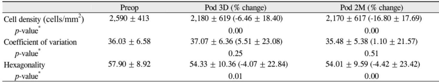
Table 2. Change of endothelial cell density, coefficient of variation and hexagonality over time
Preop Pod 3D (% change) Pod 2M (% change)
Cell density (cells/mm2) 2,590 ± 413 2,180 ± 619 (-6.46 ± 18.40) 2,170 ± 617 (-16.80 ± 17.69)
p-value* 0.00 0.00
Coefficient of variation 36.03 ± 6.58 37.07 ± 6.36 (5.51 ± 23.08) 35.48 ± 5.38 (1.10 ± 21.57)
p-value* 0.25 0.51
Hexagonality 57.90 ± 8.92 54.33 ± 10.36 (-4.07 ± 22.84) 54.01 ± 9.59 (-4.42 ± 23.42)
p-value* 0.01 0.00
Preop = preoperative measurement; Pod 3D = measurement at postoperative 3 day; Pod 2M = measurement at postoperative 2 month.
*Paired t-test, compared with preoperative value at each follow-up.
CV, and percentage change of HA = (preoperative HA-post- operative HA)×100/preoperative HA.
Statistical analysis
Statistical analysis was performed with the SPSS ver.
11.5 (SPSS Inc., Chicago, IL, USA). The associations of vari- ables were assessed by Pearson’s correlation coefficient and paired t-test. To evaluate the correlation of two methods (LOCS and Pentacam densitometry), we used Kendall’s tau-b value.
We selected seven variables (ACDpentacam, ACDsono, Pupilpreop, PupilCCC, LTsono, amount of viscoelastics, and Densitometrypentacam) that could be associated with endothe- lial cell change.
The univariate associations of the seven variables with the percentage change of CD, CV and HA were evaluated using simple regression analysis. We used a multiple linear regression model to evaluate the impact of various risk fac- tors on CD change alone. The seven variables were entered into this multiple regression analysis. A stepwise regression
was used, and a p-value of 0.50 or less was required for a variable to remain in the model.
Results
Baseline patient data are shown in Table 1. Changes of CD, CV and HA over time are shown in Table 2. Our data shows the wound-healing process of corneal endothelium after cataract surgery, and we observed CV recovery at two months postoperatively.
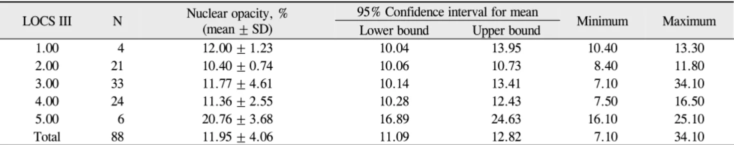
Mean Densitometrypentacam values according to grading by LOCS III classification are shown in Table 3. No eyes in our study had a nuclear opacity grade of 6 by LOCS III classification. A significant degree of concordance was found between Densitometrypentacam andLOCS III classi- fication (τb= 0.414, p = 0.000) for nuclear opacity.
Table 4 summarizes the univariate associations of the seven variables with the percentage of endothelial cell change (CD, CV and HA).
Univariate analysis demonstrated that ACV and Densito- metrypentacam were associated with the percentage decrease
Table 3. Nuclear opacity measured with Pentacam distribution according to LOCS III classification LOCS III N Nuclear opacity, %
(mean ± SD)
95% Confidence interval for mean
Minimum Maximum
Lower bound Upper bound
1.00 4 12.00 ± 1.23 10.04 13.95 10.40 13.30
2.00 21 10.40 ± 0.74 10.06 10.73 8.40 11.80
3.00 33 11.77 ± 4.61 10.14 13.41 7.10 34.10
4.00 24 11.36 ± 2.55 10.28 12.43 7.50 16.50
5.00 6 20.76 ± 3.68 16.89 24.63 16.10 25.10
Total 88 11.95 ± 4.06 11.09 12.82 7.10 34.10
LOCS III = Lens Opacities Classification System. τb(Kendall’s tau b) = 0.414, p = 0.000.
Table 4. Univariate association of our seven variables with the significance values predicting percentage change of endothelial cell density, coefficient of variation and, hexagonality at postoperative day 3 (3D) and month 2 (2M)
Follow-up Variables
Cell density Coefficient of variation Hexagonality Correlation
coefficient p-value* Correlation
coefficient p-value* Correlation
coefficient p-value*
3D Pupilpreop 0.012 0.910 -0.028 0.798 -0.133 0.218
PupilCCC -0.056 0.604 -0.016 0.879 -0.120 0.266
ACDpentacam -0.123 0.252 0.031 0.777 0.105 0.330
ACV -0.243 0.024 -0.101 0.348 0.158 0.142
LTsono -0.194 0.071 -0.069 0.523 0.022 0.841
Viscoelasticamount -0.150 0.162 0.097 0.368 -0.113 0.293
Densitometrypentacam 0.516 0.000 0.195 0.070 -0.070 0.519
2M Pupilpreop -0.085 0.431 0.028 0.795 -0.001 0.996
PupilCCC -0.079 0.465 0.011 0.919 -0.075 0.489
ACDpentacam -0.219 0.040 0.316 0.003 -0.180 0.094
ACV -0.286 0.007 0.028 0.797 0.157 0.143
LTsono -0.196 0.070 0.523 0.422 0.092 0.392
Viscoelasticsamount -0.244 0.022 0.368 0.097 -0.185 0.085
Densitometrypentacam 0.428 0.000 0.195 0.070 0.037 0.735
Pupilpreop= pupil size as measured preoperatively; PupilCCC= pupil size a measured before capsulorrhexis; ACDpentacam= anterior chamber depth measured by Pentacam; ACV = anterior chamber volume; LTsono= lens thickness measured by ultrasound; Viscoelasticamount= amount of viscoelastic; Densitometrypentacam= lens opacity measured by Pentacam densitometry.
*p<0.05, statistically significant.
of CD at postoperative day 3. ACDpentacam, ACV, amount of viscoelastics, and Densitometrypentacam were associated with the percentage decrease of CD two months postoperatively.
ACDpentacam was associated with the percentage change of CV at two months postoperatively.
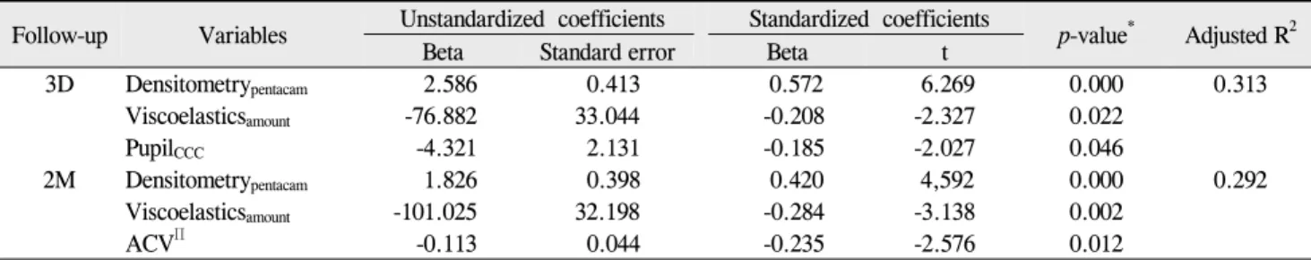
A multiple linear regression analysis was performed to identify the best set of independent predictors for the per- centage decrease of CD. These data are shown in Table 5.
The final model identified Densitometrypentacam, viscoe- lastics and PupilCCC as independent predictors of decreased CD at postoperative day 3 (adjusted R2= 31.3%) and Densitometrypentacam,viscoelastics and ACV as independent predictors of decreased CD at two months postoperatively (adjusted R2= 29.2%).
We found that intraoperative PupilCCC increased after in- jection of viscoelastics. However, we cannot compare these two values directly because PupilCCC was measured intra-
operatively at the papillary plane by an injection needle of known length, and Pupilpreop was measured using the slit-lamp biomicroscopy scale.
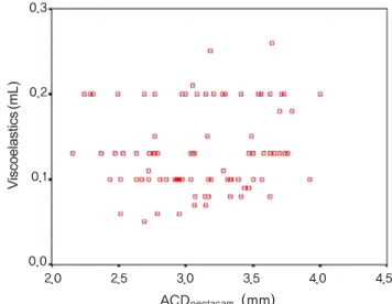
Fig. 2 shows the correlation plot between ACDpentacam and ACDsono (r = 0.823. p = 0.000). Fig. 3 shows the correlation between ACDpentacam and ACV (r = 0.650, p = 0.000). Fig. 4 shows the correlation between ACV and amount of viscoe- lastic (viscoelasticamount;r = -0.021, p = 0.85). Fig. 5 shows the correlation between ACDpentacam and viscoelasticamount
(r = 0.123, p = 0.253).
Discussion
The Pentacam allows evaluation of the entire anterior segment from the anterior corneal surface to the posterior lens surface using a rotating Scheimpflug camera [1-7]. The noncontact measuring process takes two seconds and per-
Table 5. Final stepwise, multiple regression model for predicting percentage change of endothelial cell density at postoperative day 3 (3D) and month 2 (2M)
Follow-up Variables Unstandardized coefficients Standardized coefficients
p-value* Adjusted R2
Beta Standard error Beta t
3D Densitometrypentacam 2.586 0.413 0.572 6.269 0.000 0.313
Viscoelasticsamount -76.882 33.044 -0.208 -2.327 0.022
PupilCCC -4.321 2.131 -0.185 -2.027 0.046
2M Densitometrypentacam 1.826 0.398 0.420 4,592 0.000 0.292
Viscoelasticsamount -101.025 32.198 -0.284 -3.138 0.002
ACVΠ -0.113 0.044 -0.235 -2.576 0.012
Densitometrypentacam= lens opacity measured by Pentacam densitometry; Viscoelasticsamount= amount of viscoelastics; PupilCCC= pupil size as measured before capsulorrhexis; ACVΠ= anterior chamber volume.
*p<0.05, statistically significant.
ACDsono (mm)
2.0 2.5 3.0 3.5 4.0 4.5 5.0
4.5
4.0
3.5
3.0
2.5
2.0
Fig. 2. Correlation of anterior chamber depth (mm) by Pentacam (ACDpentacam) and ultrasound (ACDsono) (r = 0.822, p<0.05).
ACDpentacam (mm)
2.0 2.5 3.0 3.5 4.0 4.5
300
200
100
0
Fig. 3. Correlation between anterior chamber depth (ACD) mea- sured by Pentacam (ACDpentacam) and anterior chamber volume (ACV) (r = 0.650, p = 0.000).
forms 12 to 50 single captures. The Pentacam has various functions, including the ability to measure ACV and nu- clear density, which can be expressed as continuous nu- meric data. Until now, we could only evaluate nuclear opac- ity by categorizing with systems such as LOCS classi- fication [7-9]. The Scheimpflug image provides the basis for objective, precise quantification of lens opacity [7]. The lens density is standardized from 0 to 100. Therefore, 0 means the lens shows no clouding, and 100 means the lens is completely opaque (Pentacam instruction manual, Oculus Inc.). Although we observed lens opacities from of LOCS III classification 1 to 5 in our study, we only obtained a rela- tively small range of Densitometrypentacam values from 7.10 to 34.10%. We might have subjectively overestimated the lens opacity using the LOCS III classification.
Since Densitometrypentacam data are not categorical data like LOCS classifications, we could more easily access stat- istical analysis using the former data type. We found a significant concordance between the LOCS III and Densitometrypentacam
methods.
Anterior chamber values are necessary for calculating in- traocular lens power when planning surgical procedures.
Precise anterior chamber measurements are essential for performing exact biometry and for surgical planning [5,6].
Several methods for measuring the anterior chamber depth are available [5,6,10]. The Pentacam and standard ul- trasound devices define ACD as the distance between the anterior surface of the cornea and the anterior surface of the lens.
Ultrasound devices have become the most commonly used methods to measure ACD. Contact devices such as the ultrasound have disadvantages, including corneal in- dentations, the risk for corneal abrasions and infections, off-axis measurement, and possible precorneal echo spikes [10]. Pentacam imaging is a relatively new, noncontact au- tomatic optical technique in ophthalmology. Although dif- ferent methods to determine ACD may yield significantly different results [3,10], mean ACD measurements of the Pentacam and ultrasound were significantly correlated in our study (r = 0.823, p = 0.000), a finding that was similar
ACV (mm3)
0 100 200 300
0.3
0.2
0.1
0.0
Fig. 4. Correlation between anterior chamber volume (ACV) and injected amount of viscoelastics before capsulorrhexis (r = -0.021, p = 0.850).
ACDpentacam (mm)
2.0 2.5 3.0 3.5 4.0 4.5
0.3
0.2
0.1
0.0
Fig. 5. Correlation between anterior chamber depth by Pentacam (ACDpentacam) and injected amount of viscoelastics before capsulor- rhexis (r=0.123, p=0.253).
to previous reports [2,6].
The Pentacam can evaluate ACV as well as ACD [4,5].
Both our study and previous reports found a good correla- tion between ACD and ACV (r = 0.650) [1,5].
We tried to evaluate LT using the Pentacam (LTpentacam) and to compare these values with those obtained by ultra- sound (LTsono) in 50 eyes. There was a significant difference between LT measured by Pentacam (mean ± SC, 2.13 ± 0.62) and by ultrasound (mean ± SD, 4.42 ± 0.79) (paired t-test, p<0.05). The Pentacam LT values in our study were lower than other reports [11,12] and it was impossible to constantly measure LTpentacam correctly. We suspect that this is because the posterior surface of the lens cannot be pre- cisely determined with Scheimpflug imaging, which de- pends on pupil dilation. Previous reports have emphasized pupil dilation for measuring LTpentacam (Pentacam in- struction manual). Based on this information, we used the value of LTsono to evaluate the effect on postoperative endo- thelial cell loss.
In our study, LTsono showed a distribution of 2.82 to 5.50 with a mean value of 4.26 ± 0.66 mm, which is comparable to findings of other reports [11,12]. Jivrajka et al. [12] re- ported that LT tended to be thicker in older patients and in shorter eyes. In our study, LTsono showed no correlation with axial length (r = -0.035, p = 0.748) and had only a weak correlation with age (r = 0.203, p = 0.058). We found that LTpentacam was unsuitable for determining true lens thickness.
Several studies that evaluated the percentage of endothe- lial cell loss after phacoemulsification have been published [13-15]. The average loss reported after phacoemulsifica- tion varies between 4% and 25% [13]. In our study, the per- centage loss of endothelial cell density at two months post- operatively was about 16%. Operative factors possibly as- sociated with corneal endothelial injury include ultrasound energy, turbulence of the irrigating solution, instrument
contact, air bubbles, touch by nucleus fragment, axial length, phacoemulsification time, nucleus grade, anterior chamber depth, and age [13-15]. Recent advances in endo- capsular phacoemulsification procedures, instruments and viscoelastic substances appear to have helped decrease the degree of endothelial damage. At first, we intended to eval- uate the relationship between seven anterior segment pa- rameters and three endothelial cell indices (CD, CV and HA), but we did not observe a correlation between either anterior segment parameters and CV or anterior segment parameters and HA, except for the relationship between ACD and CV at two months postoperatively (Table 4).
Therefore, we evaluated the correlation between anterior segment parameters and CD alone in our final regression model (Table 5).
We expected that the amount of injected viscoelastics re- quired for adequate lens-iris diaphragm backward move- ment (and thus for safe capsulorrhexis) would be correlated with ACV or ACDpentacam. However, there was no correla- tion between ACV and viscoelasticamount (r = -0.021, p = 0.85) or between ACDpentacam and viscoelasticamount (r = 0.123, p = 0.253). We suspected the reason is that viscoelasticamount
use involves pushing back the lens-iris diaphragm for ad- equate capsulotomy, which is also affected by zonular laxity and posterior (vitreous) pressure of each patient during sur- gery, not only by the real total ACV. In our study, the amount of viscoelastics used was a factor affecting endothe- lial cell density at both three days and two months postoperatively. Additionally, small pupils are an added risk with any technique for cataract extraction [16,17].
Although the endocapsular technique remains popular, anterior chamber phacoemulsification may be more ad- vantageous in specific cases, such as those with lens sub- luxation, unstable zonules or extremely hard nuclei. In these cases, there is less risk of injury to the posterior capsule if
the procedure is performed in the anterior chamber [18].
Also, in small-pupil phacoemulsification, a method can be used that manipulates part of the lens into the anterior cham- ber through the pupil to maintain the pupil in a semidilated state [19]. Therefore, in small pupil phacoemulsification, pupil size can be a factor that affects corneal endothelial cells.
Although we cannot compare PupilCCC and Pupilpreop di- rectly due to different measuring scales, our results showed that PupilCCC (rather than Pupilpreop) could have an effect on postoperative endothelial cell density.
We concluded that preoperative values measured by Pentacam were suitable for measuring all anterior segment parameters except LT, and anterior segment values mea- sured by Pentacam were good predictors for postoperative endothelial cell loss.
Conflict of Interest
No potential conflict of interest relevant to this article was reported.
References
1. Ucakhan OO, Gesoglu P, Ozkan M, Kanpolat A. Corneal ele- vation and thickness in relation to the refractive status meas- ured with the Pentacam Scheimpflug system. J Cataract Refract Surg 2008;34:1900-5.
2. Park JY, Kim SY, Jung MS. Comparison of corneal thickness and anterior chamber depth measured with Orbscan, Pentacam, and Ultrasound Pachymetry. J Korean Ophthalmol Soc 2009;50:664-9.
3. Shin YJ, Kim NH, Kim DH. Comparison of Pentacam with Orbscan. J Korean Ophthalmol Soc 2007;48:637-41.
4. Kwon SM, Oh HC, Lee DJ, et al. Comparison of anterior seg- ment parameters in angle-closure glaucoma using Scheimpflug camera. J Korean Ophthalmol Soc 2009;50:128-34.
5. Rabsilber TM, Khoramnia R, Auffarth GU. Anterior cham- ber measurements using Pentacam rotating Scheimpflug camera. J Cataract Refract Surg 2006;32:456-9.
6. Nemeth G, Vajas A, Kolozsvari B, et al. Anterior chamber
depth measurements in phakic and pseudophakic eyes:
Pentacam versus ultrasound device. J Cataract Refract Surg 2006;32:1331-5.
7. Pei X, Bao Y, Chen Y, Li X. Correlation of lens density meas- ured using the Pentacam Scheimpflug system with the Lens Opacities Classification System III grading score and visual acuity in age-related nuclear cataract. Br J Ophthalmol 2008;
92:1471-5.
8. Kubo E, Kumamoto Y, Tsuzuki S, Akagi Y. Axial length, my- opia, and the severity of lens opacity at the time of cataract surgery. Arch Ophthalmol 2006;124:1586-90.
9. Chylack LT Jr, Wolfe JK, Singer DM, et al. The Lens Opacities Classification System III. The Longitudinal Study of Cataract Study Group. Arch Ophthalmol 1993;111:831-6.
10. Koranyi G, Lydahl E, Norrby S, Taube M. Anterior chamber depth measurement: a-scan versus optical methods. J Cataract Refract Surg 2002;28:243-7.
11. Hoffer KJ. Axial dimension of the human cataractous lens.
Arch Ophthalmol 1993;111:914-8.
12. Jivrajka R, Shammas MC, Boenzi T, et al. Variability of axial length, anterior chamber depth, and lens thickness in the cata- ractous eye. J Cataract Refract Surg 2008;34:289-94.
13. Walkow T, Anders N, Klebe S. Endothelial cell loss after phacoemulsification: relation to preoperative and intra- operative parameters. J Cataract Refract Surg 2000;26:727- 32.
14. Hayashi K, Hayashi H, Nakao F, Hayashi F. Risk factors for corneal endothelial injury during phacoemulsification. J Cataract Refract Surg 1996;22:1079-84.
15. O'Brien PD, Fitzpatrick P, Kilmartin DJ, Beatty S. Risk fac- tors for endothelial cell loss after phacoemulsification sur- gery by a junior resident. J Cataract Refract Surg 2004;30:
839-43.
16. Cho YK, Kim EC, Kim MS. The surgical results of phacoe- mulsification cataract surgery according to pupil size. J Korean Ophthalmol Soc 2007;48:761-7.
17. Gimbel HV. Nucleofractis phacoemulsification through a small pupil. Can J Ophthalmol 1992;27:115-9.
18. Alio JL, Mulet ME, Shalaby AM, Attia WH. Phacoemulsi- fication in the anterior chamber. J Cataract Refract Surg 2002;28:67-75.
19. Fine IH, Packer M, Hoffman RS. Phacoemulsification in the presence of a small pupil. In: Steinert RF, editor. Cataract surgery: technique, complication and management. 2nded.
Philadelphia: Saunders; 2004. p. 211-22.