https://doi.org/10.20307/nps.2018.24.4.247
247
Comparison of Biological Activities of Korean Halophytes
Jeong Min Lee, Mi-Jin Yim, Dae-Sung Lee, Myeong Seok Lee, Yun Gyeong Park, Jae Hyuk Jeon, and Grace Choi*
Department of Applied Research, National Marine Biodiversity Institute of Korea, Seocheon 33662, Republic of Korea
Abstract − Halophytes are expected to possess abundant secondary metabolites and various biological activities because of habitat in extreme environments. In this study, we collected 14 halophytes (Asparagus oligoclonos, Calystegia soldanella, Carex pumila, Chenopodium glaucum, Elymus mollis, Glehnia littoralis, Limonium tetragonum, Messerschmidia sibirica, Rosa rugosa, Salsola komarovii, Spergularia marina, Suaeda glauca, Suaeda maritima, and Vitex rotundifolia) native to Korea and compared their total polyphenol contents, antioxidant and anti-inflammatory activities. The total polyphenol contents of R. rugosa (27.28%) and L.
tetragonum (13.17%) were significantly higher than those of the other 12 halophytes and L. tetragonum, R.
rugosa, and M. sibirica showed significantly greater antioxidant activities than the other 11 halophytes, as determined by DPPH (2,2-diphenyl-1-picrylhydrazyl). A. oligoclonos, E. mollis, and C. pumila showed significantly greater anti-inflammatory activities than the other 11, as determined by NO (Nitric oxide) and PGE
2(Prostaglandin E
2) levels. In contrast, these three extracts had normal and low total polyphenol contents among the 14 halophytes. Consequently, the total polyphenol content in the 14 studied halophytes appeared to be related to antioxidant, but not anti-inflammatory activity levels.
Keywords − Antioxidant, Anti-inflammatory, Halophyte plants, Polyphenol, NO, PGE
2Introduction
Halophyte plants that grow in saline environments (tidal flats, sand dunes, and coast) have unique adaptive mechanisms that allow them to withstand high salt concentrations and thus have a unique physiologically active substance.
1For this reason, halophyte plants have the potential to be used as edible plants, fuel, medicine, and a source of useful chemicals.
2Recently, studies on halophyte plants have been conducted to identify unique metabolites to determine whether they correlate to bioactivities of interest to industry and medicine.
3Deleterious environmental conditions such as salinity, drought, and luminosity lead to oxidative stress in halophyte plants and to the production of reactive oxygen species (ROS: the superoxide anion, hydroxyl radical, and hydrogen peroxide) and reactive nitrogen species (RNS:
nitric oxide, nitrogen dioxide, and peroxynitrite). ROS and RNS are involved in processes that cause cellular damage, metabolic disorders, senescence, aging, cancer, arthritis, arteriosclerosis, and dermatitis.
4-7Halophyte
plants also produce various secondary metabolites, including polyphenol, carotenoids, and vitamins to withstand the oxidative stress generated by severe environmental conditions.
8Indeed, halophyte plants are known to possess various biological activities such as neuroprotective, antioxidant, antibacterial, anti-inflammatory, anticancer, and hepatoprotective effects.
3, 9-13Polyphenol compounds, which are ubiquitous in nature, are characterized by an aromatic ring bearing one or more hydroxyl groups. These compounds exert beneficial health and physiological properties including antioxidant, antimicrobial, anti-inflammatory, analgesic, antipyretic, cardioprotective, antihypertensive, anticancer, anti-allergic, anti-wrinkle, and antidiabetic effects.
14-22For these reasons, interest in polyphenol compounds as health supplements is increasing. In this study, we evaluated the association between polyphenol content and anti-inflammatory and anti-oxidant activities in 14 halophyte plants (Asparagus oligoclonos, Calystegia soldanella, Carex pumila, Chenopodium glaucum, Elymus mollis, Glehnia littoralis, Limonium tetragonum, Messerschmidia sibirica, Rosa rugosa, Salsola komarovii, Spergularia marina, Suaeda glauca, Suaeda maritima, and Vitex rotundifolia) found in Korea.
*Author for correspondence
Grace Choi, Department of Applied Research, National Marine Biodiversity Institute of Korea, Jangsan-ro 101 beon-gil, Janghang- eup, Seocheon-gun, Chungcheongnam-do, 33662, Korea.
Tel: +82-41-950-0770; E-mail: [email protected]
Experimental
Plant materials and extraction – Halophyte plant samples [A. oligoclonos (H1), C. soldanella (H2), C.
pumila (H3), C. glaucum (H4), E. mollis (H5), G.
littoralis (H6), L. tetragonum (H7), M. sibirica (H8), R.
rugosa (H9), S. komarovii (H10), S. marina (H11), S.
glauca (H12), S. maritima (H13), and V. rotundifolia (H14)]
were collected from Taean-gun, Chungcheongnam-do, Korea in June 2016. The collected halophyte plants were identified by Ph. D. J. H. Lee, Korea Medicinal Resources Herbarium (KMRH). Voucher specimens were deposited at the Marine Biodiversity Institute of Korea (MABIK).
Each whole plant was lyophilized separately and extracted five times with 70% EtOH for 1 h using a sonicator (WUC-N30H, DAIHAN Scientific Co. Ltd., Korea). The resultant extracts were concentrated using a Buchi rotary evaporator R-120 system (Buchi, Switzerland) under reflux in vacuo, lyophilized, pulverized, and stored at -80
oC until use.
Instruments and materials – The absorbance was measured using a microplate reader (EL800, Bio-Tek, USA). RPMI (Roswell Park Memorial Institute) 1640, FBS (Fetal Bovine Serum), DMEM (Dulbecco Modified Eagle Medium), penicillin, and streptomycin were purchased from Gibco/BRL Life Technologies Inc. (USA), and CCKs (Cell Counting Kit-8) were purchased from Dojindo Laboratories (Japan). Gallic acid, Folin-Ciocalte reagent, Na
2CO
3(Sodium carbonate), DPPH (2,2-diphenyl- 1-picrylhydrazyl), potassium persulfate, LPS (lipopoly- saccharide), and Griess reagent were obtained from Sigma (USA). All other reagents were of analytical grade.
Determination of total polyphenol content (TPC) – The total polyphenol contents of the halophyte extracts and gallic acid (10~500 μg/mL) were determined using Folin–Ciocalteu reagent.
23Briefly, 20 μL of each extract was added to 100 µL Folin-Ciocalteu reagent and then allowed to react in the dark for 3 min at 25
oC. This mixture was added to 80 µL 7.5% Na
2CO
3and placed in the dark for 20 min at 25
oC. The absorbance was then determined in triplicate at 765 nm using a microplate reader. The total polyphenol content was calculated with a calibration curve created with a gallic acid standard and expressed as a percentage (%) of gallic acid equivalents per gram of extract (mg/GAEg).
DPPH radical scavenging assay – DPPH assays of the halophyte extracts were performed according to the method described by Lee et al..
23Briefly, 100 µL of various concentrations (1.563~400 μg/mL) of the halophyte extract was added to 100 µL of 60 μM DPPH in a 96-
well plate. The mixture was shaken vigorously and left in the dark at 25
oC for 30 min, after which absorbance was measured at 516 nm with ascorbic acid as a positive control. DPPH scavenging activity was expressed as the half maximal inhibitory concentration (IC
50) value.
Cell culture – RAW 264.7 macrophages cells were obtained from the American Type Culture Collection (USA). The cells were cultured in RPMI 1640 and supplemented with 10% FBS, penicillin (100 U/mL), and streptomycin (100 μg/mL) at 37
oC in a 5% CO
2incubator. Cells were washed with DMEM and treated in serum-free medium for at least 4 h prior to treatment. The cells were then stimulated with LPS.
Determination of cell viability – The cell growth inhibition was assessed using a CCK assay described by Cho et al..
24The cells were seeded at a density of 3 × 10
5cell/mL into 96-well plates and then the 96-well plate was treated with the indicated difference concentrations (10~500 μg/mL) of each halophyte extract. After incubation for 24 h, CCK-8 was added and incubated at 37
oC for 2 h, the 96-well plates were read at 450 nm with a microplate reader.
Measurement of NO production – NO generated by the cells was measured using the Griess reaction method.
23Briefly, RAW 264.7 cells were plated in 96- well plates at a density of 1.5 × 10
5cell/mL and then treated with the indicated different concentrations (10~500 μg/mL) of each halophyte extract. The cells were stimulated with 1 μg/mL LPS for 24 h after which 100 µL cell culture medium was added to 100 μL Griess reagent and the mixture was incubated at 25
oC for 10 min. The absorbance at 540 nm was measured using a microplate reader. The NO production was expressed as a percentage (%).
Measurement of PGE
2production – PGE
2production of the halophyte extracts was measured according to the method described by Lee et al..
23Briefly, RAW 264.7 cells were plated in 24-well plates and treated with the indicated concentrations (10~500 μg/mL) of each extract.
The cells were then stimulated with 1 μg/mL LPS for 24 h. PGE
2expression levels were measured using an enzyme immunosorbent assay (ELISA) kit (Cayman chemical, USA). The amount of PGE
2released was expressed as a percentage (%).
Result
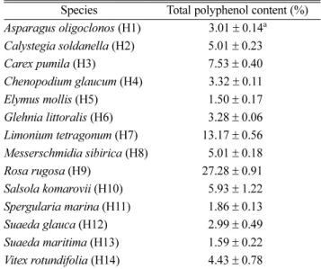
TPC – The total polyphenol content of the 14 halophyte extracts ranged between 1.50% and 27.28%
(Table 1). The highest total polyphenol content was found
in the R. rugosa extract (27.28%, H9), followed by the L.
tetragonum extract (13.17%, H7). The total polyphenol contents in the other 12 halophytes decreased as follows:
C. pumila (7.53%, H3), S. komarovii (5.93%, H10), C.
soldanella (5.01%, H2), M. sibirica (5.01%, H8), V.
rotundifolia (4.43%, H14), C. glaucum (3.32%, H4), G.
littoralis (3.28%, H6), A. oligoclonos (3.01%, H1), S.
glauca (2.99%, H12), S. marina (1.86%, H11), S.
maritima (1.59%, H13), and E. mollis (1.50%, H5). Of note is that the phenol content of R. rugosa (H9) was twice as high as that of L. tetragonum (H7) and approximately twenty times higher than that of E. mollis (H5).
Antioxidant activity – Antioxidant activity of halophytes was determined using DPPH assay (Fig. 1), and the DPPH radical scavenging activities were expressed as IC
50values. The highest antioxidant activities were found in L. tetragonum (17.28 μg/mL, H7), R. rugosa (29.73 μg/mL, H9), and M. sibirica (60.33 μg/mL, H8). V.
rotundifolia (85.11 μg/mL, H14), G. littoralis (89.82 μg/
mL, H6), C. soldanella (112.13 μg/mL, H2), and C.
pumila (235.80 μg/mL, H3) exhibited moderate antioxidant activity. C. glaucum (409.75 μg/mL, H4), A. oligoclonos (421.25 μg/mL, H1), E. mollis (H5), S. komarovii (H10), S. glauca (H12), S. marina (H11), and S. maritima (H13) showed weak or nonexistent antioxidant activities.
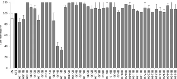
Anti-inflammatory activity – We examined cell viability after treatment with the 14 halophyte extracts via a CCK-8 assay (Fig. 2). No significant cytotoxicity was observed, although treatment with C. glaucum (H4) reduced cell viability to 60% at a dose of 50 μg/mL and 67% at a dose of 100 μg/mL.
Anti-inflammatory activity of the halophyte extracts was evaluated by measuring NO production and PGE
2levels in LPS-stimulated RAW264.7 cells after treatment with the extracts. As shown in Fig. 3., all the halophyte extracts suppressed NO production. A. oligoclonos (500 μg/mL, H1) and E. mollis (500 μg/mL, H5) inhibited NO production by 11.83 and 30.41%, respectively. In particular, C. pumila (100 μg/mL, H3) and G. littoralis (100 μg/mL, H6) inhibited NO production by 44.22 and 26.39%, respectively.
As shown in Fig. 4., A. oligoclonos (H1) and E. mollis (H5) at 300 μg/mL inhibited PGE
2release by 44.10 and 33.72%, respectively, whereas at 500 μg/mL they inhibited Table 1. Total polyphenol content of 14 halophyte extracts
expressed as percentage (%).
Species Total polyphenol content (%) Asparagus oligoclonos (H1) 3.01 ± 0.14
aCalystegia soldanella (H2) 5.01 ± 0.23
Carex pumila (H3) 7.53 ± 0.40
Chenopodium glaucum (H4) 3.32 ± 0.11
Elymus mollis (H5) 1.50 ± 0.17
Glehnia littoralis (H6) 3.28 ± 0.06 Limonium tetragonum (H7) 13.17 ± 0.56 Messerschmidia sibirica (H8) 5.01 ± 0.18
Rosa rugosa (H9) 27.28 ± 0.91
Salsola komarovii (H10) 5.93 ± 1.22 Spergularia marina (H11) 1.86 ± 0.13
Suaeda glauca (H12) 2.99 ± 0.49
Suaeda maritima (H13) 1.59 ± 0.22
Vitex rotundifolia (H14) 4.43 ± 0.78
a