Original Article
Clinical Features of Korean Patients with Congenital Aniridia
Shin Hae Park, Young Gun Park, Mee Yon Lee, Man Soo Kim
Department of Ophthalmology and Visual Science, The Catholic University of Korea School of Medicine, Seoul, Korea
Purpose: To investigate the clinical features of Korean patients with congenital aniridia.
Methods: This retrospective study focused on 60 eyes from 31 patients who were diagnosed with congenital aniridia at Kangnam St. Mary’s Hospital from 1996 to 2007. Patient age, gender, visual acuity (VA), family history, and previous ocular history were recorded. The presence of keratopathy, glaucoma, cataract, foveal hypoplasia, and other ocular or systemic anomalies were evaluated for each patient.
Results: The proportion of sporadic cases was 29.0%. Cataract (82.5%), glaucoma (51.6%), keratopathy (71.6%), and foveal hypoplasia (81.8%) commonly accompanied aniridia. Thirty-four (60.7%) eyes had VAs less than 20/200 and 20 eyes (35.7%) had VAs between 20/200 and 20/60. In patients without a past history of ocular sur- gery, the mean central corneal thickness was 643.05 ± 37.67 μm and the mean endothelial cell count was 3,349.44 ± 408.17 cells/mm2. Ocular surface surgeries were performed in 6 eyes. The clarity of the transplanted corneal graft vanished in 5 eyes with the progression of peripheral neovascularization and subepithelial fibrosis.
The mean age of cataract surgery in 8 eyes was 29.8 ± 5.9 years. Postoperative worsening of corneal clouding and glaucomatous damage were observed in 4 eyes. Two infants had bilateral congenital glaucoma. Two chil- dren with sporadic aniridia were identified to have Wilm’s tumors.
Conclusions: Congenital aniridia is a progressive congenital disorder that is commonly accompanied by complica- tions that can lead to impaired vision. Regular, careful examinations for these accompanying complications should be performed in all patients with congenital aniridia.
Key Words: Aniridic glaucoma, Aniridic keratopathy, Congenital aniridia
ⓒ2010 The Korean Ophthalmological Society
This is an Open Access article distributed under the terms of the Creative Commons Attribution Non-Commercial License (http://creativecommons.org/licenses /by-nc/3.0/) which permits unrestricted non-commercial use, distribution, and reproduction in any medium, provided the original work is properly cited.
Received: January 14, 2010 Accepted: May 11, 2010
Reprint requests to Man Soo Kim. Department of Ophthalmology and Visual Science, Seoul St. Mary’s Hospital, The Catholic University of Korea School of Medicine, #505 Banpo-dong, Seocho-gu, Seoul 137-701, Korea.
Tel: 82-2-2258-1188, Fax: 82-2-599-7405, E-mail: [email protected]
Congenital aniridia is a rare ocular malformation that af- fects the development of multiple ocular structures, this ab- normality is caused by a mutation in the PAX6 gene located on chromosome 11p13 [1-3]. Iris hypoplasia is the most ob- vious sign, but a broad spectrum of disorders can manifest from various mutations in the PAX6 gene [2-4]. Many pa- tients develop corneal opacities, cataracts, nystagmus, and foveal and optic nerve hypoplasia. Aniridia typically causes severe visual impairment; foveal hypoplasia is a major fac- tor that can decrease visual function in these patients [5].
The incidence of congenital aniridia ranges from 1:64,000 to 1:96,000 [5]. In 2/3 of cases the abnormality is inherited in an autosomal dominant fashion with almost complete penetrance and variable expressivity, 1/3 of the
cases of aniridia are sporadic [1,6,7].
The aim of this study was to investigate the clinical fea- tures of Korean patients with congenital aniridia. We ana- lyzed the influence of treatment on the accompanying com- plications to determine the visual prognosis and assess dis- ease progression.
Materials and Methods
This retrospective study focused on Korean patients who were diagnosed with congenital aniridia at Kangnam St.
Mary’s Hospital from 1996 to 2007. All cases were bi- lateral; however, 2 patients had already developed phthisis bulbi in one eye. We therefore included 60 eyes from 31 patients.
The age, gender, visual acuity (VA), family history, and previous ocular history of the patients were recorded. A thorough slit lamp microscopic examination was performed.
The sporadic cases were diagnosed with typical clinical fea- tures of congenital aniridia without a positive family history.
The presence or absence of keratopathy, glaucoma, cata-
Table 1. Classification of aniridic keratopathy
Grade Corneal signs
0 Clear cornea
1 Peripheral mudding with ingrowth of neovascular tissue not exceeding 1 mm from the limbal arch
2 Peripheral neovascularization in at least the peripheral half of the cornea, corneal clouding, and subepithelial fibrosis
3 Involvement of the central cornea
Table 2. Demographic data of patients with congenital aniridia at the initial visit
Patients with congenital aniridia Gender
Male 15 (48.4)
Female 16 (51.6)
Pattern of inheritance
Familial 22 (71.0)
Sporadic 9 (29.0)
Age distribution
≤10 5 (16.1)
10≤ <20 2 (6.5)
20≤ <30 12 (38.7)
30≤ <40 8 (25.8)
≥40 4 (12.9)
VA distribution
<0.02 8 (14.3)
0.02≤ <20/200 26 (46.4)
20/200≤ <20/60 20 (35.7)
≥20/60 2 (3.6)
Values are presented as number of patients (%).
Visual acuity (VA) could not be assessed in 4 eyes from 2 children, one was a young infant and the other was mentally retarded.
Table 3. Ocular findings accompanying congenital aniridia Ocular findings No. of eyes (%) Aniridic keratopathy 43 (71.6)
Cataract 33 (82.5)a
Glaucoma 31 (51.6)b
Foveal hypoplasia 36 (81.8)a
aRetinal examination was possible in 44 eyes that were without significant corneal or lens opacity.
bThis rate was calculated from the 40 eyes without a prior history of cataract extraction.
ract, and foveal hypoplasia were evaluated in each patient at the initial visit. Patients with uncontrolled intraocular pres- sure or a glaucomatous optic disc were regarded as having glaucoma. The extent of corneal involvement was classified into four grades (Table 1). The central corneal thickness was measured with an ultrasonic pachymeter (AL-2000;
Tomey, Erlangen, Germany). Specular microscopy was per- formed using a non-contact Konan specular microscope (Robo Ca; Konan, Hyogo, Japan). The intraocular pressure was measured with Goldmann’s applanation tonometry and a Tonopen.
Statistical analysis was performed using the t-test with SPSS ver. 15.0 (SPSS Inc., Chicago, IL, USA). A p-value
<0.05 was considered statistically significant.
Results
Patient demographics
Table 2 shows the demographic data of the 60 eyes from
31 patients at the initial visit. The male/female ratio was 0.48. The proportion of sporadic cases was 29.0%. The mean patient age at the initial visit was 27.5 ± 9.6 years (range, 0.1 to 52 years). The majority of the patients were in their twenties (38.7%) and thirties (25.8%). Common chief complaints included a progressive decrease in VA due to cataract or keratopathy and uncontrolled intraocular pressure.
More than half of the patients had VAs less than 20/200 (Table 2). Twenty eyes (35.7%) had VAs between 20/200 and 20/60. Only two eyes from 1 patient had VAs better than 20/60. In addition to iris involvement, cataract (82.5%), glaucoma (51.6%), keratopathy (71.6%), and foveal hypo- plasia (81.8%) were also observed (Table 3). The mean grade of keratopathy in 22 eyes with VAs better than 20/200 was 1.62 ± 0.89, which was statistically significantly lower than that in 34 eyes with VAs < 20/200 (2.38 ± 1.02, p = 0.035).
Aniridic keratopathy
Various degrees of corneal opacities were found in 43 eyes (71.6%). Partial mudding in the peripheral limbal cor- nea with in-growth of vessels was regarded as grade 1 aniri- dic keratopathy (Table 1 and Fig. 1B) [8,9].
In children under the age of 10 years, 5 eyes had rela- tively clear corneas. Grade 1 keratopathy was found in 3 eyes from 2 patients who were treated for congenital glau- coma and grade 3 keratopathy was observed in 2 eyes.
Grade 1 keratopathy was found in all 4 eyes from 2 patients who were in the second decade of life.
In 35 eyes without a past history of ocular surgery, the mean central corneal thickness was 643.05 ± 37.67 μm. In 27 eyes, the mean endothelial cell count was 3,349.44 ± 408.17 cells/mm2. The endothelial cell count could not be obtained in 8 eyes with significant corneal surface irregu- larities or corneal clouding.
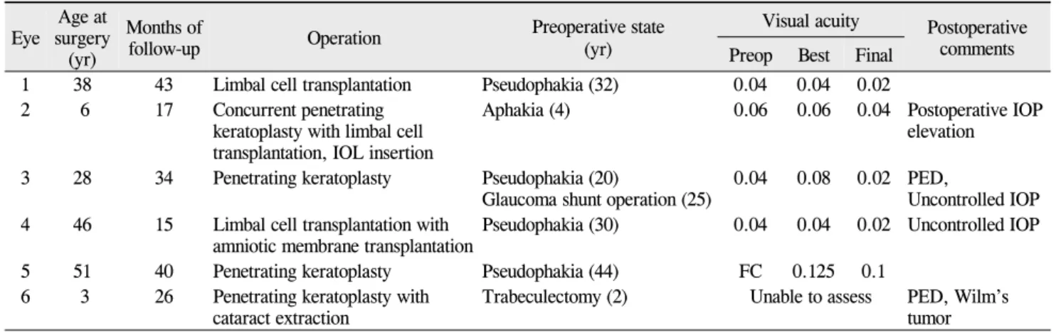
Ocular surface surgeries were performed in 6 eyes (10.0%) due to marked corneal clouding that affected VA.
The surgical results are summarized in Table 4. Postoperative intraocular pressure (IOP) elevation occurred in 3 eyes and a persistent epithelial defect was a complication in 2 eyes.
After surgery, the clarity of the transplanted corneal graft vanished with progression of peripheral neovascularization, subepithelial fibrosis, and thickening of the grafted cornea.
A B
Fig. 1. A 14-year-old female with congenital aniridia. (A) Note the total absence of the iris using retroillumination photography. This girl had early stage aniridic keratopathy manifesting as peripheral limbal neovascularization, a mild cortical cataractous lens was also found. (B) Note the advanced glaucomatous cupping with absence of the foveal reflex.
Table 4. Pre- and postoperative data for the eyes that underwent ocular surface surgery Eye Age at
surgery (yr)
Months of
follow-up Operation Preoperative state
(yr)
Visual acuity Postoperative comments Preop Best Final
1 38 43 Limbal cell transplantation Pseudophakia (32) 0.04 0.04 0.02 2 6 17 Concurrent penetrating
keratoplasty with limbal cell transplantation, IOL insertion
Aphakia (4) 0.06 0.06 0.04 Postoperative IOP elevation
3 28 34 Penetrating keratoplasty Pseudophakia (20)
Glaucoma shunt operation (25) 0.04 0.08 0.02 PED,
Uncontrolled IOP 4 46 15 Limbal cell transplantation with
amniotic membrane transplantation Pseudophakia (30) 0.04 0.04 0.02 Uncontrolled IOP 5 51 40 Penetrating keratoplasty Pseudophakia (44) FC 0.125 0.1
6 3 26 Penetrating keratoplasty with
cataract extraction Trabeculectomy (2) Unable to assess PED, Wilm’s tumor IOL = intraocular lens; IOP = intraocular pressure; FC = finger counting; PED = persistent epithelial defect.
Only one eye had an improvement in final VA.
Aniridic glaucoma
Glaucoma was found in 31 eyes (51.6%). Twelve eyes had newly detected glaucomatous optic disc changes and 17 eyes were already being treated with topical medication be- cause the diagnosis of glaucoma had been made at another clinic.
Of the 14 eyes from the 7 patients under the age of 20, 10 eyes (71.4%) from 5 patients had glaucoma. One male and one female infant had congenital glaucoma in both eyes and both had undergone bilateral trabeculectomy. Four eyes from 2 patients were diagnosed with glaucoma during the early teen years. Two eyes were complicated with glaucoma after keratoplasty and cataract surgery, respectively.
Among the 46 eyes from 24 adult patients with an age greater than 20, 21 eyes (45.7%) of 11 patients had glaucoma.
Three eyes had undergone Ahmed valve implantation and
6 eyes had undergone trabeculectomy. Twenty-two eyes had glaucoma controlled with topical anti-glaucomatous medications.
Cataract
Twenty eyes had a prior history of cataract extraction.
Their mean age at the time of cataract extraction was 25.75 years.
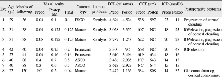
Of the remaining 40 eyes, lens opacity was found in 33 eyes (82.5%) at the initial visit. Five eyes exhibited lens subluxation combined with zonular weakness. Young pa- tients had a mild degree of anterior or posterior subcapsular lens opacity, as shown in Fig. 1A. Table 5 summarizes the clinical features of 8 eyes in which phacoemulsification and implantation of an intraocular lens were performed. The mean age at the time of cataract surgery was 29.8 ± 5.9 years and the mean preoperative endothelial cell count was 3,497.50 ± 383.11 cells/mm2. At the time of cataract sur-
Table 5. Pre- and postoperative data for the eyes that underwent cataract surgery Eye Age(yr) Months of
follow-up
Visual acuity
Cataract type Intraop.
problems
ECD (cells/mm2) CCT (μm) IOP (mmHg)
Postoperative problems Preop Postop
6M Final Preop Postop Preop Postop Preop Postop
1 29 36 0.04 0.1 0.1 PSCO Zonulysis 4,694 4,524 538 597 23 11 Progression of corneal clouding
2 31 38 0.04 0.125 0.125 Mature Zonulysis 3,058 3,355 607 NC 18 23 IOP elevation, progression of corneal clouding 3 31 38 0.08 0.125 0.125 Mature Zonulysis 3,787 1,248 622 NC 20 27 IOP elevation, progression
of corneal clouding
4 42 40 0.04 0.25 0.2 Brunescent 3,300 NC 668 NC 20 48 IOP elevation
5 27 41 0.04 0.16 0.16 Brunescent 3,610 3,496 619 634 18 16
6 40 88 0.4 0.7 0.5 ASCO 3,436 2,985 NC 643 14 15
7 40 88 0.3 0.6 0.5 ASCO 3,623 2,923 NC 644 15 15
8 22 120 FC 0.2 0.04 Mature 2,472 1,165 534 808 14 32 Glaucoma shunt op, corneal compromise M = month; ECD = endothelial cell density; CCT = central corneal thickness; IOP = intraocular pressure; PSCO = posterior subcapsular opacity; ASCO = anterior subcapsular opacity; NC = not checked; FC = finger counting.
gery, the mature and brunescent types of lens opacities predominated. Anterior vitrectomy with transscleral fixation of the intraocular lens was performed in 3 eyes with weak zonules. Postoperative 6-month VA improved in all of these eyes. Four eyes suffered from the development or deterio- ration of glaucoma. Corneal clouding and peripheral neo- vascularization became worse after cataract surgery in 4 eyes, one of which required corneal transplantation surgery.
Foveal hypoplasia
A fundus examination was possible in 44 eyes that were without significant corneal or lens opacity. Foveal hypo- plasia, defined as the absence of a foveal reflex (Fig. 1B), was found in 36 eyes (81.8%).
Combined ocular and systemic anomalies
One male infant with a positive familial history for con- genital aniridia had Peter’s anomaly and congenital glauco- ma in both eyes. One female infant with sporadic congenital aniridia was also diagnosed with congenital glaucoma. Wilm’s tumors were found in two children (6.5%) with sporadic congenital aniridia. The Wilm's tumors were diagnosed at 40 and 33 months after birth, respectively; these 2 patients were treated with nephrectomy and chemotherapy.
Discussion
Approximately 30 cases of congenital aniridia have been reported in Korea since the first report of 4 cases of con- genital aniridia in Koreans in 1977 [8-11]. Recently, Kim et al. [11] reported the presence of PAX6 gene mutations in 12 Korean aniridia patients. This study investigated Korean aniridic patients to estimate the prevalence and character- istics of associated disorders, disease course, and visual
prognosis. In summary, cataract, keratopathy, and foveal hypoplasia were present in over 70% of the Korean patients with congenital aniridia. Glaucoma was present in about half of the patients. Very few of the aniridic patients had VAs better than 20/60. The primary factor in the progressive decrease in VA is thought to be deterioration of the asso- ciated keratopathy with aging. The majority of the patients in this study were in their twenties (38.7%) and thirties (25.8%), with the most common chief complaints including a progressive decrease in VA due to cataract and kerato- pathy and uncontrolled intraocular pressure.
It is well-established that aniridic keratopathy is the result of a limbal stem cell deficiency [4,12,13]. A peripheral pan- nus with a relatively clear central cornea was also observed in the majority of the young patients in this study. With age, neovascularization gradually advances into the central cor- nea until the entire cornea is involved. Subepithelial fibrosis and stromal haze were also observed. The central corneal thickness increased to 643.05 ± 37.67 μm with a relatively normal endothelial cell count (3,349.44 ± 408.17 cells/mm2), which was comparable to other previously reported results [14,15]. A thickened cornea was presumed to be caused by developmental changes in the epithelium and stroma and not by endothelial dysfunction. In this study, more than half of the patients under the age of 20 years had worse than grade 1 keratopathy. Preservative-free lubricants and autol- ogous serum eyedrops are helpful in patients with early stage keratopathy [13,16-18].
In this study, the long term surgical results of penetrating keratoplasty with or without limbal allograft were very poor. A persistent epithelial defect caused by a limbal cell deficiency and postoperative IOP elevation were common problems following keratoplasty. The same pre-graft cor- neal changes recurred and led to graft failure. All the eyes that had undergone keratoplasty had a prior history of cata- ract or glaucoma surgery. These patients had higher grade
keratopathy in their pseudophakic or aphakic eyes com- pared to their phakic contralateral eyes. Aniridic keratop- athy can worsen following surgery that involves excessive manipulation of the limbus [6].
Cataract was the most common associated disorder in the aniridic patients and increased with age. The mean age at the time of cataract surgery was 29.8 ± 5.9 years.
There was a relatively high risk of intraoperative and postoperative complications in these aniridic cataract patients. They exhibited fragile anterior lens capsules and associated lens subluxation or weak zonules. Deterioration of glaucoma was the main postoperative problem and was observed in 4 eyes (50%). In addition, we found that corneal clouding became worse after cataract surgery in 4 eyes (50%). This clouding was caused by edematous changes due to endothelial dysfunction in 1 eye or progressive stro- mal haze unrelated to endothelial dysfunction in 3 eyes.
Aniridia has a profibrotic nature [16,19].Reinhard et al.
[20] found postoperative deterioration of corneal surface disorders in 4 eyes and chronic endothelial cell loss in 3 eyes among 19 congenital aniridic patients.The proximity of the intraocular lens to immature vessels in the rudi- mentary iris, subclinical chronic inflammation, and the unique vulnerability to progressive fibrosis caused by PAX6 gene mutations may be possible mechanisms for the observed progressive fibrosis following cataract surgery in eyes with congenital aniridia [19]. Progressive corneal clouding and glaucomatous changes can also impede final VA. Early cataract surgery is not advisable in aniridic pa- tients; however, it was difficult to definitively determine the impact of this surgery on visual prognosis because of the small number of cases in this study.
The incidence of glaucoma in cases of congenital aniridia has been reported to range from 6% to 75% [18,21,22]. In this study, 51.6% of the patients had glaucoma. Glaucoma usually develops during late childhood in patients with con- genital aniridia and is related to progressive circumferential changes in the angle, along with the gradual forward migra- tion of abnormal iris tissue and the obscuration of the poste- rior trabecular meshwork and scleral spur [18,21]. In this study, the prevalence of glaucoma (71.4%) was also high in patients under the age of 20. In addition, congenital glauco- ma was observed in 2 patients. Young patients with aniridia should be kept under close surveillance for the development of glaucoma. Increased central corneal thickness leads to a significant overestimation of the IOP in patients with con- genital aniridia. Therefore, careful and regular examina- tions of the optic disc are very important for the detection and management of glaucoma in patients of all ages with congenital aniridia. Surgeons should also be aware that glaucoma can develop or deteriorate after cataract or cor- neal transplantation surgery.
Foveal hypoplasia may partially or entirely be respon- sible for visual impairment in patients with congenital aniridia. In previous reports, the incidence of foveal hypo-
plasia in cases of congenital aniridia has been reported to range from 50 to 74% [1,5,6]. Patients with congenital anir- idia exhibit retinal dysfunction of varying degrees. This ret- inal dysfunction can be accounted for by foveal hypoplasia secondary to PAX6 mutation or phototoxicity as a result of a maldeveloped iris. Photophobia may be caused by mac- ular hypoplasia, but not by a large pupil.
Wilm’s tumors were found in two children with sporadic congenital aniridia. Because the PAX6 gene is in close proximity to the WT1 gene, deletions of 11p13 involving both the PAX6 and WT1 genes could result in the presence of a Wilm’s tumor in addition to congenital aniridia [18].
Children with congenital sporadic aniridia develop Wilm’s tumor before the age of 2 to 3 years. Ophthalmologists should be aware of this systemic association with malig- nancy, especially in sporadic cases. All children with spora- dic aniridia should undergo repeated abdominal ultra- sonography and clinical examinations.
In conclusion, congenital aniridia is a progressive con- genital disorder that is commonly accompanied by compli- cations that can lead to impaired vision, including cataract, keratopathy, glaucoma, and foveal hypoplasia. The long-term surgical results of penetrating keratoplasty with or without limbal allograft were very poor in this study. There was also a high risk of intraoperative and postoperative complica- tions in aniridic cataract patients. Progressive corneal clouding or glaucomatous damage can impede final VA fol- lowing cataract surgery. Regular and careful examinations for accompanying complications are necessary in all pa- tients who suffer from congenital aniridia.
Conflict of Interest
No potential conflict of interest relevant to this article was reported.
Acknowledgements
This research was supported by Seoul St. Mary's Clinical Medicine Research Program year of 2009 through The Catholic University of Korea.
References
1. Prosser J, van Heyninen V. PAX6 mutations reviewed. Hum Mutat 1998;11:93-108.
2. Hill RE, Hanson IM. Molecular genetics of the Pax gene family. Curr Opin Cell Biol 1992;4:967-72.
3. Macdonald R, Wilson SW. Pax proteins and eye development. Curr Opin Neurobiol 1996;6:49-56.
4. Holland EJ, Djalilian AR, Schwartz GS. Management aniri- dic keratopathy with keratolimbal allograft: a limbal stem cell transplantation technique. Ophthalmology 2003;110:125-30.
5. Tremblay F, Gupta SK, De Becker I, et al. Effects of PAX6 mutations on retinal function: an electroretinographic study.
Am J Ophthalmol 1998;126:211-8.
6. Nelson LB, Spaeth GL, Nowinski TS, et al. Aniridia. A
review. Surv Ophthalmol 1984;28:621-42.
7. Shaw MW, Falls HF, Neel JV. Congenital aniridia. Am J Hum Genet 1960;12(4 Pt 1):389-415.
8. Park YG, Suh DH, Lee HS. 4 cases of congenital aniridia. J Korean Ophthalmol Soc 1977;18:419-22.
9. Ahn SK, Kang JS, Shyn KH. A case of congenital aniridia. J Korean Ophthalmol Soc 1989;30:815-8.
10. Park SJ, Kim HT, Kim SM, Chung SK. Implantation of blackdiaphragm intraocular lens in cataract surgery with con- genital aniridia. J Korean Ophthalmol Soc 1998;39:1748-54.
11. Kim JH, Hwang BS, Lee JH, Cha SC. PAX6 mutations and clinical features of congenital aniridia. J Korean Ophthalmol Soc 2008;49:1794-800.
12. Eden U, Beijar C, Riise R, Tornqvist K. Aniridia among chil- dren and teenagers in Sweden and Norway. Acta Ophthalmol 2008;86:730-4.
13. Lopez-Garcia JS, Garcia-Lozano I, Rivas L, Martinez-Garchitorena J. Congenital aniridia keratopathy treatment. Arch Soc Esp Oftalmol 2006;81:435-44.
14. Weiss JS, Demartini D, Brown R, Forster RK. Specular mi- croscopy in aniridia. Cornea 1987;6:27-31.
15. Muir KW, Duncan L, Enyedi LB, et al. Central corneal thick-
ness: congenital cataracts and aphakia. Am J Ophthalmol 2007;144:502-6.
16. Lee H, Khan R, O'Keefe M. Aniridia: current pathology and management. Acta Ophthalmol 2008;86:708-15.
17. Lopez-Garcia JS, Rivas L, Garcia-Lozano I, Murube J. Autologous serum eyedrops in the treatment of aniridic keratopathy.
Ophthalmology 2008;115:262-7.
18. Brauner SC, Walton DS, Chen TC. Aniridia. Int Ophthalmol Clin 2008;48:79-85.
19. Tsai JH, Freeman JM, Chan CC, et al. A progressive anterior fibrosis syndrome in patients with postsurgical congenital aniridia. Am J Ophthalmol 2005;140:1075-9.
20. Reinhard T, Engelhardt S, Sundmacher R. Black diaphragm aniridia intraocular lens for congenital aniridia: long-term follow-up. J Cataract Refract Surg 2000;26:375-81.
21. Grant WM, Walton DS. Progressive changes in the angle in congenital aniridia, with development of glaucoma. Am J Ophthalmol 1974;78:842-7.
22. Swanner JC, Walton DS, Chen TC. Prevention of aniridic glaucoma with goniosurgery. Int Ophthalmol Clin 2004;44:
67-71.