605
Original ArticleKorean Circulation J 2006;36:605-607
ISSN 1738-5520
ⓒ 2006, The Korean Society of Circulation CASE REPORT
Subaortic Left Brachiocephalic Vein
Chan Kwon Park, MD
1, Ki Jun Kim, MD
2, Min Kyong Park, MD
1, Hong Jun Yang, MD
1, Jin Man Cho, MD
1, Doo Soo Jeon, MD
1and Man Young Lee, MD
11Department of Internal Medicine and 2Radiology, Our Lady of Mercy Hospital, The Catholic University of Korea, Incheon, Korea
ABSTRACT
Subaortic left brachiocephalic vein is a rare congenital anomaly that is sometimes found in the normal populat- ion. We report here on a case of subaortic left brachiocephalic vein that was detected incidentally by performing contrast transesophageal echocardiography (TEE) with using agitated saline and computed tomography (CT).
(Korean Circulation J 2006;36:605-607)
KEY WORDS:Congenital disorders;Brachiocephalic veins;Transesophageal echocardiography.
Introduction
The subaortic left brachiocephalic vein is a rare con- genital anomaly even among those individuals who are suffering with cardiac malformation, and it’s incidence is presumed to be very much lower in the population at large.
We present here a case of subaortic left brachioce- phalic vein that was diagnosed by TEE and CT, and we also offer a brief discussion of the medical literature that is related to this rare condition.
Case
A 63-year-old man was admitted to our hospital co- mplaining of severe left anterior chest pain of an aty- pical character. His initial blood pressure was 170/90 mmHg; however, his other vital signs, an electrocar- diogram and the routine lab findings were all within normal ranges. We performed a chest X-ray and trans- thoracic echocardiography(TTE). The posterioante- rior chest radiograph showed elongation of the pati- ent’s aorta, but it was otherwise normal. In addition, the TTE revealed no abnormality. We then decided to perform a test with TEE to rule out such aortic disease as aortic dissection or intramural hematoma as the cause of the patient’s chest pain. What we found was
an anomalous structure under the aortic arch. Never- theless, the diagnostic color or Doppler flow pattern didn’t display that structure, so we used an agitated mixture to obtain a clear image and to understand the anomalous structure. After injecting an agitated mix- ture of 10% air, 10% blood and 80% saline into the left antecubital vein
1)at the bedside, the structure was well enhanced and we presumed this structure to be a vein(Fig. 1). In order to precisely identify the anomal- ous structure, we performed sequential contrast-enh- anced chest CT, and this revealed both the non- visualized left brachiocephalic vein in the normal lo- cation and the presence of the contrast-filled anomal- ous brachiocephalic vein that was lateral from the aor- tic arch(Fig. 2). The vein ran downward along the la- teral border of the aortic arch, passed through the aor- ticopulmonary window, crossed the mediastinum post- eriorly to the ascending aorta and anteriorly to the trachea, and it finally joined the right brachiocephalic vein to form the superior vena cava. The mediastinum, both lungs and the other cardiovascular structures were all normal.
Discussion
Subaortic left brachiocephalic vein is a congenital anomaly that is less common in the thoracic venous systems than for anomalous positions of the superior vena cava or azygos vein. In fact, the incidence of this condition in the general population has been reported to be from 0.06-0.37%, and the detected incidence among the patients with congenital heart disease is from 0.15-0.98%.
2)It is known that the features com- monly seen in Tetralogy of Fallot, i.e., right aortic arch,
Received:January 9, 2006 Accepted:February 23, 2006
Correspondence:Ki Jun Kim, MD,Department of Radiology, Our Lady of Mercy Hospital, The Catholic University of Korea, 665 Bupyeong 6-dong, Bupyeong-gu, Incheon 403-720, Korea
Tel: 82-32-510-5531, Fax: 82-32-529-0964 E-mail: [email protected]
606
·Korean Circulation J 2006;36:605-607ventricular septal defect and right ventricular outflow obstruction, are the cardiac malformations most frequ- ently associated with subaortic left brachiocephalic vein.
3)The detection rate for this malady has increased with the advanced technological capacity of the nonin- vasive modalities.
The exact pathogenetic mechanism leading to sub- aortic left brachiocephalic vein is still unknown, but there are several hypotheses. The brachiocephalic veins and superior vena cava originate from the right and left precardinal veins. Each precardinal vein joins its ipsilateral posterior cardinal vein to form the common cardinal vein that flows into the sinus venousus. In the eighth week of fetal development, the precardinal ana- stomosis develops between the two precardinal veins.
Subsequently, the left precardinal vein disappears, ca- using the blood flow from the left head and neck regi- ons to be carried mainly via the precardinal vein. This anastomosis develops into the left brachiocephalic vein.
The normal precardinal anastomosis develops ventrally to the arterial structures. Anomalous left brachioce- phalic vein is thought to result from the precardinal
anastomosis being situated posteriorly to the truncus arteriosus, and double left brachiocephalic vein is the consequence of both ventral and dorsal precardinal anastomosis.
4)Minami et al.
5)and Kim et al.
6)have suggested that subaortic left brachiocephaic vein can form seconda- rily when the elongation of the aortic arch prevents the normal precardinal anastomosis; the vein then develops either anteriorly or posteriorly to the aortic sac, wherever more space is available. Elongation of the aortic arch results in narrowing of the prevascular space and widening of the subaortic space between the aortic arch and the pulmonary artery; this widening increases the chance for the development of precardi- nal anastomosis posteriorly to the aortic sac.
Morhy Borges Leal et al.
7)have described their expe- rience with 14 patients who had this lesion identified via echocardiography, and 12 of them experienced right ventricular obstruction. With the advent of cross- sectional echocardiography, the anomalous course of the left brachiocephalic vein can readily be identified non-invasively from the suprasternal notch.
A B
Fig. 1. Trasnesophageal echocardiography shows the left brachiocephalic vein (arrows) under the aortic arch (A). Enhanced by the injection of agitated saline (B).
A B
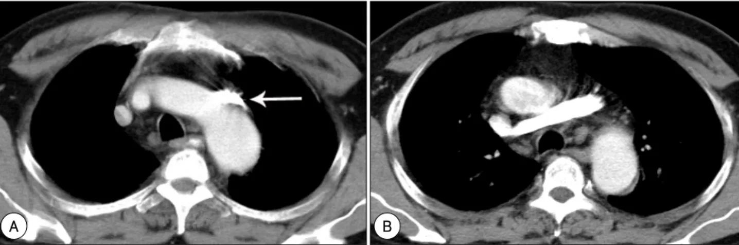
Fig. 2. Sequential contrast-enhanced chest CT scans show a contrast-filled anomalous brachiocephalic vein (arrows) lateral to the aortic arch (A).
The vein (arrows) passes through the aorticopulmonary window, traverses the mediastinum posteriorly to the ascending aorta and anteriorly to the trachea, and joins the right brachiocephalic vein to form the superior vena cava (B).
Chan Kwon Park, et al:Subaortic BCV·607
Although the subaortic brachiocephalic vein ano- maly usually has no clinical implications, it must be distinguished from other major vessels and especially on a preoperative cardiovascular examination because it is located beneath the aortic arch. On chest CT, it must be differentiated from a persistent left SVC, an atrophic right pulmonary artery and other vascular structures. This is easily accomplished by performing CT with using contrast medium.
4)A radiographic cha- racteristic of subaortic left brachiocephalic vein is that it mimicks the characteristics of mediastinal hemato- ma. Mediastinal widening and an apical pleural cap seen on chest radiography may indicate the presence of a mediastinal hematoma, which is caused by major vessel injury, and so further investigation is essential in the case of acute chest trauma.
8)With using echocar- diography, the anomalous vein is most likely to be id- entified as the right pulmonary artery. However, with the physician exercising proper awareness and care, the use of an air-blood-saline mixture makes it possible for the echocardiographer to visualize the anomalous ve- ssel beneath the aortic arch, parallel and anterior to the right pulmonary artery and the other vascular stru- ctures, as we were easily able to do.
9)The subaortic left brachiocephalic vein is a rare congenital anomaly in the general population. It is important for physicians to be well informed about this condition so that they do not to fail to detect it nor mistake it for other anomalous cardiovascular st- ructures. Detection can be easily achieved by ascertain-
ing the exact venal pathway via contrast transesophag- eal echocardiography with using agitated saline and CT.
REFERENCES
1) Jeon DS, Luo H, Iwami T, et al. The usefulness of a 10% air- 10% blood-80% saline mixture for contrast echocardiography:
Doppler measurement of pulmonary artery systolic pressure. J Am Coll Cardiol 2002;39:124-9.
2) Chern MS, Ko JS, Tsai A, Wu MH, Teng MM, Chang CY. Ab- errant left brachiocephalic vein: CT imaging findings and em- bryologic correlation. Eur Radiol 1999;9:1835-9.
3) Gerlis LM, Ho SY. Anomalous subaortic position of the brachio- cephalic (innominate) vein: a review of published reports and report of three new cases. Br Heart J 1989;61:540-5.
4) Takada Y, Narimatsu A, Kohno A, et al. Anomalous left brachio- cephalic vein: CT findings. J Comput Assist Tomogr 1992;16:
893-6.
5) Minami M, Noda M, Kawaguchi N, et al. Postaortic left in- nominate vein: radiological assessment and pathogenesis. Clin Radiol 1993;48:52-6.
6) Kim SH, Chung JW, Im JG, Choi YW, Choe YH, Han MC.
Subaortic left innominate vein: radiologic findings and consi- deration of embryogenesis. J Thorac Imaging 1999;14:142-6.
7) Morhy Borges Leal S, Andrade JL, de Soza M, et al. Anomalous subaortic course of the left brachiocephalic (innominate) vein:
echocardiographic diagnosis and report of an unusual associ- ation. Cardiol Young 2002;12:159-3.
8) Yama N, Nara S, Itoh Y, et al. A case of anomalous subaortic left brachiocephalic vein mimicking mediastinal hematoma. J Tra- uma 2005;58:419-20.
9) Shaffer EW, Snider R, Peters J, Farnsworth R. Echocardiogra- phic detection of anomalous course of the left innominate vein.
Int J Card Imaging 1985;1:167-9.