• 검색 결과가 없습니다.

Prognostic Value of Initial Echocardiographic Features in Patients With Tuberculous Pericarditis

N/A
N/A
Protected

Academic year: 2022

Share "Prognostic Value of Initial Echocardiographic Features in Patients With Tuberculous Pericarditis"

Copied!
10
0
0

로드 중.... (전체 텍스트 보기)

전체 글

(1)

377

Open Access

Prognostic Value of Initial Echocardiographic Features in Patients With Tuberculous Pericarditis

Hyung Oh Choi, MD

1

, Jong-Min Song, MD

1

, Tae Sun Shim, MD

2

, Sang-Hyun Kim, MD

1

, In-Hyun Jung, MD

1

, Duk-Hyun Kang, MD

1

and Jae-Kwan Song MD

1

1

Division of Cardiology and

2

Pulmonary and Critical Care Medicine, Asan Medical Center, University of Ulsan College of Medicine, Seoul, Korea ABSTRACT

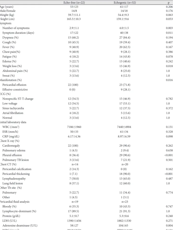
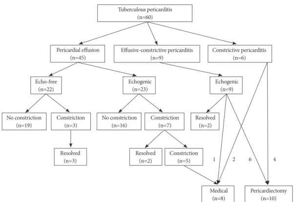
Background and Objectives: Tuberculous (TB) pericarditis is a major cause of constrictive pericarditis requiring pericar- diectomy. We sought to determine initial prognostic factors in patients with TB pericarditis. Subjects and Methods: We evaluated initial presentation and clinical outcomes (mean follow-up 32 ± 27 months) in 60 consecutive patients newly diag- nosed with TB pericarditis. Results: Initial presentations were pericardial effusion (PE), effusive-constrictive pericarditis, and constrictive pericarditis in 45 (75%), 9 (15%), and 6 (10%) patients, respectively. Of the 54 patients without initial constric- tive pericarditis, 32 (59%) showed echogenic materials in PE, including frond-like exudative coating and fibrinous strands.

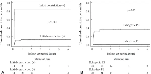
These patients had a longer disease duration before diagnosis, were initially more symptomatic, in a more advanced state, showed more persistent pericardial constrictions (38% vs. 0%, p<0.001) despite anti-TB medications, and tended to require pericardiectomy more often (19% vs. 0%, p=0.07, p<0.05 by Kaplan-Meier). All patients with effusive-constrictive pericardi- tis showed echogenic PE. Of the 60 total patients, 10 (17%) underwent pericardiectomies during follow-up. All of these pa- tients showed initial pericardial constrictions, whereas no patient without initial pericardial constriction underwent pericar- diectomy (p<0.001). Seven patients showed transient pericardial constrictions that resolved without pericardiectomy.

Conclusion: Initial pericardial constriction and echogenic PE are poor prognostic signs for persistent pericardial constric- tion and pericardiectomy in patients with newly diagnosed TB pericarditis. These results suggest that early diagnosis and prompt anti-TB medication may be critical. (Korean Circ J 2010;40:377-386)

KEY WORDS: Pericarditis; Tuberculosis; Echocardiography; Prognosis.

Received: December 16, 2009 Accepted: March 2, 2010

Correspondence: Jong-Min Song, MD, Division of Cardiology, Asan Medical Center, University of Ulsan College of Medicine, 86 Asanbyeong- won-gil, Songpa-gu, Seoul 138-736, Korea

Tel: 82-2-3010-3168, Fax: 82-2-486-5918 E-mail: [email protected]

cc

This is an Open Access article distributed under the terms of the Cre- ative Commons Attribution Non-Commercial License (http://creativecom- mons.org/licenses/by-nc/3.0) which permits unrestricted non-commer- cial use, distribution, and reproduction in any medium, provided the ori- ginal work is properly cited.

tures, including pericardial effusion (PE), effusive-constricti- ve pericarditis, and constrictive pericarditis.

2)3)

Pericardial con- strictive scarring, the most serious complication requiring pericardiectomy, occurs in 18-46% of patients with TB PE de- spite anti-TB chemotherapy and treatment with corticoste- roids.

3-5)

The predictors of TB constrictive pericarditis have been suggested in several small studies,

6-12)

but remain to be clearly established.

3)

Echogenic materials seen in PE on 2-di- mensional echocardiography, such as exudative coating and fibrinous strands, can predict pericardial complications irre- spective of underlying disease etiologies,

13)

and are more spe- cific to TB PE.

14-18)

However, the clinical prognostic value of such echogenic materials in TB PE has not been clearly dem- onstrated. Therefore, we evaluated clinical outcomes of new- ly-diagnosed TB pericarditis patients and determined initial prognostic predictors for the development of constrictive pericarditis requiring pericardiectomy.

Introduction

Tuberculosis (TB) continues to be a leading cause of mor-

bidity and mortality from infectious disease worldwide,

1)

and TB pericarditis is arguably the leading cause of pericardi-

tis in the world, despite a low incidence in developed coun-

tries. TB pericarditis patients initially present with diverse fea-

(2)

Subjects and Methods

From 1995 to 2007, 60 consecutive patients (mean age 58±

19 years, 30 females) were newly diagnosed with TB pericardi- tis at the Asan Medical Center, Seoul, Korea, and treated with anti-TB chemotherapy. Sixteen patients were also included in our previous report.

13)

No patient had any evidence of hu- man immunodeficiency virus infection. For each patient, a complete clinical history was taken, and physical examina- tion, chest X-ray, and 2-dimensional Doppler echocardiogra- phy were performed. Symptom onset time could be identified in 54 patients. Of the 60 patients, 47 underwent pericardio- centesis and/or pericardiostomy. Pericardial fluid and biopsy specimens were analyzed for chemistry, cytology, histopathol- ogy examination, acid-fast bacilli staining, culture for Myco- bacterium tuberculosis, adenosine deaminase (ADA) mea- surement, and polymerase chain reaction (PCR) detecting M.

tuberculosis.

19)

Diagnosis of TB pericarditis was categorized as definite or probable, as previously reported.

3)4)

A “definite” di- agnosis was made when M. tuberculosis was found in PE fluid or pericardial samples by acid-fast bacilli staining, TB culture, or positive TB-PCR; or when granuloma was seen. All bacte- riologically confirmed cases were pan-susceptible to anti-TB drugs. A “probable” diagnosis was made when there was proof of TB elsewhere in a patient with unexplained pericar- ditis or when PE showed a lymphocyte-dominant exudate with a high ADA (>40 U/L) level.

All patients underwent transthoracic 2-dimensional echo- cardiography (Sonos 7500, Philips Medical Systems, Andover, MA; Vivid 7, General Electric, Waukesha, WI; Acuson Sequo- ia, Mountain View, CA, USA), and all data were analyzed by a single expert. Patients with PE were divided into two groups by differences in initial echocardiographic characteristics of PE and pericardium. Echo-free PE was defined as PE with- out any echogenic materials (Fig. 1A),

20)

whereas echogenic PE was defined as PE showing echogenic materials in PE or on the pericardium (Fig. 1B).

15)17)20)

Effusive-constrictive pericarditis was diagnosed when con- strictive physiology was not resolved by PE removal, or when constrictive physiology was observed together with only a mild to moderate amount of PE. Constrictive physiology was defin- ed as a significant respiratory variation in mitral inflow E wave velocity (≥25%) and a prominent increase in diastolic flow re- versal with expiration in the hepatic vein on 2-dimensional Dop- pler echocardiography.

21)22)

Chest contrast-enhanced comput- ed tomography (CT) was performed in 39 patients.

Clinical and echocardiographic observations were conduct- ed for 32±27 months (median: 25 months) on patients taking adequate anti-TB medications for at least 6 months. Pericardial constriction was diagnosed when constrictive physiology (as described above) and diffuse pericardial thickening without PE were found on 2-dimensional Doppler echocardiogra-

phy.

21)23)

Referral for pericardiectomy was determined by at- tending physicians’ discretion.

Numeric variables are expressed as means±SD. Statistical analysis of between-groups differences was performed using Student’s unpaired t-test. The χ

2

test and Fisher’s exact test were used to compare frequency ratios between groups. A Kaplan- Meier analysis and the log rank test were employed to compa- re event-free survival rates. A p of <0.05 was considered signifi- cant.

Results

Initial presentations were PE, effusive-constrictive pericar- ditis, and constrictive pericarditis in 45 (75%), 9 (15%), and 6 (10%) patients, respectively. Of the 45 patients who present- ed with PE, 9 patients (15%) showed initial cardiac tampon- ade defined as a large amount of PE, significant respiratory variations in mitral inflow E velocity, and diastolic collapse of the right atrium or ventricle. Of the 9 patients with effusive- constrictive pericarditis, constrictive physiology was not re- solved by PE removal in 4 patients and constrictive physiolo- gy was observed together with only mild to moderate PE in 5 patients. Of the 54 patients without initial constrictive peri- carditis, 32 (59%) showed echogenic PE, whereas 22 showed echo-free PE. Clinical features of both groups are summariz- ed in Table 1. Weight loss was more prevalent in patients with echogenic PE than in those with echo-free PE, and the form- er patients presented with a greater number of symptoms, of longer duration, than did the latter cohort. All nine patients with effusive-constrictive pericarditis showed echogenic PE.

Pleural effusion on initial chest X-ray and pericardial thick- Fig. 1. Representative examples of echo-free pericardial effusion (PE) (A) and echogenic PE (B). PE: pericardial effusion.

A

B

(3)

Table 1. Comparison of clinical characteristics of echo-free and echogenic pericardial effusion patients

Echo-free (n=22) Echogenic (n=32) p

Age (years) 53±23 61±170 0.206

Male/Female 14/8 14/180 0.176

Weight (kg) 59.7±11.1 54.4±9.300 0.063

Height (cm) 165.5±10.3 159.1±9.6000 0.053

Symptom

Number of symptoms Symptom duration (days) Dyspnea (%)

Cough (%) Fever (%) Chest pain(%) Fatigue (%) Edema (%) Weight loss (%) Abdominal pain (%) Sweat (%)

2.9±1.1 17±22 15 (68.2)0 10 (45.5)0 9 (40.9) 9 (40.9) 4 (18.2) 5 (22.7) 3 (13.6) 5 (22.7) 3 (13.6)

4.0±1.50 40±380 27 (84.4) 19 (59.4) 20 (62.5) 09 (28.1) 14 (43.8) 13 (40.6) 15 (46.9) 08 (25.0) 04 (12.5)

0.003 0.011 0.194 0.407 0.167 0.386 0.078 0.242 0.018 1.000 1.000 Manifestation (%)

Pericardial effusion Effusive-constrictive

22 (100)0.

0 (0)00.

23 (71.8) 09 (28.1)

0.016

ECG (%)

Nonspecific ST-T change Low voltage

Sinus tachycardia Atrial fibrillation Normal

12 (54.5)0 12 (54.5)0 5 (22.7) 4 (18.2) 3 (13.6)

15 (46.9) 17 (53.1) 12 (37.5) 05 (15.6) 04 (12.5)

0.782 1.000 0.372 1.000 1.000 Initial laboratory data

WBC (/mm

3

) ESR (mm/h) CRP (mg/dL)

7186±1960 50±33 6.17±4.36

7440±49040 61±340 8.97±6.590

0.131 0.328 0.098 Chest X-ray (%)

Cardiomegaly Pulmonary edema Pleural effusion Pulmonary TB lesion

22 (100).0 1 (4.5)0 8 (36.4) 3 (13.6)

29 (90.6) 2 (9.4) 29 (90.6) 07 (21.9)

0.262 0.638

<0.001 0.501 Chest CT (%)

Pericardial calcification Pericardial thickening Lymphadenopathy Lung field lesion

0n=14 2 (14.3) 1 (7.1)0 7 (50.0) 8 (57.1)

n=20 .00 (0)0.0.

18 (90.0) 13 (65.0) 12 (60.0)

0.162

<0.001 0.487 1.000 Other Tb site (%)

Pulmonary Other

5 (22.7) 1 (4.5)0

11 (34.4) 1 (3.1)

0.774 Pericardial fluid analysis

Bloody (%)

Lymphocyte-dominant (%) Protein (g/dL)

LDH (U/L)

Adenosine deaminase (U/L) WBC (/mm

3

)

0n=19 6 (35.3) 17 (89.5)0

5.1±0.7 1390±1456

58±27 2868±2674

n=23 10 (43.5) 21 (91.3) 5.3±0.60 1882±13300

104±6500 7780±14425

0.747

1.000

0.240

0.271

0.004

0.131

ECG: electrocardiogram, WBC: white blood cell, ESR: erythrocyte sedimentation rate, CRP: C-reactive protein, TB: tuberculosis, LDH: lac-

tate dehydrogenase

(4)

ening on chest CT were significantly more prevalent in pa- tients with echogenic PE than in those with echo-free PE.

ADA activity in pericardial fluid was higher in the former pa- tients than in the latter group. Definite diagnoses were more often possible in patients with echogenic PE than in those with echo-free PE (Table 2), and TB-specific pericardial patho- logy results, such as stained acid-fast bacilli and granuloma, were more frequently positive in echogenic patients. TB-PCR on PE fluid was more often positive in echogenic PE patients than in echo-free PE patients. Durations of anti-TB chemothe- rapy and proportions of patients treated with adjunctive cor- ticosteroids did not differ between the two groups (Table 3).

During follow-up, new pericardial constrictions developed in 10 patients, of whom 7 had shown echogenic PE on initial echocardiography (Fig. 2). Of these 10 patients, 100% (3/3) of echo-free PE patients and 29% (2/7) of those with initial echo- genic PE showed transient pericardial constrictions that re- solved during longer follow-up. No patient with initial echo- free PE but 5 patients (22%) with initial echogenic PE with- out initial pericardial constrictions, developed unresolved pericardial constrictions. In nine patients with initial effusive- constrictive pericarditis, two (22%) showed transient constric- tions that resolved during anti-TB therapy, whereas six (67%) required pericardiectomies. The overall incidence of pericar- dial constriction on both initial and follow-up echocardio- graphic evaluations was higher in patients with initial echo- genic PE than in those with initial echo-free PE (50.0% vs.

13.6%, p<0.01). During follow-up, pericardiectomies were

Table 2. Comparison of diagnostic criteria of tuberculous pericar- ditis between echo-free and echogenic pericardial effusion patients Echo-free Echogenic p TB in pericardial fluid (%)

TB culture-positive (%) TB PCR-positive (%) Bacilli or granuloma (%)*

TB in other sites (%) High ADA (%) Criterion count Definite diagnosis (%)

01/19 (5.3)0 02/19 (10.5) 02/17 (11.8) 04/15 (26.7) 06/22 (27.3) 14/19 (73.7) 1.32±0.560 06/22 (27.3)

0/26 (0)00 08/26 (30.8) 10/21 (47.6) 17/20 (85.0) 12/32 (37.5) 19/24 (79.2) 2.06±0.98 24/32 (75.0)

0.422 0.154 0.034 0.001 0.560 0.728 0.001 0.002

*Echo-free: TB (0), Granuloma (4); Echogenic: TB (2), Granuloma (15). TB: tuberculosis, PCR: polymerase chain reaction, ADA: ad- enosine deaminase

Table 3. Treatments and clinical events in patients with echo-free and echogenic pericardial effusion

Echo-free (n=22)

Echogenic

(n=32) p

Anti-tuberculosis drugs Medication duration (months) Secondary drugs (%)

13±16 2 (9.1)0

10±5 3 (9.4)0

0.434 1.000 Steroids (%)

Use

Initial dose (mg/day)

8 (36.4) 56±12

18 (56.3)0 050±11

0.176 0.271 Events (%)

Transient constriction Persistent constriction Pericardiectomy

3 (13.6) 0 (0)00.

0 (0)00.

4 (12.5) 12 (37.5)0

6 (18.8) 1.000 0.001 0.071

Fig. 2. Clinical outcomes of tuberculous pericarditis patients according to clinical and echocardiographic classifications.

Tuberculous pericarditis (n=60)

Echogenic

(n=23) Echogenic

(n=9)

Resolved (n=2) Echo-free

(n=22)

Constrictive pericarditis (n=6) Effusive-constrictive pericarditis

(n=9) Pericardial effusion

(n=45)

No constriction

(n=19) No constriction

(n=16) Constriction

(n=3) Constriction

(n=7)

Resolved

(n=3) Resolved

(n=2)

Medical

(n=8) Pericardiectomy (n=10) Constriction

(n=5) 1 2 6 4

(5)

performed on six patients with initial echogenic PE, but in no patient with initial echo-free PE. Of six patients who pre- sented with initial constrictive pericarditis, four (67%) un- derwent pericardiectomies. None of these 6 patients showed pericardial constriction resolution despite the use of anti-TB medications.

Of the 60 patients, 18 (30%) showed persistent constrictive pericarditis during follow-up. The incidence of unresolved pericardial constriction was higher in patients with initial con- strictions than in those without, and was greater in patients with echogenic PE than in those with echo-free PE, as shown by Kaplan-Meier analyses of the unresolved constrictive peri- carditis development rate (Fig. 3). Unresolved constrictive pe- ricarditis developed in about 38% of patients who presented with echogenic PE, and in no patient with initial echo-free PE. Of 18 patients with persistent constrictive pericarditis, 8 patients were followed-up medically because their symptoms were tolerable with medications, including diuretics.

Of 60 patients, 10 (17%) underwent pericardiectomies dur- ing follow-up. On initial echocardiography, these patients showed a higher prevalence of pericardial constriction, thick- ening, and calcification, than did patients who did not requi- re pericardiectomy (Table 4). The former patients showed a higher prevalence of pleural effusion and pericardial thick- ening by chest X-ray and CT, and had longer symptom dura- tion, than did patients who did not require pericardiectomy.

The durations of anti-TB medication and proportions of pa- tients treated with steroids did not differ significantly between the two groups. Patients with pericardial constrictions on ini- tial echocardiography showed a higher pericardiectomy rate than did those without constriction, by Kaplan-Meier analy- sis (p<0.001) (Fig. 4A). Similarly, patients with initial echoge- nic PE showed a higher pericardiectomy rate than did those with echo-free PE (p<0.05) (Fig. 4B). No mortality from peri-

carditis or surgery was observed in our study population. For 38 patients with initial constrictive physiology or echogenic PE (Table 5), Kaplan Meier analysis revealed that rate of per- sistent constriction during follow-up was lower in patients with than without steroid therapy (p=0.011) (Fig. 5).

Discussion

Predictors of constrictive pericarditis and pericardiectomy

Echogenic substances in PE are suggestive of TB pericardi- tis and signs of pericardial inflammation and a hypersensi- tivity reaction.

14-17)

However, echogenic substances are also found in the PE of other etiologies as risk factors for the de- velopment of constrictive pericarditis, irrespective of cause, in patients with PE.

13)24)25)

We previously reported on 178 con- secutive patients with moderate to severe PE of any etiology, including 16 patients with TB PE.

13)

This study of 60 patients diagnosed with TB pericarditis was designed to evaluate the clinical outcome of TB pericarditis, and identify poor prog- nostic indicators for permanent constriction and pericardi- ectomy. Initial constrictive physiology was the major poor prognostic factor in TB pericarditis, which we did not find in our previous study as we only included PE patients. In this study, 59% of patients with TB pericarditis accompanied by PE showed echogenic PE, consistent with previous reports.

15)17)

In PE patients, intrapericardial echogenic materials were as- sociated with a greater level of symptomatic presentation and a more advanced disease state. All patients who presented with effusive-constrictive pericarditis showed echogenic PE. Pa- tients with initial echogenic PE developed more unresolved pericardial constrictions and required pericardiectomy despite the use of anti-TB medications, than did those with echo-free PE. In previous studies, echogenic PE was regarded as specific

A B

Follow-up period (year) Follow-up period (year)

Patients at risk Patients at risk

Initial constriction (+) Echogenic PE

Initial constriction (-) Echo-free PE

Initial constriction (-)

Echo-Free PE p<0.001

p<0.05 Initial constriction (+)

Echogenic PE

Un re so lv ed co ns tri ct iv e p er ica rd iti s Un re so lv ed co ns tri ct iv e p er ica rd iti s

1 1

2 13

26 21

0 0

14 31

44 22

0.8 0.8

1.0 1.0

0.6 0.6

0.4 0.4

0.2 0.2

0.0 0.0

3 3

2 2

12

19 14

4 4

4 4

7 7

5 6 5 6

1 2

2 2

Fig. 3. Kaplan-Meier analyses of developing persistent constrictive pericarditis in patients with or without initial pericardial constriction (A), and

in patients with echogenic pericardial effusion (PE) or echo-free PE (B). PE: pericardial effusion.

(6)

Table 4. Comparison of initial clinical and echocardiographic characteristics between patients who required pericardiectomies and those who did not

Pericardiectomy (+) (n=10) Pericardiectomy (-) (n=50) p

Age (years) 50±13 59±12 0.177

Male/Female 4/6 26/24 0.731

Weight (kg) 55.7±10.4 56.1±10.4 0.900

Height (cm) 161.2±10.80 161.0±10.20 0.901

Symptom

Number of symptoms Symptom duration (days) Dyspnea (%)

Cough (%) Fever (%) Chest pain (%) Fatigue (%) Edema (%) Weight loss (%) Abdominal pain (%) Sweating (%)

4.2±1.2 105±280

9 (90.0) 7 (70.0) 6 (60.0) 3 (30.0) 3 (30.0) 4 (40.0) 4 (40.0) 3 (30.0) 3 (30.0)

3.5±1.5 24±23 38 (76.0) 28 (56.0) 27 (54.0) 18 (36.0) 18 (36.0) 16 (32.0) 15 (30.0) 12 (24.0) 05 (10.0)

0.191

<0.001 0.436 0.499

0.718 0.711 0.700 0.120 Manifestation (%)

Pericardial effusion Effusive-constrictive Constrictive pericarditis

0 (0)00.

6 (60.0) 4 (40.0)

45 (90.0) 3 (6.0) 2 (4.0)

<0.001

Initial laboratory data WBC (/mm

3

) ESR (mm/h) CRP (mg/dL)

6360±2491 57±53 4.61±4.93

7422±4081 55±34 7.95±5.96

0.432 0.919 0.168 Chest X-ray (%)

Cardiomegaly Pulmonary edema Pleural effusion Pulmonary TB lesion

5 (50.0) 0 (0)00.

10 (100)0.

2 (20.0)

48 (96.0) 06 (12.0) 33 (66.0) 10 (20.0)

0.001 0.577 0.049 1000.

Chest CT (%)

Pericardial calcification Pericardial thickening Lymphadenopathy Lung field lesion

n=8 1 (12.5) 8 (100) 4 (50.0) 4 (50.0)

0n=31 2 (6.5) 16 (51.6) 17 (54.8) 18 (58.1)

0.505 0.015 1000.

0.709 Diagnostic criteria (%)

TB in pericardial fluid TB culture-positive TB PCR-positive Bacilli or granuloma TB in other sites High ADA Criteria count Definite diagnosis

1 (33.3) 0 (0)00.

3 (60.0) 8 (80.0) 6 (60.0) 0 (0)00.

1.80±0.79 9 (90.0)

1 (2.3) 10 (23.3) 10 (28.6) 15 (51.7) 17 (34.0) 33 (78.6) 1.72±0.93

24 (48.0)

0.128 1000.

0.307 0.152 0.161 0.058 0.800 0.017

Echogenic PE (%) 100 (6/6)000 54.2 (26/48)00 0.071

Anti-tuberculosis drugs Medication duration (months) Secondary drugs (%)

15±14 2 (20.0)

11±11 05 (10.0)

0.360

0.330

(7)

for TB PE,

14-18)

but the high prevalence of echogenic PE makes its value in TB PE patients unclear. Here we have demonstrat- ed that echogenic PE is a predictor of constrictive pericarditis in patients with TB pericarditis.

Only some patients who initially presented with effusive- constrictive pericarditis or constrictive pericarditis required pericardiectomy during follow-up, whereas no patient without initial pericardial constriction underwent pericardiectomy.

Some patients with pericardial thickening by initial CT need- ed pericardiectomy. Patients who present with pericardial thickening and constriction and/or echogenic PE have poor prognoses and may develop persistent constrictive pericar- ditis requiring pericardiectomy. TB pericarditis patients initial- ly presenting without constriction and with echo-free PE may not develop permanent pericardial constriction nor require pericardiectomy, provided anti-TB medication is adequate.

Initial pericardial constriction and echogenic PE may reflect a more advanced disease state and a higher degree of pericar- dial inflammation. Symptom duration before diagnosis was greater in echocardiographically advanced patients, defined as those showing fibrinous strands and thickened pericardia,

18)

in agreement with our present data showing that patients with echogenic PE had been symptomatic for longer than echo-free PE patients. Patients who presented with constrictive pericar- ditis showed an even greater symptom duration (average 83±

35 days) than did those who presented with PE. These results

demonstrate that initial echocardiographic characteristics are informative with respect to both disease duration and inflam- mation severity. The fact that patients initially diagnosed with advanced disease are more likely to require pericardiectomy than patients with less severe illness emphasizes that TB peri- carditis needs to be detected early. Timely diagnosis, followed by appropriate treatment, is essential to prevent constrictive pericarditis and pericardiectomy. We observed a beneficial ef- fect of steroid therapy on preventing persistent pericardial con- striction in patients with initial echogenic PE or constrictive physiology in the present study. However, randomized con- trolled study will be needed to verigy this finding, because more patients with PE were included in the steroid therapy group, whereas more patients with constrictive physiology were in the non-steroid group.

Transient pericardial constriction

We also found that 7 patients (11.7% of the total) developed transient pericardial constrictions that resolved during lon- ger follow-up (6.3±6.3 months, range 1-18 months) with use of anti-TB medications. Data from the Mayo Clinic showed that about 17% of constrictive pericarditis patients developed transient constrictions, the common etiology being prior car- diovascular surgery, whereas only 1 case of TB pericarditis was transient.

26)

However, TB pericarditis seems to be a primary etiology of transient constrictive pericarditis in Korea, where Table 4. Continued

Pericardiectomy (+) (n=10) Pericardiectomy (-) (n=50) p

Steroids Use (%)

Initial dose (mg/day)

5 (50.0) 47±13

23 (46.0) 53±11

1.000 0.299 WBC: white blood cell, ESR: erythrocyte sedimentation rate, CRP: C-reactive protein, TB: tuberculosis, PCR: polymerase chain reaction, ADA: adenosine deaminase, PE: pericardial effusion

A B

Follow-up period (year) Follow-up Period (year)

Patients at risk Patients at risk

Initial constriction (+) Echogenic PE

Initial constriction (-) Echo-free PE

Initial constriction (-)

Echo-free PE p<0.001

p<0.05 Initial constriction (+)

Echogenic PE

Pe ric ar di ec to m y Un re so lv ed co ns tri ct iv e p er ica rd iti s

1 1

7 17

28 15

0 0

15 32

44 22

0.8 0.8

1.0 1.0

0.6 0.6

0.4 0.4

0.2 0.2

0.0 0.0

3 3

2

5

2

15

21 9

4 4

2 5

8 5

5 6 5 6

1 2

2 2

Fig. 4. Kaplan-Meier analyses of pericardiectomy in patients with or without initial pericardial constriction (A), and in patients with echogenic

pericardial effusion (PE) or echo-free PE (B). PE: pericardial effusion.

(8)

Table 5. Comparisions of clinical characteristics for treatment with or without steroids in patients with echogenic pericardial effusion or con- strictive physiology

No steroid (n=18) Steroid (n=20) p

Age (years) 61.2±18.5 59.8±15.1 0.797

Male/Female 7/11 9/11 0.752

Weight (kg) 53.2±11.4 54.6±7.40 0.659

Height (cm) 158.3±10.90 159.6±8.600 0.683

Symptom

Number of symptoms Symptom duration (days) Dyspnea

Cough Fever Chest pain Fatigue Edema Weight loss Abdominal pain Sweat

4.7±1.6 47.6±37.1 16 (88.9) 12 (66.7) 11 (61.1) 7 (38.9) 10 (55.6) 9 (50.0) 10 (55.6) 7 (38.9) 2 (11.1)

3.6±1.1 45.1±43.9 16 (80.0)0 13 (65.0)0 13 (65.0)0 5 (25.0) 7 (35.0) 6 (30.0) 6 (30.0) 3 (15.0) 3 (15.0)

0.021 0.859 0.663 1.000 1.000 0.489 0.328 0.320 0.188 0.144 1.000 Manifestation

Pericardial effusion Effusive-constrictive Constrictive

9 (50.0) 5 (27.8) 4 (22.2)

14 (70.0)0 4 (20.0) 2 (10.0)

0.663

ECG

Nonspecific ST-T change Low voltage

Sinus tachycardia Atrial fibrillation Normal

11 (61.1) 12 (66.7) 7 (38.9) 3 (16.7) 0 (0.0)

10 (50.0)0 7 (35.0) 6 (30.0) 3 (15.0) 4 (20.0)

0.532 0.103 0.734 1.000 0.107 Initial laboratory data

WBC (/mm

3

) ESR (mm/h) CRP (mg/dL)

8122±6559 52±39 6.18±6.70

6520±1536 64±39 9.44±6.51

0.325 0.472 0.216 Chest X-ray

Cardiomegaly Pulmonary edema Pleural effusion Pulmonary TB lesion

14 (77.8) 5 (27.8) 15 (83.3) 4 (22.2)

17 (85.0)0 0 (0.0)0 20 (100)0.

5 (25.0)

0.687 0.017 0.097 1.000 Chest CT

Pericardial calcification Pericardial thickening Lymphadenopathy Lung field lesion

1 (8.3) 11 (91.7)

5 (41.7) 7 (58.3)

0 (0.0)0 12 (92.3)0

9 (69.2) 7 (53.8)

0.480 1.000 0.238 1.000 Other Tb site

Pulmonary Other

9 (50.0) 1 (5.6)

7 (35.0) 0 (0.0)0

0.257

Pericardial fluid analysis Bloody

Lymphocyte-dominant Protein (g/dL) LDH (U/L)

Adenosine deaminase (U/L) WBC (/mm

3

)

3 (37.5) 7 (87.5) 5.4±0.5 2477±1062

89.7±79.1 4115±3180

7 (43.8) 14 (93.3)0

5.2±0.7 1562±1387 112.0±57.00 09874±17825

1.000

1.000

0.501

0.147

0.444

0.381

(9)

Table 5. Continued

No steroid (n=18) Steroid (n=20) p

Diagnostic criteria TB in pericardial fluid TB culture-positive TB PCR-positive Bacilli or granuloma TB in other sites High ADA Criteria count Definite diagnosis

1 (9.1) 5 (45.5) 4 (44.4) 6 (66.7) 10 (55.6) 5 (55.6) 1.7±1.1 11 (61.1)

0 (0.0)0 3 (18.8) 7 (50.0) 13 (86.7)0

7 (35.0) 14 (87.5)0

2.2±0.8 16 (80.0)0

0.407 0.206 1.000 0.326 0.328 0.142 0.132 0.288 Anti-tuberculosis drugs

Medication duration (months) Secondary drugs

11.3±11.3 1 (5.6)

10.5±4.90 4 (20.0)

0.766 0.344

Pericardiectomy 5 (27.8) 5 (25)0. 1.000

ECG: electrocardiogram, WBC: white blood cell, ESR: erythrocyte sedimentation rate, CRP: C-reactive protein, LDH: lactate dehydrogenase, TB: tuberculosis, ADA: adenosine deaminase

TB is more prevalent than in the United States.

27)

The relative- ly high incidence of transient pericardial constrictions in TB pericarditis patients suggests observation for 6 months with adequate anti-TB medication and echocardiography follow- up, especially in patients initially presenting without overt con- strictive pericarditis.

Study limitations

This study was a retrospective observational study, and hen- ce has the limitations inherent to such studies. Not all pa- tients met definitive TB PE diagnosis criteria. Steroid treat- ment was not controlled, being at the discretion of attending physicians, and this may affect the incidence of pericardial

constriction and pericardiectomy. However, neither the pro- portions of steroid-treated patients, nor initial steroid thera- py doses, differed between patient groups (Table 3 and 4). The lack of invasive catheterization data for understanding the hemodynamic aspects of disease evolution, particularly in patients with constrictive pericarditis, was another limita- tion of this study. Constrictive physiology determined by a sig- nificant respiratory variation in mitral inflow E wave velocity (≥25%) could have lead to false negative in diagnosing con- striction, especially in patients with increased preload.

28)

The clinical outcomes and predictors for constriction demonstrat- ed in this study were limited to TB pericarditis. We could not demonstrate independent determinants of outcomes using multivariate regression analysis, because no variable with sta- tistical significance was identified, probably due to relatively small number of patients and large number of variables. Al- though the incidence of constrictive pericarditis in TB pericar- ditis was higher than in other types of pericarditis such as ma- lignant or idiopathic pericarditis,

13)

further studies would be necessary to evaluate clinical outcomes and poor prognostic predictors in these specific categories.

Conclusions

In conclusion, initial echocardiographic and clinical pre- sentations are of substantial prognostic value in patients with newly diagnosed TB pericarditis, and initial pericardial con- striction and echogenic PE seem to be poor prognostic signs for persistent constrictive pericarditis and pericardiectomy.

These results suggest that early TB diagnosis and prompt an- ti-TB therapy may be critical.

REFERENCES

1) Zumla A, Mwaba P, Huggett J, Kapata N, Chanda D, Grange J. Reflec- tions on the white plague. Lancet Infect Dis 2009;9:197-202.

Un re so lv ed co ns tri ct iv e p er ica rd iti s

0.0 1.0 2.0 3.0 4.0 5.0

Steroid therapy (-)

Steroid therapy (+) p=0.011

Follow-up period (year) Patients at risk Steroid therapy (-)

Steroid therapy (+) 1

4 9 0

18 20

3

2 5 2

7

4

1 3

5

2 6

0

1

Fig. 5. Kaplan-Meier analyses of persistent constrictive pericardi-

tis in patients with or without steroid therapy in patients with initial

echogenic pericardial effusion or constrictive physiology (n=38).

(10)

2) Mayosi BM, Wiysonge CS, Ntsekhe M, et al. Clinical characteristics and initial management of patients with tuberculous pericarditis in the HIV era: the Investigation of the Management of Pericarditis in Africa (IMPI Africa) registry. BMC Infect Dis 2006;6:2.

3) Syed FF, Mayosi BM. A modern approach to tuberculous pericarditis.

Prog Cardiovasc Dis 2007;50:218-36.

4) Mayosi BM, Burgess LJ, Doubell AF. Tuberculous pericarditis. Circu- lation 2005;112:3608-16.

5) Mayosi BM, Volmink JA, Commerford PJ. Pericardial Disease: an Evi- dence-Based Approach to Diagnosis and Treatment. 2nd ed. London:

BMJ Books; 2003.

6) Suwan PK, Potjalongsilp S. Predictors of constrictive pericarditis after tuberculous pericarditis. Br Heart J 1995;73:187-9.

7) Hageman JH, D Esopo ND, Glenn WW. Tuberculosis of the pericardi- um: a long-term analysis of forty-four proved cases. N Engl J Med 1964;

270:327-32.

8) Williams IP, Hetzel MR. Tuberculous pericarditis in south-west Lon- don: an increasing problem. Thorax 1978;33:816-7.

9) Komsuoglu B, Goldeli O, Kulan K, Gedik Y. Tuberculous pericarditis in north-east Turkey: an echocardiographic study. Acta Cardiol 1994;49:

157-63.

10) Long R, Younes M, Patton N, Hershfield E. Tuberculous pericarditis:

long-term outcome in patients who received medical therapy alone. Am Heart J 1989;117:1133-9.

11) Ng TT, Strang JI, Wilkins EG. Serodiagnosis of pericardial tuberculo- sis. QJM 1995;88:317-20.

12) Komsuoglu B, Goldeli O, Kulan K, Komsuoglu SS. The diagnostic and prognostic value of adenosine deaminase in tuberculous pericar- ditis. Eur Heart J 1995;16:1126-30.

13) Kim SH, Song JM, Jung IH, Kim MJ, Kang DH, Song JK. Initial echo- cardiographic characteristics of pericardial effusion determine the pericardial complications. Int J Cardiol 2009;136:151-5.

14) Chia BL, Choo M, Tan A, Ee B. Echocardiographic abnormalities in tuberculous pericardial effusion. Am Heart J 1984;107:1034-5.

15) George S, Salama AL, Uthaman B, Cherian G. Echocardiography in differentiating tuberculous from chronic idiopathic pericardial effu- sion. Heart 2004;90:1338-9.

16) Ku CS, Chiou KR, Lin SL, Liu CP, Chaing HT. Echocardiographic

features of tuberculous pericarditis. J Chin Med Assoc 2003;66:613-6.

17) Liu PY, Li YH, Tsai WC, et al. Usefulness of echocardiographic intra- pericardial abnormalities in the diagnosis of tuberculous pericardial effusion. Am J Cardiol 2001;87:1133-5.

18) Yang CC, Lee MH, Liu JW, Leu HS. Diagnosis of tuberculous peri- carditis and treatment without corticosteroids at a tertiary teaching hos- pital in Taiwan: a 14-year experience. J Microbiol Immunol Infect 2005;

38:47-52.

19) Park SY. The usefulness of pericardial biopsy to evaluate the causes of pericardial disease. Korean Circ J 1999;29:517-22.

20) Hinds SW, Reisner SA, Amico AF, Meltzer RS. Diagnosis of pericar- dial abnormalities by 2D-echo: a pathology-echocardiography corre- lation in 85 patients. Am Heart J 1992;123:143-50.

21) Oh JK, Hatle LK, Seward JB, et al. Diagnostic role of Doppler echo- cardiography in constrictive pericarditis. J Am Coll Cardiol 1994;23:

154-62.

22) Jung HG. Respiratory variations of Doppler echocardiographic pa- rameters in cardiac tamponade. Korean Circ J 1998;28:412-24.

23) Dal-Bianco JP, Sengupta PP, Mookadam F, Chandrasekaran K, Tajik AJ, Khandheria BK. Role of echocardiography in the diagnosis of con- strictive pericarditis. J Am Soc Echocardiogr 2009;22:24-33.

24) Come PC, Miklozek CL, Riley MF, Carl LV, Morgan JP. Echocardio- graphic changes in rapidly developing pericardial constriction. Am He- art J 1985;109:1385-7.

25) Sinha PR, Singh BP, Jaipuria N, Rao KD, Shetty GG, Avasthey P. In- trapericardial echogenic images and development of constrictive peri- carditis in patients with pericardial effusion. Am Heart J 1996;132:

1268-72.

26) Haley JH, Tajik AJ, Danielson GK, Schaff HV, Mulvagh SL, Oh JK.

Transient constrictive pericarditis: causes and natural history. J Am Coll Cardiol 2004;43:271-5.

27) Yang HS, Song JK, Song JM, et al. Clinical characteristics of constric- tive pericarditis diagnosed by echo-Doppler technique in Korea. J Ko- rean Med Sci 2001;16:558-66.

28) Oh JK, Tajik AJ, Appleton CP, Hatle LK, Nishimura RA, Seward JB.

Preload reduction to unmask the characteristic Doppler features of

constrictive pericarditis: a new observation. Circulation 1997;95:796-9.

수치

Table 1. Comparison of clinical characteristics of echo-free and echogenic pericardial effusion patients Echo-free (n=22) Echogenic (n=32) p Age (years) 53±23 61±170 0.206 Male/Female 14/8 14/180 0.176 Weight (kg) 59.7±11.1 54.4±9.300 0.063 Height (cm) 165
Fig. 2. Clinical outcomes of tuberculous pericarditis patients according to clinical and echocardiographic classifications
Fig. 3. Kaplan-Meier analyses of developing persistent constrictive pericarditis in patients with or without initial pericardial constriction (A), and  in patients with echogenic pericardial effusion (PE) or echo-free PE (B)
Table 4. Comparison of initial clinical and echocardiographic characteristics between patients who required pericardiectomies and those  who did not
+4

참조

관련 문서

However, more data are clearly required in larger populations to determine whether LAVI can be applied clinically in the management of patients with progressive MS. Furthermore,

Objective: To evaluate the prognostic value of volume-based metabolic parameters measured with 18 F-fluorodeoxyglucose ( 18 F-FDG) positron emission tomography (PET) in patients

Patients with the following criteria were included: (1) a previous initial curative surgery for stage I–III breast cancer; (2) recurrence only in one regional area (ALN, IMN,

The first randomized Placement of Aortic Transcatheter Valves (PARTNER) trial presented the value of TAVI as a durable and lifesaving treatment in high risk patients with

Increased brah- ma-related gene 1 expression predicts distant metastasis and shorter survival in patients with invasive ductal carcinoma of the breast. Sphingosine kinase 1 is

Prognostic Value of Immunohistochemical Biomarkers at Different Cut-off Values in Patients with Diffuse Large B-cell Lymphoma Treated with CHOP Chemotherapy.. Many predictive

Initial sampling (i) was done before SIT and follow-up sampling (f) was done during SIT (31.2 ± 25.7 months after maintenance SIT). All nine patients were treated with pollen-SIT

Conclusions: PNIp was significantly related to biologically aggressive tumor patterns but was not a prognostic factor for biochemical serum PSA recurrence or cancer-specific