ABSTRACT
Purpose: This study aimed to evaluate survival outcomes and identify prognostic factors for regional oligo-recurrence in breast cancer patients who received salvage local treatment.
Methods: In the breast cancer registry of our institution, 18,790 patients received curative surgery for stage I–III breast cancer between January 1995 and June 2016. Of those patients, only 87 (0.5%)underwent salvage local treatment for isolated nodal recurrence on the axillary lymph nodes (ALNs) (n = 58), supraclavicular lymph nodes (SCNs) (n = 17), or internal mammary lymph nodes (IMNs) (n = 12).
Results: The median follow-up duration after regional oligo-recurrence was 49 months (range: 6–194 months). For patients with recurrence of ALN, SCN, or IMN, the 5-year progression-free survival (PFS) and overall survival (OS) rates were 40.0%, 32.1%, and 25.0%, respectively (p = 0.3) and 62.7%, 70.0%, and 58.3%, respectively(p = 0.97). In the multivariable analysis for PFS, age at recurrence ≥ 65 years, disease-free interval < 24 months, non-luminal A subtype, and in-field failure (marginally significant) were found to be risk factors (RFs). However, the location of the tumor was not a significant factor for PFS (p = 0.71). When we stratified patients by the number of RFs, the 5-year PFS rates were 67.5% for patients with ≤ 1 RF and 7.3% for those with > 1 RF (p < 0.01). For patients with ≤ 1 RF, the 5-year PFS rates were 73.5% in the ALN group and 51.1% in the SCN/IMN group (p = 0.09).
For patients with > 1 RF, the 5-year PFS rates were 7.3% in the ALN group and 7.1% in the SCN/IMN group (p = 1.00).
Conclusion: In breast cancer patients with regional oligo-recurrence, clinical outcomes after salvage treatment were favorable in patients with ≤ 1 RF, while patients with > 1 RF had poor prognoses irrespective of the location of recurrence.
Keywords: Breast neoplasms; Local therapy; Lymph nodes; Prognosis; Recurrence
INTRODUCTION
Locoregional recurrence is common after curative treatment for breast cancer. However, isolated regional recurrence, which is a recurrence only in regional lymphatics without concurrent local recurrence (LR) and distant metastasis (DM), is a rare event [1-8]. Generally, 3 types of isolated regional recurrences have been reported, namely isolated axillary lymph
Original Article
Received: Apr 24, 2020 Accepted: Oct 15, 2020 Correspondence to Doo Ho Choi
Department of Radiation Oncology, Samsung Medical Center, Sungkyunkwan University School of Medicine, 81 Irwon-ro, Gangnam-gu, Seoul 06351, Korea.
E-mail: [email protected]
© 2020 Korean Breast Cancer Society This is an Open Access article distributed under the terms of the Creative Commons Attribution Non-Commercial License (https://
creativecommons.org/licenses/by-nc/4.0/) which permits unrestricted non-commercial use, distribution, and reproduction in any medium, provided the original work is properly cited.
ORCID iDs Jong Yun Baek
https://orcid.org/0000-0003-4701-4904 Doo Ho Choi
https://orcid.org/0000-0002-0524-3883 Won Park
https://orcid.org/0000-0003-4686-2071 Haeyoung Kim
https://orcid.org/0000-0003-1808-7573 Won Kyung Cho
https://orcid.org/0000-0002-4736-8270 Gyu Sang Yoo
https://orcid.org/0000-0002-5542-5263 Conflict of Interest
The authors declare that they have no competing interests.
Jong Yun Baek , Doo Ho Choi , Won Park , Haeyoung Kim , Won Kyung Cho , Gyu Sang Yoo
Department of Radiation Oncology, Samsung Medical Center, Sungkyunkwan University School of Medicine, Seoul, Korea
Survival and Prognostic Factors for
Breast Cancer Patients with Regional
Oligo-Recurrence
Author Contributions
Conceptualization: Baek JY, Choi DH, Park W, Kim H, Cho WK, Yoo GS; Data curation: Baek JY, Park W, Kim H, Cho WK; Formal analysis:
Baek JY, Yoo GS; Supervision: Choi DH; Writing - original draft: Baek JY, Park W; Writing - review & editing: Choi DH, Kim H, Cho WK, Yoo GS.
node (ALN), supraclavicular lymph node (SCN), and internal mammary lymph node (IMN) recurrences, and the incidence of isolated regional recurrence is very rare [4-7,9-16].
Currently, oligo-recurrence, which is considered a curable disease, is receiving a lot of attention [17]. Each isolated regional recurrence is also an oligo-recurrence. Few studies have shown the relationship between regional oligo-recurrence and survival outcomes for each isolated regional recurrence site probably because of the low incidence rates. Prognostic factors have not been sufficiently analyzed in previous studies. Nevertheless, variables related to better local control have been frequently associated with better survival [6,9-11,15,16].
Therefore, salvage local treatment has commonly been used in these patients.
We aimed to evaluate survival outcomes and identify additional prognostic factors for breast cancer patients with regional oligo-recurrences who received salvage local treatments such as surgery or radiotherapy (RT).
METHODS
Patients
According to the breast cancer registry at our institution, 18,790 patients underwent surgery for non-metastatic breast cancer between January 1995 and June 2016. Patients with the following criteria were included: (1) a previous initial curative surgery for stage I–III breast cancer; (2) recurrence only in one regional area (ALN, IMN, or SCN) without simultaneous LR or DM; (3) a distant work-up, such as bone scintigraphy, computed tomography (CT), and positron emission tomography-computed tomography (PET/CT) at the time of regional oligo-recurrence; and (4) salvage local treatment. Overall, 98 patients experienced recurrence in 1 regional area. Eight patients were excluded because distant radiologic examination test results were insufficient. Therefore, all patients diagnosed with regional oligo-recurrences before 2000 were excluded. One patient did not re-visit our institution after regional oligo- recurrence, and 2 patients received only systemic treatment. After the exclusion, 87 patients were included in this study.
All patients received initial treatments between 1995 and 2016, and no patients had ALN dissection omission according to the ACOSOG Z0011 trial. The standard bilateral tangential field without an additional axillary field was used for routine breast/chest wall RT. The upper margin of the routine field of the breast/chest wall, however, contained the middle of the clavicle head; hence, some parts of this axillary area were irradiated in patients treated with RT.
Eighty-one of 87 patients (93.1%) underwent PET/CT at the time of first recurrence.
In patients who did not undergo PET/CT, more than 2 imaging modalities, such as ultrasonography, bone scintigraphy, and CT, were performed to determine the presence of other metastases. Pathologic confirmation was performed in all patients except one. This patient had an apparent recurrent IMN lesion that was abnormal on both CT and PET/CT but that responded well to salvage RT.
Salvage treatments
At our institution, salvage treatment for regional oligo-recurrence generally comprises local
therapy with adequate systemic treatment. Salvage local treatment consists of surgery, RT,
or combined treatments. The surgeries were dissection or excisional biopsy, which were
difficult to distinguish clearly in many cases. The overall method for salvage treatment was determined after considering numerous factors such as patients' and clinicians' opinions, initial treatment, location of the lesion, pathology, molecular subtype, completeness of surgical resection, and comorbidities.
Definitions
Disease-free interval (DFI) was defined as the time between the start of the initial treatment and the first diagnosis of recurrence. In-field failure was defined as all recurrent lesions inside the initial RT field area. For molecular subtypes, luminal A breast cancer was defined as estrogen receptor (ER) (+) and/or progesterone receptor (PR) (+) with Ki-67 1+ and human epidermal growth factor receptor 2 (HER2) (−), with the Ki-67 data categorized as 1+ (0%–
25%), 2+ (25%–50%), 3+ (50%–75%), or 4+ (75%–100%) in our institution. Luminal B breast cancer was defined as ER (+) and/or PR (+) with Ki-67 1+ and HER2 (+) or ER (+) and/or PR (+) with Ki-67 ≥ 2+. HER2 breast cancer was defined as ER (−), PR (−), and HER2 (+). The ER (−), PR (−), and HER2 (−) cases were grouped as triple-negative breast cancer. The molecular subtype of recurrent lesions was tested in 64.4% (56/87) of patients. For 15 patients who had data on initial molecular subtype but not on recurrent lesions, the initial molecular subtype was used to decrease the proportion of missing data in the univariable and multivariable analyses. The initial lymph node was biopsy-confirmed clinical node positive or pathologic node positive. All patients treated with neoadjuvant chemotherapy were initially in clinical stage III, and 12 of the 13 patients had biopsy-confirmed lymph node metastasis. Any systemic therapy used to treat disease progression after salvage local therapy was not considered salvage systemic therapy. Progression-free survival (PFS) was defined as the time between the diagnosis date of the first recurrence and a second recurrence or death. Overall survival (OS) was defined as the time between the diagnosis date of the first recurrence and death.
Statistical analyses
Patients were stratified according to age as ≥ 65 and < 65 years to evaluate the effect of old age on prognosis. There was a significant difference in clinical outcomes between stages I and II and between stages I and III, but no difference was observed between stages II and III; thus, the stages were divided into stages I and II–III. Although the initial lymph node (negative vs. positive) may have been important in terms of regional recurrence, comparing stage I to stages II–III had a lower p-value and was therefore selected for final analysis. Similarly, since there was no significant difference between the subtypes except for luminal A subtypes, they were grouped into the luminal A and non-luminal A subtypes (Supplementary Figure 1).
Twelve of 18 patients who were initially HER2 (+) did not use targeted agents during the first treatment; however, three of them used targeted agents for salvage treatment. They were not analyzed separately because the number of patients was too small.
Patient characteristics were described with respect to location using a frequency table. The
association among categorical factors was analyzed using Fisher's exact test or Pearson's
χ
2test. The Kaplan-Meier method was used to estimate survival rates, and significant
differences between the groups were determined using the log-rank test. Cox proportional
hazards regression was used for multivariable analysis to obtain hazard ratios (HRs) and
confidence intervals (CIs). Variables with p < 0.1 in the univariable analysis for either the OS
or PFS and the recurrence location were included in our multivariable model. The backward
stepwise elimination method was also tested; however, the final model included the same
variables except recurrence location, which was the last factor excluded. SPSS version 25
(SPSS Inc., Chicago, IL, USA) was used for all statistical analyses.
Ethical statement
The Institutional Review Board of our institution approved this study (IRB file No. 2019-11- 093). Since this study was retrospective in nature, patient consent was not required.
RESULTS
Patients
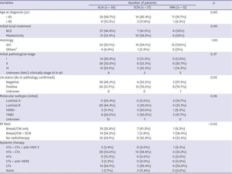
The 87 women with regional oligo-recurrences were classified into 3 groups according to the location of the recurrence: ALN (n = 58), SCN (n = 17), or IMN (n = 12). The crude regional oligo-recurrence rates were 0.3% (ALN), 0.1% (SCN), and 0.1% (IMN). Table 1 shows the patient characteristics at the time of initial treatment. Histology results were as follows:
invasive ductal carcinoma (n = 82), micropapillary carcinoma (n = 2), metaplastic carcinoma (n = 2), and mucinous carcinoma (n = 1). In the SCN group, the proportion of patients who
Table 1. Patient characteristics at initial diagnosis (n = 87)
Variables Number of patients p
ALN (n = 58) SCN (n = 17) IMN (n = 12)
Age at diagnosis (yr) 0.60
< 65 52 (89.7%) 14 (82.4%) 11 (91.7%)
≥ 65 6 (10.3%) 3 (17.6%) 1 (8.3%)
Initial local treatment 0.90
BCS 27 (46.6%) 7 (41.2%) 6 (50%)
Mastectomy 31 (53.4%) 10 (58.8%) 6 (50%)
Histology 1.00
IDC 54 (93.1%) 16 (94.1%) 12 (100%)
Others
*4 (6.9%) 1 (5.9%) 0 (0%)
Initial pathological stage 0.37
I 14 (26.9%) 2 (13.3%) 0 (0.0%)
II 26 (50.0%) 8 (53.3%) 6 (85.7%)
III 12 (23.1%) 5 (33.3%) 1 (14.3%)
Unknown (NAC) clinically stage III in all 6 2 5
LN status (Bx or pathology confirmed) 0.05
Negative 28 (48.3%) 4 (23.5%) 3 (27.3%)
Positive 30 (51.7%) 13 (76.5%) 8 (72.7%)
Unknown 0 0 1
Molecular subtype (initial) 0.26
Luminal A 11 (24.4%) 0 (0.0%) 2 (16.7%)
Luminal B 20 (44.4%) 3 (30.0%) 4 (33.3%)
HER2+ 5 (11.1%) 2 (20.0%) 1 (8.3%)
TNBC 9 (20.0%) 5 (50.0%) 5 (41.7%)
Unknown 13 7 0
RT field < 0.05
Breast/CW only 19 (32.8%) 7 (41.2%) 1 (8.3%)
Breast/CW + SCN 14 (24.2%) 1 (5.9%) 7 (58.4%)
No radiotherapy 25 (43.1%) 9 (52.9%) 4 (33.3%)
Systemic therapy
HTx + CTx + anti-HER-2 2 (3.4%) 0 (0.0%) 1 (8.3%)
HTx + CTx 29 (50.0%) 10 (58.8%) 4 (33.3%)
HTx 9 (15.5%) 0 (0.0%) 0 (0.0%)
CTx + anti-HER2 3 (5.2%) 0 (0.0%) 0 (0.0%)
CTx 14 (24.1%) 5 (29.4%) 6 (50.0%)
None 1 (1.7%) 2 (11.8%) 0 (0.0%)
Values are presented as number (%).
ALN = axillary lymph node; SCN = supraclavicular lymph node; IMN = internal mammary lymph node; BCS = breast-conserving surgery; IDC = invasive ductal carcinoma; NAC = neoadjuvant chemotherapy; LN = lymph node; Bx = biopsy; HER2 = human epidermal growth factor receptor 2; TNBC = triple-negative breast cancer; RT = radiotherapy; CW = chest wall; HTx = hormone therapy; CTx = chemotherapy.
*
Micropapillary carcinoma (n = 2), metaplastic carcinoma (n = 2), and mucinous carcinoma (n = 1).
received RT on the chest wall + SCN was significantly low (24.9% in the ALN group, 5.9% in the SCN group, 58.4% in the IMN group; p < 0.05). No patient with IMN recurrence received RT in the IMN field initially.
The clinical and biological features of regional oligo-recurrences are shown in Table 2. The median patient age was 52 years (range: 30–79 years). The median DFI was 34 months (range:
4–189 months). Regional recurrences occurred in non-suspicious lymph nodes at the initial diagnosis or newly appearing lymph nodes. In-field failure occurred in 15 of 43 (25.9%) patients in the ALN group, which was significantly higher than that in the SCN group (1/17 [5.9%] patients) and the IMN group (0/12 [0.0%] patients) (p < 0.05). All patients in the ALN group received salvage surgery, which was a significantly higher percentage than the SCN group (12/17 [60.6%] patients) and the IMN group (4/12 [33.3%] patients) (p < 0.01).
Salvage RT was performed in 18.8% (3/16) of patients with in-field failure and 77.5% (55/71) of patients with out-field failure. Thus, salvage RT could be significantly affected by whether Table 2. Patient characteristics at first recurrence (n = 87)
Variables Number of patients p
ALN (n = 58) SCN (n = 17) IMN (n = 12)
F/U period (after recurrence) 0.62
Median, range (months) 51.5 (6–194) 39 (11–163) 40 (7–134)
Age at recurrence (yr) 0.56
< 65 50 (86.2%) 13 (76.5%) 10 (83.3%)
≥ 65 8 (13.8%) 4 (23.5%) 2 (16.7%)
DFI (mo) 0.15
< 24 20 (34.5%) 4 (23.5%) 7 (58.3%)
≥ 24 38 (65.5%) 13 (76.5%) 5 (41.7%)
Pathologic confirmation 1.00
Yes 58 (100%) 17 (100%) 11 (91.7%)
No 0 (0.0%) 0 (0.0%) 1 (8.3%)
PET/CT 0.83
Yes 53 (91.4%) 16 (94.1%) 12 (100%)
No 5 (8.6%) 1 (5.9%) 0 (0.0%)
In-field failure < 0.05
Yes 15 (25.9%) 1 (5.9%) 0 (0.0%)
No 43 (74.1%) 16 (94.1%) 12 (100%)
Molecular subtype (recur) 0.76
Luminal A 14 (30.4%) 2 (28.6%) 2 (66.6%)
Luminal B 18 (39.1%) 3 (42.9%) 0 (0.0%)
HER2+ 4 (8.7%) 1 (14.3%) 0 (0.0%)
TNBC 10 (21.7%) 1 (14.3%) 1 (33.3%)
Unknown 12 10 9
Salvage local therapy < 0.05
Surgery 27 (46.6%) 2 (11.8%) 0 (0.0%)
RT 0 (0.0%) 5 (29.4%) 8 (66.7%)
Surgery + RT 31 (53.4%) 10 (58.8%) 4 (33.3%)
Dose of salvage RT
Median, range (EQD2, Gy) 50 (42–60) 54 (44–68) 57 (44–68)
Salvage systemic therapy
HTx + CTx + anti-HER-2 1 (1.7%) 2 (11.8%) 0 (0.0%)
HTx + CTx 18 (31.0%) 2 (11.8%) 0 (0.0%)
HTx 21 (36.2%) 6 (35.3%) 5 (41.7%)
CTx + anti-HER-2 2 (3.4%) 0 (0.0%) 1 (8.3%)
CTx 10 (17.2%) 3 (17.6%) 4 (33.3%)
Anti-HER-2 2 (3.4%) 1 (5.9%) 0 (0.0%)
None 4 (6.9%) 3 (17.6%) 1 (8.3%)
ALN = axillary lymph node; SCN = supraclavicular lymph node; IMN = internal mammary lymph node; F/U = follow-up; DFI = disease-free interval; PET/CT
= positron emission tomography-computed tomography; HER2 = human epidermal growth factor receptor 2; TNBC = triple-negative breast cancer; RT =
radiotherapy, EQD2 = equivalent dose in 2 Gy fractions; HTx = hormone therapy; CTx = chemotherapy.
the case involved in-field failure (p < 0.01). Chemotherapy or hormone therapy for salvage treatment was delivered to 90.8% (79/87) of patients.
With respect to molecular subtypes, 43 patients had data on both initial and recurrent lesions. Of these patients, 76.7% (33/43) had the same molecular subtypes for both lesions.
When patients were grouped into the luminal A or non-luminal A subtypes, 81.4% (35/43) had the same subtype. The effect of subtype change could not be evaluated because of the small number of patients (Supplementary Table 1).
Clinical outcomes
The median follow-up duration after regional oligo-recurrence was 49 months (range:
6–194 months). During the follow-up period, 56 of the 87 (64.4%) patients had a second recurrence and 50 of those 56 (89.3%) patients had failures with DM (Figure 1). Of 56 patients with second recurrences, 45 (80.4%) experienced a second recurrence within 2 years. Of 31 patients who did not experience a second recurrence during follow-up, 20 had > 5 years of follow-up. Thirty-four patients died after the second recurrence and three died without recurrence. Overall, the 5-year PFS rate was 36.7%, and the 5-year OS rate was 63.6%. In the ALN, SCN, and IMN groups, the 5-year PFS and OS rates were 40.0%, 32.1%, and 25.0% (p = 0.30), and 62.7%, 70.0%, and 58.3% (p = 0.97), respectively (Supplementary Figure 2).
Prognostic factors
The results of the univariable analysis are shown in Table 3. Patients with initial stage II–III, initially positive lymph node, DFI < 24 months, in-field failure, and non-luminal A subtype had significantly lower PFS. The age at the time of recurrence (≥ 65 years) had borderline significance for PFS (p = 0.07). For OS, patients who were aged ≥ 65 years and had a non- luminal A subtype at the time of recurrence had significantly worse outcomes.
Same regional area
(17 patients) Other regional area (19 patients)
Distant metastasis (50 patients) (2/56) 3.6%
(4/56) 7.1%
(5/56) 8.9% 14.3%
(8/56) 16.1%
(9/56)
50.0%
(28/56)
2nd recurrence: 56 of 87 (64.4%) patients
Figure 1. Pattern of failure after regional oligo-recurrences.
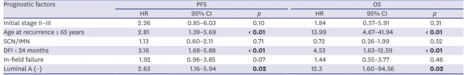
In the multivariable analysis, age at recurrence of ≥ 65 years (HR, 2.81; 95% CI, 1.39–5.69; p <
0.01), DFI < 24 months (HR, 3.16; 95% CI, 1.68–5.88; p < 0.01), and non-luminal A subtype (HR, 2.63; 95% CI, 1.16–5.94; p = 0.02) were significant poor prognostic factors for PFS. In-field failure also had borderline significance for PFS (HR, 1.92; 95% CI, 0.96–3.85; p = 0.07). However, the location of recurrence did not affect PFS (p = 0.52). When we performed the backward stepwise elimination method, the location of the recurrence was the last factor excluded for the final model, and it was still not significant. After excluding patients initially with stage I disease and/or with in-field failure, to reduce a possible confounding effect, the location of recurrence was still not a significant factor in the multivariable analysis for PFS (Supplementary Table 2).
For OS, age at recurrence of ≥ 65 years (HR, 13.99; 95% CI, 4.67–41.94; p < 0.01), DFI < 24 months (HR, 4.53; 95% CI, 1.63–12.59; p < 0.01), and non-luminal A subtype (HR, 12.30; 95% CI, 1.60–94.56; p = 0.02) were significantly poor prognostic factors (Table 4).
The patients were stratified according to the number of risk factors (RFs), such as age at recurrence of ≥ 65 years, DFI < 24 months, in-field failure, and a non-luminal A subtype. The 5-year PFS rates were 67.5% in patients with ≤ 1 RF (n = 38) and 7.3% in those with > 1 RF (n
= 41) (p < 0.01). Patients with ≤ 1 RF also had significantly better PFS within the ALN group and the SCN or IMN groups (p < 0.01 and p = 0.02, respectively). In addition, those in the ALN group with ≤ 1 RF tended to have a better prognosis than those in the SCN or IMN group Table 3. Prognostic factors for PFS and OS in the univariable analysis
Variables No. 5-year PFS p 5-year OS p
Initial stage (I vs. II vs. III) 0.04 0.53
I 16 66.2% 82.5%
II 40 37.5% 63.1%
III 18 27.8% 59.3%
Initial stage (I vs. II–III) < 0.01 0.27
I 16 66.2% 82.5%
II–III 70 32.4% 60.3%
Initial lymph node 0.01 0.54
Negative 35 56.1% 71.0%
Positive 51 25.1% 59.9%
Age at recurrence (yr) 0.07 < 0.01
< 65 73 43.1% 72.4%
≥ 65 14 14.3% 19.0%
DFI (mo) < 0.01 0.15
< 24 31 19.4% 49.6%
≥ 24 56 46.3% 70.9%
Location 0.13 0.87
ALN 58 40.0% 62.7%
SCN/IMN 29 29.0% 64.8%
In-field failure 0.02 0.31
Yes 16 12.5% 50.7%
No 71 42.6% 66.5%
Luminal A 0.01 < 0.01
(+) 18 65.8% 92.3%
(−) 61 26.3% 55.5%
Salvage local therapy 0.15 0.26
Surgery 29 26.8% 51.2%
RT 13 30.8% 50.0%
Combined 45 44.9% 74.5%
Salvage systemic therapy 0.43 0.72
Yes 76 34.0% 61.5%
No 8 27.3% 70.7%
PFS = progression-free survival, OS = overall survival; ALN = axillary lymph node; SCN = supraclavicular lymph node; IMN = internal mammary lymph node; DFI =
disease-free interval; RT = radiotherapy.
with ≤ 1 RF (5-year PFS: 73.5% in the ALN group vs. 51.1% in the SCN/IMN group; p = 0.09).
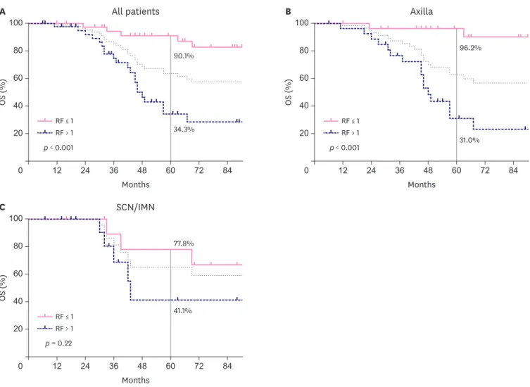
For those with > 1 RF, such a difference by location was not observed (Figure 2). There was also a significant difference according to the number of RFs for OS, with 90.9% of those with 5-year OS belonging to the group with ≤ 1 RF and 34.3% belonging to the group with > 1 RF (p
< 0.01) (Figure 3).
Table 4. Prognostic factors for PFS and OS in the multivariable analysis
Prognostic factors PFS OS
HR 95% CI p HR 95% CI p
Initial stage II–III 2.26 0.85–6.03 0.10 1.84 0.57–5.91 0.31
Age at recurrence ≥ 65 years 2.81 1.39–5.69 < 0.01 13.99 4.67–41.94 < 0.01
SCN/IMN 1.13 0.60–2.11 0.71 0.72 0.26–1.99 0.52
DFI < 24 months 3.16 1.68–5.88 < 0.01 4.53 1.63–12.59 < 0.01
In-field failure 1.92 0.96–3.85 0.07 1.44 0.55–3.77 0.46
Luminal A (−) 2.63 1.16–5.94 0.02 12.3 1.60–94.56 0.02
PFS = progression-free survival; OS = overall survival; HR = hazard ratio; CI = confidence interval; SCN = supraclavicular lymph node; IMN = internal mammary lymph node; DFI = disease-free interval.
Months All patients
p < 0.001 73.3%
24.4%
7.3%
67.4%
0
PF S (%)
40 80 100
60 48 36 24
A
60
20
84 72 12
RF ≤ 1 RF > 1
Months SCN/IMN
p = 0.02 61.4%
21.4%
7.1%
51.1%
0
PF S (%)
40 80 100
60 48 36 24
C
60
20
84 72 12
RF ≤ 1 RF > 1
Months Axilla
p < 0.001 77.8%
25.9%
7.3%
73.5%
0
PF S (%)
40 80 100
60 48 36 24
B
60
20
84 72 12
RF ≤ 1 RF > 1
Figure 2. PFS according to the number of RFs.
(A) PFS according to the number of RFs in all patients, (B) PFS according to the number of RFs in patients with recurrence on axilla, and (C) PFS according to the number of RFs in patients with recurrence on SCN or IMN.
PFS = progression-free survival; RF = risk factor; SCN = supraclavicular lymph node; IMN = internal mammary lymph node.
DISCUSSION
In this study, we compared the clinical outcomes of 87 patients with regional oligo- recurrences of breast cancer. Overall, the 5-year PFS and OS rates were 36.7% and 63.6%, respectively. There was no significant difference in PFS or OS according to the location of recurrence. Age at recurrence of ≥ 65 years, DFI < 24 months, non-luminal A subtype, and in-field failure (marginally significant) were significant RFs for PFS according to the multivariable analysis. The 5-year PFS and OS rates were significantly different according to the number of RFs (5-year PFS and OS = 67.5% and 90.9% with ≤ 1 RF vs. 7.3% and 34.3%
with > 1 RF, respectively; both p < 0.01).
Isolated regional recurrences without local metastasis and DM are rare in breast cancer patients [1-8]. Isolated ALN recurrence is relatively frequent but occurs in only 0.5%–4.6%
of patients [7,9-14]. SCN, another site of regional recurrence, has an isolated SCN recurrence rate of 0.4%–1.8% [4,15,16]. Isolated IMN recurrence is very rare, with an incidence rate of only 0.1%–0.5% [5,6].
Months Axilla
31.0%
96.2%
0
OS (%) 40
80 100
60 48 36 24
B
60
20
84 72 12
p < 0.001 RF ≤ 1 RF > 1
Months All patients
34.3%
90.1%
0
OS (%) 40
80 100
60 48 36 24
A
60
20
84 72 12
p < 0.001 RF ≤ 1 RF > 1
Months SCN/IMN
41.1%
77.8%
0
OS (%) 40
80 100
60 48 36 24
C
60
20
84 72 12
p = 0.22 RF ≤ 1 RF > 1
Figure 3. OS according to the number of RFs.
(A) OS according to the number of RFs in all patients, (B) OS according to the number of RFs in patients with recurrence on axilla, and (C) OS according to the number of RFs in patients with recurrence on SCN or IMN.
OS = overall survival; RF = risk factor; SCN = supraclavicular lymph node; IMN = internal mammary lymph node.
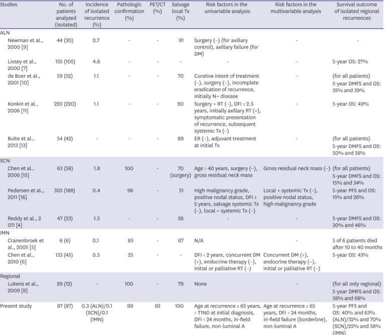
Although regional oligo-recurrence (isolated ALN, SCN, and IMN recurrence) is considered curable, the number of studies on these recurrences is limited owing to rarity, and most studies are focused on isolated ALN recurrences. In patients with isolated ALN recurrences, the 5-year OS rates were 27%–58%. [7,9-11,13]. For patients with isolated SCN recurrences, an OS of 26%–46% has been reported in a few studies [4,15,16]. Only one study reported a 5-year OS of 43% for patients with isolated IMN recurrences [6].
In our study, for patients with isolated recurrence of ALN, SCN, or IMN, the 5-year PFS and OS were 40.0%, 32.1%, and 25.0% (p = 0.3), and 62.7%, 70.0%, and 58.3% (p = 0.97), respectively, which were higher than those previously reported. PET/CT was performed to diagnose true regional oligo-recurrences in most patients, and all patients received salvage local treatment, which has been shown to be associated with better survival in previous studies. Considering these, the outcomes were reasonable (Table 5).
Generally, patients with isolated ALN recurrences were thought to have better prognosis than those with IMN or SCN recurrences, and the location of recurrence was the first indicator to identify prognostic factors. However, there were no significant differences in PFS or OS based on the location of recurrence in the univariable and multivariable analyses. There was only a trend showing better PFS in the ALN group with ≤ 1 RF (p = 0.09). However, age at recurrence of ≥ 65 years, DFI < 24 months, in-field failure, and non-luminal A subtype were the more important prognostic factors in both the univariable and multivariable analyses. All these have been significant factors in previous studies, and we re-identified the significance in regional oligo-recurrences (Table 5). We also found that there was a difference of > 50% in terms of both the 5-year PFS and OS between the groups with ≤ 1 and > 1 RF.
The fundamental limitation of this study is that it was a retrospective study on rare recurrent cases from a single institution. We evaluated only patients with isolated regional recurrences, while most previous studies included patients with regional recurrences with or without simultaneous local metastasis or DM. Therefore, this study was based on a small and heterogeneous group of patients. Our study has a major strength of including only isolated regional recurrences. This strength is enhanced by the confirmation of isolated regional recurrence without distant metastasis via PET/CT at the time of recurrence. In the future, multicenter large-scale research is necessary to verify our findings.
In conclusion, we found that for patients with regional oligo-recurrence of breast cancer who had undergone salvage treatment, including local therapy, the prognosis was primarily affected by the following RFs: age at recurrence of ≥ 65 years, DFI < 24 months, in-field failure, and non-luminal A subtype, rather than the location of the recurrence. Whether the recurrence location was the axilla or IMN/SCN, regional oligo-recurrences with ≤ 1 RF had better outcomes when salvage therapy was delivered with sufficient local treatment.
In patients with > 1 RF, more effective systemic treatments to decrease DM should be
considered even if the location of recurrence is the axilla.
SUPPLEMENTARY MATERIALS
Supplementary Table 1
Relevance of molecular subtypes of initial and recurrent disease (p < 0.001) Click here to view
Table 5. Recent studies including analysis on patients with isolated regional recurrences
Studies No. of
patients analyzed (isolated)
Incidence of isolated recurrence
(%)
Pathologic confirmation
(%)
PET/CT (%) Salvage
local Tx (%)
Risk factors in the
univariable analysis Risk factors in the
multivariable analysis Survival outcome of isolated regional
recurrences ALN
Newman et al.,
2000 [9] 44 (30) 0.7 - - 91 Surgery (−) (for axillary
control), axillary failure (for DM)
- -
Livsey et al.,
2000 [7] 155 (105) 4.6 - - - - - 5-year OS: 27%
de Boer et al.,
2001 [10] 59 (52) 1.1 - - 70 Curative intent of treatment
(−), surgery (−), incomplete eradication of recurrence, initially N+ disease
- (for all patients) 5-year DMFS and OS:
35% and 39%
Konkin et al.,
2006 [11] 220 (220) 1.1 - - 90 Surgery + RT (−), DFI ≤ 2.5
years, initially axillary RT (+), symptomatic presentation of recurrence, subsequent systemic Tx (−)
- 5-year OS: 49%
Bulte et al.,
2013 [13] 54 (42) - - - 89 ER (−), adjuvant treatment
at initial Tx - (for all patients)
5-year DMFS and OS:
50% and 58%
SCN
Chen et al.,
2006 [15] 63 (58) 1.8 100 - 70
(surgery) Age > 40 years, surgery (−),
gross residual neck mass Gross residual neck mass (−) (for all patients) 5-year DMFS and OS:
15% and 34%
Pedersen et al.,
2011 [16] 305 (188) 0.4 96 - 51 High malignancy grade,
positive nodal status, DFI ≤ 2 years, salvage systemic Tx (−), local + systemic Tx (−)
Local + systemic Tx (−), positive nodal status, high malignancy grade
5-year PFS and OS:
15% and 26%
Reddy et al., 2
011 [4] 47 (23) 1.5 - - 26 - - 5-year DMFS and OS:
30% and 46%
IMN
Cranenbroek et
al., 2005 [5] 6 (6) 0.1 83 - 67 N/A - 5 of 6 patients died
after 10 to 40 months Chen et al.,
2010 [6] 133 (45) 0.5 25 - - DFI < 2 years, concurrent DM
(+), endocrine therapy (−), initial or palliative RT (−)
Concurrent DM (+), endocrine therapy (−), initial or palliative RT (−)
5-year OS: 43%
Regional Lukens et al.,
2009 [8] 29 (12) - 100 - 79 None - (for all only regional)
5-year DMFS and OS:
56% and 68%
Present study 87 (87) 0.3 (ALN)/0.1 (SCN)/0.1
(IMN)
99 93 100 Age at recurrence ≥ 65 years,
> T1N0 at initial diagnosis, DFI < 24 months, in-field failure, non-luminal A
Age at recurrence ≥ 65 years, DFI < 24 months, In-field failure (borderline), non-luminal A
5-year PFS and OS: 40% and 63%
(ALN)/32% and 70%
(SCN)/25% and 58%
(IMN)
PET/CT = positron emission tomography-computed tomography; Tx = treatment; ALN = axillary lymph node; DM = distant metastasis; OS = overall survival; DMFS
= distant metastasis-free survival; RT = radiotherapy; DFI = disease-free interval; ER = estrogen receptor; SCN = supraclavicular lymph node; PFS = progression-
free survival; IMN = internal mammary lymph node.
Supplementary Table 2
In patients with out-field failure and initial stage II−III, multivariable analysis on prognostic factors for PFS
Click here to view
Supplementary Figure 1
PFS by the different molecular subtypes.
Click here to view
Supplementary Figure 2
PFS and OS by the location of the recurrence.
Click here to view
REFERENCES
1. Wadasadawala T, Vadgaonkar R, Bajpai J. Management of isolated locoregional recurrences in breast cancer: a review of local and systemic modalities. Clin Breast Cancer 2017;17:493-502.
PUBMED | CROSSREF
2. Harris EE, Hwang WT, Seyednejad F, Solin LJ. Prognosis after regional lymph node recurrence in patients with stage I-II breast carcinoma treated with breast conservation therapy. Cancer 2003;98:2144-51.
PUBMED | CROSSREF
3. Jeong Y, Kim SS, Gong G, Lee HJ, Ahn SH, Son BH, et al. Treatment results of breast cancer patients with locoregional recurrence after mastectomy. Radiat Oncol J 2013;31:138-46.
PUBMED | CROSSREF
4. Reddy JP, Levy L, Oh JL, Strom EA, Perkins GH, Buchholz TA, et al. Long-term outcomes in patients with isolated supraclavicular nodal recurrence after mastectomy and doxorubicin-based chemotherapy for breast cancer. Int J Radiat Oncol Biol Phys 2011;80:1453-7.
PUBMED | CROSSREF
5. Cranenbroek S, van der Sangen MJ, Kuijt GP, Voogd AC. Diagnosis, treatment and prognosis of internal mammary lymph node recurrence in breast cancer patients. Breast Cancer Res Treat 2005;89:271-5.
PUBMED | CROSSREF
6. Chen L, Gu Y, Leaw S, Wang Z, Wang P, Hu X, et al. Internal mammary lymph node recurrence: rare but characteristic metastasis site in breast cancer. BMC Cancer 2010;10:479.
PUBMED | CROSSREF
7. Livsey JE, Magee B, Stewart AL, Swindell R. Axillary recurrence following conservative surgery and radiotherapy in early breast cancer. Clin Oncol (R Coll Radiol) 2000;12:309-14.
PUBMED | CROSSREF
8. Lukens JN, Vapiwala N, Hwang WT, Solin LJ. Regional nodal recurrence after breast conservation treatment with radiotherapy for women with early-stage breast carcinoma. Int J Radiat Oncol Biol Phys 2009;73:1475-81.
PUBMED | CROSSREF
9. Newman LA, Hunt KK, Buchholz T, Kuerer HM, Vlastos G, Mirza N, et al. Presentation, management and outcome of axillary recurrence from breast cancer. Am J Surg 2000;180:252-6.
PUBMED | CROSSREF
10. de Boer R, Hillen HF, Roumen RM, Rutten HJ, van der Sangen MJ, Voogd AC. Detection, treatment and outcome of axillary recurrence after axillary clearance for invasive breast cancer. Br J Surg 2001;88:118-22.
PUBMED | CROSSREF
11. Konkin DE, Tyldesley S, Kennecke H, Speers CH, Olivotto IA, Davis N. Management and outcomes of isolated axillary node recurrence in breast cancer. Arch Surg 2006;141:867-72.
PUBMED | CROSSREF
12. van der Ploeg IM, Nieweg OE, van Rijk MC, Valdés Olmos RA, Kroon BB. Axillary recurrence after a tumour-negative sentinel node biopsy in breast cancer patients: a systematic review and meta-analysis of the literature. Eur J Surg Oncol 2008;34:1277-84.
PUBMED | CROSSREF
13. Bulte JP, van Wely BJ, Kasper S, Kuijt G, van den Wildenberg FJ, Strobbe LJ, et al. Long-term follow-up of axillary recurrences after negative sentinel lymph node biopsy: effect on prognosis and survival. Breast Cancer Res Treat 2013;140:143-9.
PUBMED | CROSSREF
14. Houvenaeghel G, Classe JM, Garbay JR, Giard S, Cohen M, Faure C, et al. Survival impact and predictive factors of axillary recurrence after sentinel biopsy. Eur J Cancer 2016;58:73-82.
PUBMED | CROSSREF
15. Chen SC, Chang HK, Lin YC, Leung WM, Tsai CS, Cheung YC, et al. Prognosis of breast cancer after supraclavicular lymph node metastasis: not a distant metastasis. Ann Surg Oncol 2006;13:1457-65.
PUBMED | CROSSREF
16. Pedersen AN, Møller S, Steffensen KD, Haahr V, Jensen M, Kempel MM, et al. Supraclavicular recurrence after early breast cancer: a curable condition? Breast Cancer Res Treat 2011;125:815-22.
PUBMED | CROSSREF
17. Niibe Y, Hayakawa K. Oligometastases and oligo-recurrence: the new era of cancer therapy. Jpn J Clin Oncol 2010;40:107-11.
PUBMED | CROSSREF