J O U R N A L O F
Veterinary Science
J. Vet. Sci. (2005), 6(4), 349–352
Comparison of an enzyme-linked immunosorbent assay with serum neutralization test for serodiagnosis of porcine epidemic diarrhea virus infection
Jin Sik Oh, Dae Sub Song, Jeong Sun Yang, Ju Young Song, Hyoung Joon Moon, Tae Yung Kim, Bong Kyun Park*
Department of Veterinary Medicine, College of Veterinary Medicine and School of Agricultural Biotechnology, Seoul National University, Seoul 151-742, Korea
An indirect porcine epidemic diarrhea (PED) virus (PEDV) enzyme-linked immunosorbent assay (ELISA) was compared with the serum neutralization (SN) test by testing 46 samples from experimentally infected sows, 73 samples from naïve sows, and 1,024 field sow samples from 48 commercial swine farms of undefined PED status.
The SN test and the ELISA were performed using PEDV, KPEDV-9 strain. Viral proteins as a coating antigen of PEDV ELISA were extracted from the cytoplasm of PEDV-infected Vero cells using a non-ionic detergent, Triton X-100, and a simple protocol of PEDV ELISA was followed. The presence of antibodies in these experimental samples was confirmed by SN and ELISA in which the sensitivity of the ELISA was 89.1%, and the corresponding specificity was 94.5%. On testing 1,024 field samples, an overall agreement of 84.2% was generated between the SN and ELISA. This study demonstrates that the PEDV ELISA is a useful serodiagnostic screening test at herd level for detecting swine antibodies against PEDV.
Key words: ELISA, porcine epidemic diarrhea virus, serum neutralization test
Porcine epidemic diarrhea virus (PEDV), a member of the coronavirus group I [17], causes watery diarrhea, dehydration, and high mortality in suckling pigs through the destruction of epithelial cells of the small intestine [4,14]. A serological diagnosis for porcine epidemic diarrhea (PED) has been made through the demonstration of antibodies against PEDV. Currently, the serum neutralization (SN) test is the serological method used most commonly in veterinary diagnostic laboratories for detecting swine serum antibodies against PEDV. The SN test in this study is conducted
routinely at our laboratory with the cutoff value of 1 : 4.
Titers at 1 : 2 may be true positive but may also be false positive due to nonspecific reactions. Some of the known positive and negative samples collected from experimentally infected sows had the titer of 1 : 2 (data not shown). Serum neutralization assay is a time-consuming and labor-intensive immunoassay requiring serial dilution of serum samples, preparation of cells, titration of virus, and manual reading of plates [11,12]. In addition, it is not useful for mass screening of swine sera for diagnosis of PED. However, SN test shows high specificity [9,10,18].
In the previous reports, an indirect enzyme-linked immunosorbent assay (ELISA) with a modification of ELISA antigen preparation was developed for detecting swine antibodies against PEDV [9,10,18]. However, these ELISAs showed highly nonspecific background when routinely applied to swine sera and the sensitivity of the ELISA should be improved with this antigen [3,9,10]. The objective of this study was to establish a reliable indirect ELISA and to compare the ELISA with the SN test to assess the agreement between the tests using experimental and field-serum samples.
Previously PEDV antibody- and PEDV-free, a commercial sow population was inoculated intramuscularly (IM) with 1 ml of KPEDV-9 virus (10
3.5TCID
50/0.1 ml). Positive sera (SN titer; ≥ 1:4) were obtained from 46 sows at 4 weeks postinoculation (PI) and negative sera (SN titer; <1:4) were also obtained from 73 commercial sows. Sow serum samples of 1,024 from 48 commercial swine farms of undefined PED status were obtained from submissions of CVMVL- SNU. All samples were tested on the PEDV SN [11,12]
according to the standard protocol routinely performed at our laboratory. Briefly, Vero cells (ATCC CCL-81) were regularly maintained in α -MEM (minimal essential medium) supplemented with 5% fetal bovine serum, and 2%
antibiotic-antimycotic agent mixture (Life Technologies, USA). PEDV, KPEDV-9 strain [13] was propagated in Vero cells with maintenance medium of α -MEM supplemented
*Corresponding author
Tel: +82-2-880-1255; Fax: +82-2-885-0263 E-mail: [email protected]
Short Communication
350 Jin Sik Oh et al.
with 0.02% yeast extract, 0.3% tryptose phosphate broth, and 2 µ g/ml of trypsin [8,11,13]. All swine sera were inactivated at 56
oC for 30 minutes and stored at − 20
oC. The sera were diluted two-folds, and PEDV of 200 TCID
50/ 0.1 ml was mixed with an equal volume of diluted sera.
Mixture was incubated for 1 hour at 37
oC. Subsequently, 0.1 ml of each virus-serum mixture was inoculated onto Vero cell monolayers of 96-well tissue culture plates which was washed 3 times with phosphate buffered saline (PBS, pH7.2). After adsorption for 1 hr at 37
oC, inocula were discarded, and plates were washed 3 times with PBS.
Maintenance medium containing trypsin (2 µ g/ml) was then added into each well, and plates were incubated for 5 days at 37
oC. SN titers were expressed as the reciprocals of the highest serum dilution resulting in the inhibition of cytopathic effect.
To prepare ELISA antigen, Vero cells were grown in 490 cm
2roller bottles and confluent monolayers were rinsed 3 times with PBS. Subsequently, 10 ml of PEDV (10
4.5TCID
50/ml) was inoculated into each roller bottle, the cells were incubated for 1 hour at 37
oC, and the maintenance medium was replaced. After incubation of the cells for 48 hours, the infected cells were washed 3 times with 0.85%
saline, harvested using a cell scraper, and resuspended to be 1/10 of its original medium volume in 0.85% saline. Triton X-100 was then added to the suspension to be a final concentration of 0.2%. The suspension was stirred overnight at 4
oC, and then for 1 hour at 37
oC. Soluble preparation was clarified at 12,000 × g for 30 min. Supernatant was collected, and excess Triton X-100 was removed using Bio-Bead SM- 2 Absorbent (Bio-Rad, USA). Mock-infected Vero cell cultures were also processed according to the same method used for the virus-infected cells. Protein contents of prepared antigens were determined using the BCA kit (Pierce, USA).
The ELISA antigens were prepared with Vero cell cultures infected with PEDV [8]. Triton X series of nonionic detergents were known to bind preferentially to hydrophobic proteins [2,16]. Binding affinity of ELISA antigens containing Triton X-100 to microtiter wells was low due to inhibition of antigen binding by Triton X-100 [5,7]. Thus, to harvest high quality viral antigen for PEDV ELISA, Triton X-100-bound high molecular-weight cellular protein was removed by Bio- beads SM2.
Optimal concentration of mock-infected cell and PEDV ELISA antigens were determined by checkerboard titration.
Hyperimmune PEDV reference serum was prepared from an antibody-free and PEDV-free 2-week-old pig inoculated IM with 1 ml of PEDV, KPEDV-9 strain (10
5.5TCID
50/ 0.1 ml) and then the serum was collected at 4 weeks post inoculation. Negative reference serum was also obtained from a mock-infected pig of the same age. Maximum difference between positive and negative reference sera was observed when the sera were diluted to be 1 : 50 and the antigen concentrations were adjusted to be 0.1 µ g/well. The
maximal dilution of anti-swine HRP-conjugated goat antibody and the optimal reaction time have been determined previously.
To prepare PEDV ELISA plates, mock-infected cell and PEDV ELISA antigens were diluted to be the concentration of 1.0 µ g/ml in a coating buffer (50 mM bicarbonate buffer, pH 9.6). Alternate 8-well rows of 96-well microtiter plates (Nalge Nunc International, USA) were coated by adding 100 µ l/well of each ELISA antigen, and the plates were incubated overnight at 4
oC. Antigens were poured off, and the plates were then washed 5 times with PBST (PBS, 0.05% Tween 20). After washing the plates, the remaining free-binding sites were blocked with 200 µ l of blocking solution (5% rabbit serum (Gemini Bioproducts, USA) in PBS) for 1 hour at 37
oC. Two positive and negative reference each, and 44 sample swine sera were diluted to be 1 : 50 in PBST, 100 µ l of which was transferred to a pair of well in each plate, and plates were then incubated for 1 hour at 37
oC. After washing plates, 100 µ l of HRP-conjugated goat anti-pig IgG (KPL, USA) (1 : 2,000 dilution) was added, and plates were further incubated for 1 hour at 37
oC. The color was developed using ABTS (KPL, USA) for 30 minutes at room temperature in the dark. Optical density (OD) was measured at 405 nm. Corrected OD and corrected value (CV) of each swine serum were calculated as follow.
Corrected OD = OD of a sample swine serum on PEDV ELISA antigen − OD of a sample swine serum on mock- infected cell ELISA antigen
CV = (OD of a sample swine serum on PEDV ELISA antigen − OD of a sample swine serum on mock-infected cell ELISA antigen) ÷ (Mean OD of negative control swine serum on PEDV ELISA antigen − Mean OD of negative control swine serum on mock-infected cell ELISA antigen)
Sera from sows vaccinated with Vero cell culture-derived PEDV may react with the remaining cellular components of PEDV ELISA antigen, inducing high background and low specificity. Thus, a formula for corrected OD was designed for removing high background reactions and for improving specificity. In an ELISA for the antibody detection against porcine reproductive and respiratory syndrome virus (PRRSV), the ELISA antigens against eight different
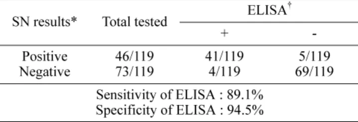
Table 1. Comparison of porcine epidemic diarrhea virus ELISA with serum neutralization (SN)
SN results* Total tested ELISA
†+ -
Positive
Negative 46/119
73/119 41/119
4/119 5/119
69/119 Sensitivity of ELISA : 89.1%
Specificity of ELISA : 94.5%
*The cutoff value for SN was 1:4.
†
ELISA results were expressed as corrected value and ≥2.0 was
considered as positive.
ELISA for serodiagnosis of PED 351
PRRSV isolates showed different sensitivities [6]. However, because there was no report indicating the presence of different PEDV serotypes [15], the authors did not consider PEDV ELISA antigens derived from other strains.
The sensitivity and specificity of the ELISA were determined at cutoff value of CV 2.0. The optimal pair of sensitivity and specificity was 89.1% and 94.5% as shown in Table 1. To evaluate cross-reactivity with antibody against other swine viral pathogens in the ELISA, it was investigated with transmissible gastroenteritis virus (TGEV) specific sera obtained from experimentally infected pigs, positive swine sera against swine influenza virus (SIV) (Serotype H3N2 & H1N1) provided by ISU-VDL (Iowa State University’s Veterinary Diagnostic Laboratory, USA) and positive swine sera against pseudorabies virus (PrV).
The antisera to SIVs, PrV, and TGEV had negative CV, suggesting no cross reactivity with PEDV ELISA antigen.
In an established PEDV ELISA, only PEDV antiserum was reacted to the PEDV antigen. In addition, correlation of PEDV ELISA to SN was measured to evaluate the reliability of ELISA, and R values were calculated using the Microcal Origin 6.0 program (Microcal Software, USA). As shown in Fig. 1, the R-value between corrected OD and SN titer, or between CV and SN titer was 0.837 and 0.857, respectively, indicating that the ELISA is the same reliable as SN test. However, some degree of discrepancy may be expected between these assays and they may not detect antibodies against PEDV. No comparative data was available regarding on the correlation between the SN test and the serological ELISA against PEDV. When a new diagnostic test is evaluated to a “gold standard”, two tests need to be biologically independent [1].
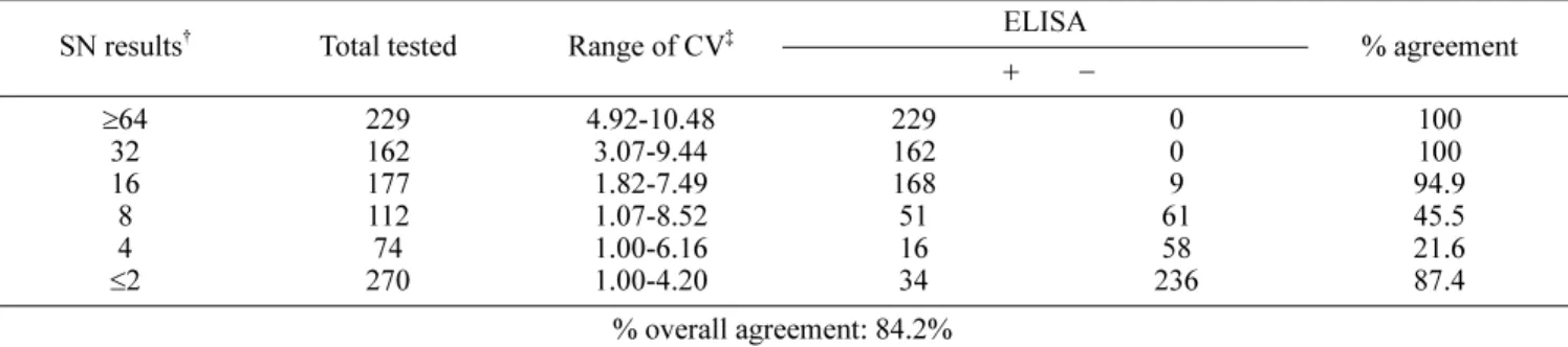
On testing 1,024 field sow samples from 48 swine farms, this comparison generated an overall agreement of 84.2%
between these tests as shown in Table 2. These results suggest that if the SN is used as the “gold standard”, the ELISA would generate 17.0% false-negative results by missing 128 of 754 SN-positive samples. On the other hand, the ELISA would produce 12.6% false-positive results by
Fig. 1. Correlation between a corrected OD and a corrected value of PEDV ELISA to serum neutralization (SN) test. Each closed circle ( ● ) represents a corrected OD (A) and a corrected value (B) of the reference serum against a known SN titer.
Table 2. Comparison of porcine epidemic diarrhea virus ELISA with serum neutralization (SN) by testing 1,024 field sow serum samples*
SN results
†Total tested Range of CV
‡+ ELISA − % agreement
≥ 64 32 16
8 4
≤ 2
229 162 177 112 270 74
4.92-10.48 3.07-9.44 1.82-7.49 1.07-8.52 1.00-6.16 1.00-4.20
229 162 168 51 16 34
0 0 61 9 236 58
100 100 94.9 45.5 21.6 87.4
% overall agreement: 84.2%
*All samples were submitted to CVMVL-SNU for porcine epidemic diarrhea virus SN tests without known PED status.
†
The SN test titers were expressed as the reciprocal dilution of serum samples.
‡