흰쥐에서 고용량의 스테로이드 투여가 혈액 응고 및 혈전 용해에 미치는 영향
가톨릭대학교 의과대학 정형외과학교실
송 주 현・김 용 식
- Abstract -
Effects of High-dose Steroid Treatment on Coagulopathy and Hypofibrinolysis in Rats
Joo-Hyun Song, M.D. and Yong-Sik Kim, M.D.
Department of Orthopaedic Surgery, College of Medicine, The Catholic University of Korea, Seoul, Korea
This study was conducted to investigate the effects of high-dose glucocorticoid administration on coagulation and fibrinolysis in rats. Sixty healthy Wistar rats were divided into three groups. Group A consisted of 20 control animals that received 5ml/kg isotonic saline for 1 weeks. Group B consisted of 20 animals that received 5㎎/㎏of methylprednisolone intraperitoneally for 1 week. Group C comprized of 20 animals that received 10㎎/㎏of methylprednisolone intraperitoneally for 1 week. After the com- pletion of treatment, the letels of protein C antigen, protein C activity, protein S antigen and protein S activity, tissue type plasminogen activator(tPA), lipoprotein(a)(Lp(a)) and plasminogen activator inhibitor-1(PAI-1) were measured. The PAI-1 protein expression was evaluated with Western-blotting.
There was no significant difference in the plasma level of protein C antigen and activity, protein S antigen and activity, tPA and Lp(a) among the three groups. However, blood level of PAI-1 and pro- tein expression of PAI-1 were significantly increased in the rats treated with high-dose glucocorticoid.
In light of the above results, we postulated that increased secretion of PAI-1 plays a major role in the pathogenesis of osteonecrosis of the femoral head in rats treated with high-dose steroid.
Key Words : Femoral head, Osteonecrosis, High-dose steroid, Hypofibrinolysis, PAI-1
※통신저자: 김 용 식
서울특별시영등포구여의도동6 2번지
가톨릭대학교의과대학성모병원정형외과학교실 Tel : 02) 3779-1192, Fax : 02) 783-0252
서 론
대퇴골두무혈성괴사증의원인이나병인은아직확실 히밝혀지지않고있으나스테로이드의사용이가장흔한 원인으로알려져오고있다. 일반적으로는스테로이드를
장기간복용하거나사용용량이많을수록대퇴골두무혈 성괴사증의병발위험성도큰것으로알려져있으나6 , 1 2 , 2 8 , 3 0 )
일부학자들은단기간의사용으로도무혈성괴사증이발 생하며처음1개월간의사용이대퇴골두무혈성괴사증을 발생케하는데가장중요하다고보고하고있다. 스테로이 드의사용으로인한대퇴골두무혈성괴사증의발병병인
중현재까지가장널리받아들여지고있는학설은스테로 이드의사용으로인해혈중지질이증가되고이에따라지 방색전이발생하거나5 , 1 4 )골수의지방세포가증가되어주 위의 혈관을 압박함으로서혈관이폐쇄된다는것인데3 3 ) 병인이무엇이든간에결국은대퇴골두에혈액을공급하 는혈관의폐쇄가주원인이다1 3 ). Glueck 등9 )은특별한원인 을밝혀내지못했던특발성대퇴골두무혈성괴사증환자 의대부분에서정상인에비해혈액응고이상과섬유소용 해의장애가있다고보고하고있으며이차적으로발생한 대퇴골두무혈성괴사증의환자에서도혈액응고및섬유 소용해의장애를보고하고있다. 또한최근Sartori 등2 9 )은 스테로이드를 장기간 사용한 심장 이식 환자에서 혈중 P A I - 1이증가한다고보고하고있다. 그러나아직까지스테 로이드의사용으로어느정도의혈액응고이상과혈전용 해장애가발생하는지, 용량에따른차이가있는지그리고 혈전응고및섬유소용해이상이실제로대퇴골두에서발 생하는지에대해서는밝혀지지않고있다. 이연구에서는 대퇴골두무혈성괴사증의흔한유발인자로받아들여지 고있는스테로이드가혈액응고억제에관여하는C 단백 과S 단백그리고섬유소용해이상과관련된tPA, PAI-1, L p ( a )의분비와조직내P A I - 1의활성도및발현량에어떠한 영향을끼치는지관찰함으로써스테로이드의사용에의 한대퇴골두무혈성괴사증의발병병인을알아보고자하 였다.
방 법 1. 연구대상
건강한 생후 6개월 이전(체중 2 5 0 ~ 3 0 0 g )의 W i s - t a r계 흰쥐숫컷 6 0마리를대상으로 2 0마리는대조군( A군), 40 마리는실험군으로하였으며실험군은스테로이드의사 용용량에따라각각B군과C군으로나누었다.
2. 연구방법
1) 스테로이드의투여
대조군 2 0마리에는 isotonic saline 5㎖/㎏을 1일 1회 1 주간 복강내 주사하였고 실험군 4 0마리중 B 군 2 0마리 에는 methylprednisolone acetate 5㎎/㎏를 1일 1회 1주간 복강내주사하였고나머지C 군 2 0마리에는 m e t h y l p r e d- nisolone acetate 10㎎/㎏를 역시같은방법으로1주간주 사하였다. 고용량의 스테로이드 사용으로 인한 화농성 감염발생을 예방하고자Lincomycin 2㎎/㎏를각 동물에
이틀에한차례씩근육주사하였다.
2) 혈액및대퇴골두의채취
실험 3주에 각 군의 모든 흰쥐들을 ketamine chlo- r i d e ( 5 0㎎/㎏) 마취하에 복강을 종으로 절개하고 하대정 맥에서6㏄의혈액을채취한후3.8% sodium citrate가처 리된 시험관에 3㏄ 씩 나누어 주입한 뒤 4 , 0 0 0 r p m에서 1 0분간원심분리하여혈장을채취하였다. 그후양측고 관절을전방으로탈구시키고대퇴골경부가포함되도록 대퇴골두를절취하였다. 양측대퇴골두는 P A I - 1에대한 Western blotting을위하여관절연골을제거한후영하 7 0
℃에급속냉동저장하였다.
3) 혈액응고이상의측정
혈액응고이상을측정하기위하여채취한혈장으로C 단백의 항원 및 활성도를측정하고 S단백의 항원 및활 성도를측정하였다.
가. C 단백항원의측정
실험대상에서얻은혈장을1:102 배로희석한뒤각흡 착 w e l l에 2 0 0㎕씩나누어넣고실온에2시간방치하였다.
세척후 c o n j u g a t e를2 0 0㎕첨가하고다시실온에2시간방 치한후세척하여과산화수소기질을 2 0 0㎕넣은뒤정확 히3분후3M 황산 5 0㎕를첨가하였다. 10분후에측정시 약인Asserachrom Protein C(Diagnostica Stago, France)를첨 가시키고ELS 600 ELISA Reader(Bio-Tec, U.S.A.)를이용하 여발색도를측정하였다(단위% ) .
나. C 단백의활성도측정
실험대상에서얻은혈장을100, 50, 25, 12.5%로계대 희석시키고응고시험관에 0 . 1㎖Protein C deficient plas- m a와 0 . 1㎖standard dilution test plasma를혼합하였다. 37
℃에서 2분간 가온한 후 0 . 1㎖ Bioclot Protein C activa- tor(Biopool, U.S.A.)를 넣고 ACL 3000(Instrumentation Laboratory Co., Italy)을이용하여응고시간을측정하였다 (단위% ) .
다. S 단백항원의측정
실험대상에서얻은혈장을1:102 배로희석하고각흡 착 w e l l에희석된혈장을 2 0 0㎕씩넣은후실온에2시간 방치하였다. 세척후 c o n j u g a t e를 2 0 0㎕넣고다시실온에 2시간 방치한 뒤 세척하고 과산화수소기질 2 0 0㎕를 넣 고나서정확히3분후에3M 황산 5 0㎕를첨가하였다. 10 분 후에 측정 시약인 Asserachrom Protein S(Diagnostica Stago, France)를첨가시키고ELS 600 ELISA Reader (Bio- Tec, U.S.A.)를이용하여발색도를측정하였다(단위% ) .
라. S 단백의활성도측정
실험대상에서얻은혈장을100, 50, 0%로계대희석시 키고응고시험관에 0 . 1㎖Protein S deficient plasma와 0 . 1
㎖standard dilution test plasma를혼합하였다. 37℃에서2분 간가온후 0 . 1㎖Bioclot Protein S activator(Biopool, U.S.A.)를 넣고ACL 3000(Instrumentation Laboratory Co., Italy)을이용 하여응고시간을측정하였다(단위% ) .
4) 혈전용해이상의측정 가. tPA 항원량측정
실험대상에서얻은혈장을5 0㎕P E T - b u f f e r가분주된w e l l 에 2 0㎕씩분주하고약6 0 0 r p m의plate shaker에올려1시간 동안반응시켰다. 4회세척하고과산화수소기질1 0 0㎕을 추가한다음1 5분간600rpm shaker에서반응을유도한뒤 1 0 0㎕의반응정지액을첨가하여반응을정지시켰다. 10분 경과후측정시약인TrintElize tPA(Biopool, U.S.A.)를첨가시키 고ELS 600 ELISA Reader(Bio-Tec, U.S.A.)를이용하여발색도 를측정하였다(단위n g /㎖) .
나. PAI-1 항원량측정
1 0 0㎕ P E T - b u f f e r를 각 w e l l에 분주하고1분간 방치한 뒤실험동물에서얻은혈장을2 0㎕씩w e l l에분주한후 5 0
㎕의c o n j u g a t e용액을5 0 0 ~ 6 0 0 r p m의plate shaker에올려2 시간동안반응시켰다. 4회세척하고과산화수소기질을 2 0 0㎕넣은후 1 5분간600rpm shaker에서반응을유도했 다. 그후 5 0㎕의반응정지액을첨가하여반응을정지시 켰다. 10분경과 후 측정시약인 TrintElize PAI-1(Biopool, U . S . A . )를첨가한뒤ELS 600 ELISA Reader (Bio-Tec, U.S.A.) 를이용하여발색도를측정하였다(단위n g /㎖) .
다. Lipoprotein(a)의측정
실험대상에서얻은혈장 2 0㎕에 P E T - b u f f e r로희석시 킨sample buffer를섞고 p l a t e에 P E T - b u f f e r를 5 0㎕씩분 주하여혼합하였다. plate를덮고500rpm 실온에서 1 시 간동안1차반응을유도한후 c o n j u g a t e를 5 0㎕씩분주한 다. 500rpm에서 3 0분간2차반응을유도하고나서 P E T - b u f f e r로4회세척하고 s u b s t r a t e를 1 0 0㎕씩분주한다. 다 시 p l a t e를덮고500rpm 실온에서 1 5분간3차반응을유 도한후반응정지액3M 황산을50 ul 분주하고492 ㎚에 서판독한다(단위㎎/㎗) .
5) PAI-1에대한Western blotting
실험대상에서채취한대퇴골두를질소탱크에급속냉 각시킨 후 영하 7 0℃의 냉장고에 보관시켰다가 T r i P u r e
isolation reagent(Boehringer Man-nheim, Germany) 용액을넣 고Polytron 균질기를이용하여균질화시켜총단백질을 분리한 후DC microplate protein assay(Bio-Rad, Richmond, CA, U.S.A.)를이용하여단백질을 정량하였다. 10㎍에해 당하는단백질을12% SDS-polyacrylamide gel에서전기영 동한 후polyvinylidene difluoride(DVDF) membrane(Bio-Rad, Richmond, CA, U.S.A.)에전이시킨다음불특정반응을없 애기위해non-fatty dry milk(Bio-Rad, Richmond, CA, U.S.A.) 로blocking 하였다. 1:250 배로희석한affinity purified goat anti-mouse plasminogen activator inhibitor(Santa Cruz Biotechnology, U.S.A.)를일차항체로2 시간동안반응시킨 뒤 1 : 3 , 0 0 0으 로 희 석 한 anti-goat IgG alkaline phosphatase(Santa Cruz Biotechnology, U.S.A.)를이차항체 로 2 시간 동안 반응 시켰다. membrane을 세척한 후 Immun-Star Chemiuminescence(Bio-Rad, Richmond, CA, U . S . A . )와반응시킨뒤방사선필름에발광시켜띠를확인 하였고 Image Master VDS(Parma-cia Biotech, Uppsala, S w e d e n )를이용하여띠의발광도를정량화하였다.
6) 통계처리
결과는평균±표준편차로표시하였고각군에따른측 정치의비교는ANOVA test와 F i s c h e r의LSD (least signifi- cant difference)를이용한다중비교법을사용하였으며통 계학적유의수준은0.05 미만( P<0 . 0 5 )으로하였다.
결 과
실험 1주에 대조군(A 군)과 5㎎/㎏의 m e t h y l p r e d- nisolone acetate를투여한군(B 군)의흰쥐들은모두생존 하였으나 1 0㎎/㎏의methylprednisolone acetate를투여한 군(C 군)에서는4마리가사망하여실험대상에서제외시 켰다.
1) 혈액응고이상의측정
대조군과실험군모두에서C 단백과S 단백의항원은 각각 10% 미만이었으며 각 단백질의 활성도는 검출되 지않아통계학적의의를관찰할수없었다.
2) 혈전용해이상의측정 가. tPA 항원량
A 군에서는 5 . 2 9±1 . 0 8 n g /㎖, B 군에서는 5 . 6 8±
1 . 1 0 n g /㎖ 그리고 C 군에서는 6 . 3 2±7 . 2 9 n g /㎖로 측정되 었다. 당질 코르티코이드의 용량 증가에 따라 항원량도
증가 하였지만 각군 사이에통계학적유의성은 없었다 ( P>0 . 0 5 ) .
나. Lp(a)
A 군에서는 0 . 8 8±0 . 3 5㎎/㎗, B 군에서는 0 . 7 8±0 . 3 1
㎎/㎗그리고C 군에서는 1 . 2 1±0 . 2 8㎎/㎗로측정되었다.
고용량의 당질 코르티코이드 투여시 L p ( a )가 증가 하였 으나통계적으로유의하지는않았다( P>0 . 0 5 ) .
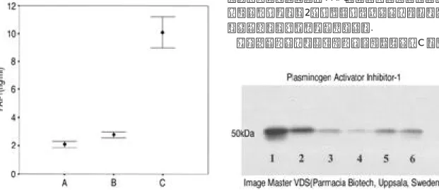
다. PAI-1 항원량
A 군에서는 2 . 1 0±1 . 0 8 n g /㎖, B 군에서는 2 . 7 7±0 . 4 8 n g /
㎖그리고C 군에서는1 0 . 0 9±2 . 2 3 n g /㎖로측정되어고용 량의당질코르티코이드투여시P A I - 1의농도가현저하게 증가하였다( P<0.05)(Fig. 1).
3) PAI-1에대한Western blotting
PAI-1 단백질의 발현을 보기위한 blotting 결과 고용량 의당질코르티코이드를투여한C 군에서50 kDa의면역 반응 띠가 관찰되었는데 C 군에서의 면역반응 띠의값 을1로보았을때, A 군은 0 . 3 6과0.19 그리고B 군은 0 . 5 6 과 0 . 5 9로당질코르티코이드투여시PAI-1 단백질의발 현이증가하였으며, 특히C군에서는단백질의발현이더 욱현저함을알수있었다(Fig. 2).
고 찰
대퇴 골두 무혈성괴사증 발생의 유발인자 혹은 위험 인자에대해서는많은보고가있으나질환의병인, 특히 초기의 병리기전은 아직까지 확실히 밝혀지지 않고 있 다. 그러나대퇴골두의혈액공급장애가주원인이라는 것은 모두가 인정하고있으며현재까지알려진 학설중 에서는대퇴골두내압력상승으로인한혈관의압박2 2 , 3 4 ), 혈중지질증가와그에따른지방색전에의한혈관의폐
쇄5 , 1 4 )그리고혈관내혈전형성에의한혈관의폐쇄9 , 1 3 )가
가장널리받아들여지고있다.
특히혈전형성에의한혈관폐쇄는최근대두되고있 는이론으로Glueck 등9 )은 특발성대퇴골두무혈성괴사 증환자에서는스테로이드나알코홀과같은선행요인에 의해 발생하는 이차적 대퇴 골두 무혈성괴사증 환자에 비해서혈전형성증가와섬유소용해의저하가뚜렷하였 다고보고하였다. 그들은혈전형성증가의원인으로자 연항응고제인C 단백과S 단백의저하와활성화된C 단 백에대한체내저항성증가를꼽았으며, 섬유소용해저 하의원인으로는형성된섬유소의용해를담당하는 t P A 의저하와 t P A의선택적억제제인P A I - 1의이상증가를제 시하였다. 그러나Kim 등1 6 )은특발성과2차성무혈성괴사 증 모두에서 혈액 응고 장애는거의 없었고 섬유소용해 의억제를나타내는혈중 P A I - 1의증가를관찰할수있었 으며특발성보다는2차성무혈성괴사증에서그농도가 더증가한다는상반된보고를하였다.
이연구결과에서도대조군과실험군모두에서C 단백
Fig. 1. Plasma concentration of plasminogen activator inhibitor-1(PAI-1) after one-weeks administra- tion of methylprednisolone acetate(group A ; control, group B ; 5㎎/㎏ of methylprednisolone acetate, group C ; 10㎎/㎏ of methylpred- nisolone acetate).
Fig. 2. Western blot analysis of PAI-1 protein in femoral head after one-weeks administration of methylprednisolone acetate. group A ; control(lane 3 & 4), group B ; 5㎎/㎏ of methyl- prednisolone acetate(lane 5 & 6), group C ; 10㎎
/㎏ of methylprednisolone acetate(lane 1 & 2).
과S 단백의항원농도가10% 미만이었고C 단백과S 단 백의 활성도 또한 검출되지 않아 혈액응고 이상소견은 관찰할수 없었다. 또한 p l a s m i n o g e n이 p l a s m i n으로 활성 화되는섬유소용해의첫단계를담당하는t P A의경우스 테로이드가혈관내피세포에서의tPA 형성을저하시킨 다고보고되고있으나1 9 )이연구에서는통계학적으로유 의한 수준은 아니었지만 오히려 당질 코르티코이드 투 여량이증가할수록t P A의농도가약간씩증가하였다.
섬유소용해를저하시키는물질로알려진L i p o p r o - t e i n ( a ) 은 세포 표면 수용체에서 p l a s m i n o g e n과 경쟁하여 p l a s- m i n o g e n의활성화를방해하고fibrin 결합부위에경쟁적으 로작용하는데1 0 , 1 1 , 1 5 , 1 8 ), 스테로이드의 사용으로 그농도가 증가된다고 보고되고 있다1 4 ). 이 연구 결과에서는 체중 1 K g당5㎎의 m e t h y l p r e d n i s o l o n e을투여한실험군(B 군) 에 서는특이할만한농도증가가없었고 1 0㎎의m e t h y l p r e d- n i s o l o n e이투여된실험군(C 군)에서는농도의증가를보이 기는하였지만통계학적으로유의한수준은아니었다.
반면t P A의선택적억제인자로알려진3 1 )P A I - 1는B 군 에서는농도증가나조직내발현이대조군에비해큰차 이가없었지만C 군에서는대조군에비해평균약5배의 농도 증가를 보였으며 대퇴 골두에 대한 P A I - 1의 Western blotting에서도 발현량이 현저히 증가하여 혈액 과 조직내 P A I - 1의 분비가더 크게 증가하는 것을알 수 있었다. PAI-1는주로간장에서합성되는단백질로서혈 관내피세포, 혈소판, 대식세포, 근육세포그리고섬유모 세포 등에 존재한다고 알려져 있으며3 1 ), 여러 가지 혈관 내 혈전 형성 질환의 주된 병리인자로 보고 되고있다
4 , 3 2 , 3 5 ).
지금까지알려진스테로이드에의한대퇴골두무혈성 괴사증의 병리기전으로는 스테로이드에 의한 지방간이 나증가된혈장지질단백으로부터지방색전이유리되어 혈관을폐쇄시킨다는지방색전이론1 4 ), 대퇴골두내지 방성골수변형및지방세포의수와크기증가로인한혈
관압박3 3 ), 스테로이드에의해조장된골다공증에발생되
는피로골절5 , 2 1 )그리고스테로이드의혈관내피근육세포 에 대한세포 독성으로 초래되는 혈관염2 0 , 2 4 , 3 6 )등이 있으 나, 원인이라고 주장되는 소견들이 스테로이드로 인한 대퇴골두무혈성괴사증의원인이아니라결과라는논란
이 많다1 , 2 ). 그러나 일반적으로 대퇴 골두 무혈성괴사증
환자의대퇴골두를조직학적으로관찰하면뚜렷한지방 색전을관찰하기어렵고골수내에서지방세포에의해서 주위혈관이압박되는소견을발견하기어렵다.
최근에는C u s h i n g씨병과같은hypercortisonism 환자에 서 P A I - 1이증가되어있고증가된 P A I - 1이이들환자에서 발생하는 섬유소용해 이상의 주 원인 이라고알려져 있
다2 6 ). 또한심장이나신장이식과같은장기이식후사용
되는 스테로이드가 P A I - 1의 증가를초래하여술후 심각 한합병증인혈전증을유발시킨다고보고2 5 , 2 6 , 2 7 , 2 9 )되는등 스테로이드와PAI-1 사이의밀접한상관관계가제시되 고있다. 이연구결과에서도스테로이드를사용한군이 대조군에 비해서 혈중내 P A I - 1의 증가가 뚜렷하였으며 사용용량이증가될수록혈중내농도도증가되는것으로 미루어 스테로이드의사용이 P A I - 1의 분비 증가에 직접 적인영향을미치는것으로생각된다.
스테로이드가 P A I - 1의 분비에 어떠한 영향을 미치는 지에 대해서는 아직 확실히 밝혀진바가 없으나 지방세
포2 3 )나 간세포1 7 )그리고 골모세포8 )에서 PAI-1 합성을 증
가 시킨다고 알려져있고 스테로이드가 PAI promoter에 존재하는Glucocorticoid Response Element(GRE)에결합하 여PAI-1 mRNA의전사와안정성을증가시킨다는가설3 ) 이제시되고있다.
대퇴 골두 무혈성괴사증을 유발할 수 있는 스테로이 드의양과사용기간에 대해서는 아직까지 많은 논란이 있다. 일반적으로사용된양이많을수록위험성이높아 진다고보고하고있으나6 , 1 2 , 2 8 , 3 0 ), Foley-Nolan 등7 )은용량과 무혈성괴사증의발생과는연관관계가없다고주장하였 다. 이 연구에서는 스테로이드의 투여 용량이 많을수록 혈액내PAI-1 의증가가현저하였는데이로미루어볼때 스테로이드의투여량이클수록대퇴골두무혈성괴사증 의발생위험성은크다고생각된다.
이연구 결과 고용량의스테로이드를일정기간 투여 하면혈중및조직내 P A I - 1의농도가증가됨으로서섬유 소용해 장해를 초래하는 것이 대퇴 골두 무혈성괴사증 의병인의하나로될수있을것으로생각된다.
REFEREBCES
01) Arlet J : Nontraumatic avascular necrosis of femoral head. Clin Orthop, 277:12-21, 1992.
02) Atsumi T, Kuroki Y and Yamano K : A microan- giographic study of idiopathic osteonecrosis of femoral head. Clin Orthop, 246:186-194, 1989.
03) Bator JM, Cohen RL and Chambers DA : Hy- drocortisone regulates the dynamics of plasminogen activator and plasminogen activator inhibitor ex- pression in cultured murine keratinocytes. Exp Cell
Res, 242:110-119, 1998.
04) Booth NA, Simpson AJ, Croll A, Bennett B and Mc Gregor IR : Plasminogen activator inhibitor (PAI-1) in plasma and platelets. Br J Haematol, 70:327-333, 1988.
05) Cruess RL : Osteonecrosis of bone; Current con- cepts as to etiology and pathogenesis. Clin Orthop, 208:30-39, 1986.
06) Feslon DT and Anderson JJ : Across-study evalu- ation of association between steroid dose and bolus steroid and avascular necrosis of bone. Lancet, 18:
902-906, 1987.
07) Foley-Nolan D, Daly C, Barry C, Woods A, Neli- gan M and Coughlan RJ : Avascular necrosis of the femoral head after post heart-transplantation and steroid dosage. Ir Med J, 85:138-139, 1992.
08) Fukumoto S, Allan EH, Zeheb R, Geleherter YT and Martin TJ : Glucocorticoid regulation of plas- minogen activator inhibitor-1 messenger ribonucle- ic acid and protein in normal and malignant rat os- teoblasts. Endocrinology, 130:797-804, 1992.
09) Glueck CJ, Freiberg R, Tracy T, Stroop D and Wang P : Thrombophilia and hypofibrinolysis. Pa- thophysiology of osteonecrosis. Clin Orthop, 334:
43-56, 1997.
10) Hajjar KA, Gavish D, Breslow J and Nachman R : Lipoprotein(a) modulation of endothelial cell sur- face fibrinolysis and its potential role in artheroscle- rosis. N a t u r e, 339:303-305, 1989.
11) Harpel PC, Gordon B and Parker TS : Plasmin catalyzes binding lipoptotein(a) to immobilzed fib- rinogen and fibrin. Proc Natl Acad, 86:3847 - 3853, 1989.
12) Hurel SJ and Kendall-Taylor P : Avascular necrosis secondary to postoperative steroid therapy.
Br J Neurosurg, 11;356-358,1997.
13) Jones JP : Intravascular coagulation and osteone- crosis. Clin Orthop, 277:41-53, 1992.
14) Jones JP : Fat embolism, intravascular coagulation and osteonecrosis. Clin Orthop, 292:294-308, 1993.
15) Karadi I, Kostner GM and Gries A, Nimpf J, Roics L and Malle E : Lipoprotein(a) and plasmino- gen are immunochemically and immunologically related. Biochem Biophysis Acta, 960:91-97, 1988.
16) Kim YS and Sun DH : Plasminogen activator inhi- bitor-1 in secondary avascualr necrosis of femoral head. Presented at 66th annual meeting of American Academy of Orthopaedic Surgeons, Feb. 1999, Anaheim, CA, U.S.A.
17) Konkle BA, Schuster SJ, Kelly MD, Hassett DE, Bohrer M and Tavassoli M : Plasminogen activa- tor inhibitor-1 messenger RNA expression is induc- ed by rat hepatocytes in vivo by dexamethasone.
Blood, 79:2636-2642, 1992.
18) Lau HKF, Teitel JM, Cheung T, Kung SKP and Garvey MB : Hypofibrinolysis in patients with hy- percoagulability ; the role of urokinase and plasmi- nogen activator inhibitor. Am J Hematol, 44:260- 265, 1993.
19) Laug WE : Glucocorticoids inhibits plasminogen activator production by endothelial cell. T h r o m b Haemostas, 50:888-892, 1983.
20) Matsui M, Saito S, Ohzono K, Saito M, Takaoka K and Ono K : Experimental steroid-induced osteo- necrosis in adult rabbit with hypersensitivity vas- culitis. Clin Orthop, 277:61-72, 1992.
21) Meyers MH : Osteonecrosis of femoral head, patho- genesis and long-term results of treatment. C l i n O r t h o p, 231:51-61, 1988.
22) Michelsen K : Pressure relationships in the bone marrow vascular bed. Acta Physiol Scand, 71:16- 21, 1967.
23) Morange PE, Aubert J, Peiretti F, Lijnen HR, Va- gue P, Verdier M, Negrel R, Juhan-Vague I and Alessi MC : Glucocorticoids and insulin promote plasminogen activator inhibitor-1 production by hu- man adipose tissue. D i a b e t e s, 48:890-895, 1999.
24) Nishimura T, Matsumoto T, Nishimo M and To- mita K : Histopathologic study of veins in steroid treated rabbits. Clin Orthop, 334:37-42, 1997.
25) Patrissi G, Sartori MT, Livi U, Casonato A, Do- nesin C, Vettore S and Girolami A : Impairment of fibrinolytic potential in long-term steroid treat- ment after heart transplantation. Transplantation, 64:1610-1614, 1997.
26) Patrissi G, Sartori MT, Riggotti P, Landro DD, Theodoridis P, Fioretti M, Capalbo M, Saggio- rato G, Boeri G and Girolami A : Reduced fibri- nolytic potential one year after kidney transplanta- tion. Transplantation, 59:1416-1420, 1995.
27) Patrassi GM, Sartori MT, Viero ML, Scaranol L, Boscaro M and Girolami A : The fibrinolytic poten- tial in patients with Cushing’s disease; a clue to their hypercoagulable state. Blood Coagul Fibrinolysis, 3:789-793, 1992.
28) Rodillo E, el-Meleigy D and Heckmatt JZ : Mul- tifocal avascular necrosis following high dosage steroid treatment of juvenile dermatomyositis. Neu-
romuscul Disord, 1;55-57, 1991.
29) Sartori MT, Maurizo PG, Sara P, Ugolino L, An- nalisa A, Panagiotis T, Massimo F and Antonio G : Relation between long-term steroid treatment after heart transplantation, hypofibrinolysis and myocardial microthrombi generation. J Heart Lung Transplant, 18 : 693-700, 1999.
30) Schlumpf U and Vonwil T : Bilateral femur head necrosis following high dose corticosteroid therapy for cretinism. Schweiz Med Wochenschr, 115:488- 494, 1985.
31) Simpson AJ, Booth NA, Moore NR and Bennett B : Distribution of plasminogen activator inhibitor (PAI-1) in tissue. J Clin Pathol, 44:139-143, 1991.
32) Thogerson AM, Jansson JH, Boman K, Nilsson TK, Weinehall L, Huhtasaari F and Hallmans G : High plasminogen activator inhibitor and tissue plas- minogen activator levels in plasma precede a first acute myocardial infarction in both men and women;
evidence for the fibrinolytic system as an impairment primary risk factor. C i r c u l a t i o n, 24:2241-2247, 1998.
33) Wang GJ, Sweet DE, Reger SI and Thompson RC : Fat cell changes as a mechanism of avascular necrosis of the femoral head in cortisone-treated rabbits. J Bone Joint Surg, 59A:729-735, 1977.
34) Wilkes CH and Visscher MB : Same physiologi- cal aspects of bone marrow pressure. J Bone Joint Surg, 57A:49 -57, 1975.
35) Wiman B : Plasminogen activator inhibitor 1 in thrombotic disease. Curr Opin Hematol, 3:372-378, 1998.
36) Yamamoto T, Hirano K, Tsutsui H, Sugioka Y and Sueishi K : Corticosteroid enhances the experi- mental induction of osteonecrosis in rabbits with Schwartzman reaction. Clin Orthop, 316:235-243, 1995.