대한소화기학회지 2002;39:304 - 308
INTRODUCTION11)
Chronic pancreatitis has been defined as progressive inflammatory disease of the pancreas. It is characterized by irreversible morphologic changes and permanent loss of exocrine function. Recent advances in molecular, genomic and imaging technologies lead to the adjustment of defi- nition and classification of chronic pancreatitis. For example,
Received: 8 October 2001, Accepted: 7 March 2002 Correspnodence to: Myung Hwan Kim, 138-736,
388-1 Pungnap-dong, Songpa-gu Seoul Korea Department of Internal Medicine, University of Ulsan College of Medicine
Tel: (02) 2224-3183/3180, Fax: (02) 476-0824 E-mail: mhkim@www.amc.seoul.kr
non-alcoholic pancreatitis is a newly emerged entity. Moreover, such studies suggest new ways of treatment for some classes.1 Recently, autoimmune chronic pancreatitis has been increased and reported to respond dramatically to oral steroid therapy alone in the clinical, imaging, and laboratory findings.2-9 However, the histological improvement of pancreatic paren- chyma after steroid therapy has not yet been reported.
In this report, we describe a 58-year-old man who had characteristic features of autoimmune chronic pancreatitis and was cured effectively with steroid therapy. To our knowledge, this is the first case report of autoimmune chronic pancreatitis in which the histological regression of parenchymal fibrosis as well as the improvement of ductal stenoses was observed after oral steroid therapy alone.
A Case of Autoimmune Chronic Pancreatitis Improved with Oral Steroid Therapy
Jin Young Kim, M.D., Hye Sook Chang, M.D., Myung Hwan Kim, M.D., Kwi Sook Choi, M.D., In Kim, M.D., Ho Hyoung Kang, M.D., Sang Soo Lee, M.D., Dong Wan Seo, M.D., Sung Koo Lee, M.D, Young Il Min, M.D., Jung Sun Kim, M.D.*, and Eun Sil Yu, M.D.*
Departments of Internal Medicine and Diagnostic Pathology*, University of Ulsan College of Medicine, Asan Medical Center, Seoul, Korea
스테로이드 복용으로 호전된 자가면역성 만성 췌장염 1예
울산대학교 의과대학 서울중앙병원 내과학교실, 진단병리과학교실*
김진영·장혜숙·김명환·최귀숙·김 인·강호형·이상수·서동완 이성구·민영일·김정선*·유은실*
국내 처음으로 자가면역성 만성 췌장염 환자 증례를 보고하는 바이다. 58세 남자 환자로서 CT상 췌장의 전반적 인 종대, 혈청 감마 글로불린의 증가, 자가면역 항체의 존재, 췌관조영술상 주체관의 전반적인 불규칙적 협착, 조 직학적 소견으로 림프구 침윤을 동반한 췌장 실질의 섬유화 변화 등, 자가면역성 췌장염의 진단기준에 필요한 특 징들을 모두 갖고 있었다. 특히 스테로이드를 경구 복용시켜 주췌관의 협착이 호전되었고 췌장 실질이 정상 구조 로 거의 복원되는 현상을 관찰할 수 있었다. (Korean J Gastroenterol 2002;39:304-308)
Key Words:
자가면역성 췌장염, 스테로이드
김진영 외 11인. 스테로이드 복용으로 호전된 자가면역성 만성 췌장염 1예
CASE REPORT
A 58-year-old male complained of mass-like feeling on the upper abdomen. He had been in good health until 2 months before onset. During the development of the above symptom, weight loss of 3 kg was noted without other symptoms such as epigastric pain, back pain, steatorrhea, low-grade fever, thirst, polyuria, and sicca syndrome. He denied smoking or drinking alcohol. There was no history of pancreatitis, hepatitis, gallbladder disease, diabetes mellitus, or collagen disease. The family history was not contributory.
Physical examination upon admission was as follows. He appeared well nourished. His blood pressure was 120/80 mmHg, the heart rate 80 beats/min, and the body temperature 36.5℃. The condition of his neck and chest was normal.
Organomegaly was also absent. There was no peripheral lymphadenopathy. The result of neurologic examination was also negative.
The results of laboratory studies were as follows;
hemoglobin 12.7 g/dL, white blood cell count 4,100/mm3, serum total protein 9.9 g/dL, albumin 3.6 g/dL, amylase 187 U/L, lipase 718 U/L, fasting plasma glucose 97 mg/dL, CA 19-9 6.5 U/mL, carcinoembryonic antigen 3.4 ng/mL.
Immunological findings showed that serum IgG was elevated to 4,100 mg/dL, while IgM and IgA were normal. The results of tests for antinuclear antibody, anti-DNA antibody, anti-SS-A antibody, anti-SS-B antibody, anti-smooth muscle antibody, anti-mitochondrial antibody, anti-lactoferrin antibody and rheu- matoid factor (RF) were all negative. However, anti-carbonic anhydrase II (anti-CA II) antibody was positive.
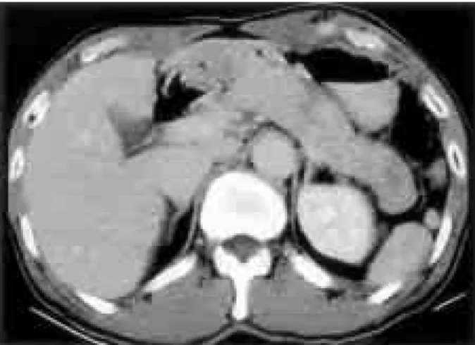
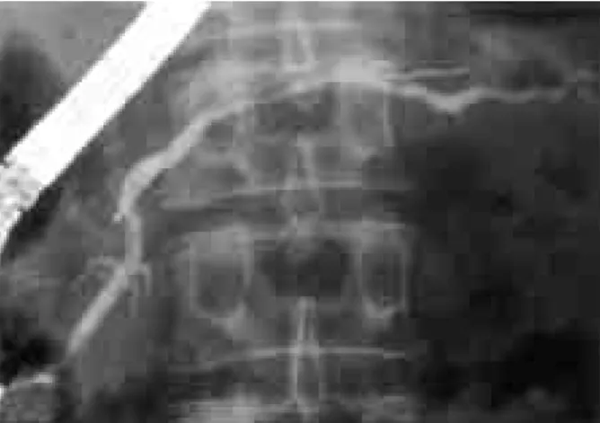
An abdominal computed tomography (CT) revealed diffuse enlargement of the whole pancreas without paraaortic lymphadenopathy or peripancreatic involvement (Fig. 1). An endoscopic retrograde pancreatography (ERP) done at our hospital demonstrated a diffuse irregular narrowing of the main pancreatic duct (Fig. 2). However, endoscopic retrograde cholangiography showed no abnormality of the bile duct.
Percutaneous core needle biopsy with 20-gauge needle was performed targeting the more swollen head portion of the pancreas under guidance of ultrasonography. The biopsy specimen revealed fibrotic changes with infiltration of inflam- matory cells and acinar cell depletion (Fig. 3). The infiltrated inflammatory cells were found to be mainly lymphocytes by immunochemical staining for leukocyte common antigen.
Fig. 1. Abdominal CT finding at admission. It shows diffuse enlargement of the pancreas.
Fig. 2. Endoscopic retrograde pancreatography at admission. It reveals a diffuse irregular narrowing of the main pancreatic duct.
Fig. 3. Histologic finding of pancreas biopsy at admission. Photo- micrograph of the biopsy specimen reveals parenchymal fibrosis, infiltration of inflammatory cells, and destruction of acinar cells (H&E, ×100).
305
The Korean Journal of Gastroenterology: Vol. 39, No. 4, 2002
The patient was strongly suspected to have chronic autoimmune pancreatitis, and then he was treated with prednisolone (40 mg/day). After two months of oral steroid treatment, he was reevaluated for pancreatitis. The patient had no complaint of any associated symptoms including mass-like feeling on the abdomen. Hyperamylasemia and hyperlipasemia were corrected. The serum ɣ-globulin and IgG levels returned to normal ranges. Anti-CA II antibody was improved to a negative value. The swelling of the whole pancreas returned to nearly normal size on CT. ERP revealed obvious improvement: diffuse irregular narrowing of the main pancreatic duct in the whole pancreas reverted to almost normal (Fig. 4). Core needle biopsy, again targeting the head portion of the pancreas, was carried out to confirm any histological improvement. Surprisingly, the result of the biopsy showed a marked regression of parenchymal fibrosis
Fig. 4. Endoscopic retrograde pancreatography after steroid therapy.
A diffuse ductal narrowing with irregularity reverted to almost normal.
Fig. 5. Histologic finding of the pancreas biopsy after steroid therapy. A photomicrography of biopsy shows regression of fibrosis and restoration of acinar cells (H&E, ×100).
with diminution of inflammatory cells and restoration of normal architecture of islet cells as well as acinar cells (Fig.
5). Subsequently, the steroid was tapered. The patient is now six months after cessation of medication and doing well without the recurrence of any abdominal symptoms and signs.
DISCUSSION
Chronic pancreatitis is usually defined as a syndrome of destructive, inflammatory conditions that encompasses many sequelae of long-standing pancreatic injury with histologic changes including irregular fibrosis, acinar cell loss, islet cell loss, and inflammatory cell infiltrates.1,10 The gold standard for the diagnosis of chronic pancreatitis is the tissue diagnosis for pathologic changes in the pancreas although CT or ERP would more likely be used to confirm the diagnosis.1 Recently, percutaneous core needle biopsy is reported as an accurate and relatively safe method for sampling the pancreas.11,12 Once the diagnosis of chronic pancreatitis is made, the prognosis is ominous because the fibrosis has generally been considered to be irreversible.
Autoimmune pancreatitis was reported first by Sarles et al.13 They described a special form of chronic pancreatitis with hypergammaglobulinemia and diffuse destructive sclerosis with inflammatory character caused by self-immunization.
Since then, many authors have reported chronic pancreatitis caused by an autoimmune mechanism.2-9 Yoshida et al proposed that the disease should be referred to as auto- immune pancreatitis if patients show the common following characteristics; increased serum gammaglobulin or IgG levels, presence of autoantibodies, diffuse enlargement of the pancreas on US or CT, diffuse irregular narrowing of the main pancreatic duct on ERP, fibrotic change with lymphocyte infiltration histopathologically, no symptoms or only mild symptoms, absence of pancreatic calcification or cysts, occasional association with other autoimmune disease, and effective steroid therapy. The present case showed the above-mentioned characteristics.
It is well known that autoimmune pancreatitis can be improved dramatically with oral steroid therapy. However, the reported improvements are only based on the radiological and laboratory findings. Most reports present the improvement of an irregular narrowing of the main pancreatic duct after steroid therapy. We also observed such improvement.
306
Kim, et al. Autoimmune chronic pancreatitis
At that time, we considered that the pancreas is a gland, and thus examined the parenchymal change as well as the ductal change after steroid therapy. To the best of our knowledge, this is the first attempt in which the histological findings were compared before and after steroid therapy for autoimmune chronic pancreatitis. Histologic findings revealed lymphoplasmacytic infiltration, parenchymal fibrosis, and atrophy of the acinar tissue besides the destruction of the ducts with marked cellular infiltration.
However, the pancreatic fibrosis was significantly regress- ed and normal architecture of the pancreatic parenchyma was almost restored after steroid therapy. It was particularly interesting that the phrase of fibrosis referred in chronic disease could have the reversibility in some clinical conditions. Similar regression has been reported in several cases of hepatic fibrosis after effective treatment. Examples are the case of primary biliary cirrhosis treated with ursodiol14 or methotrexate,15 chronic hepatitis C after treat- ment with interferon-α,16 secondary hepatic fibrosis caused by biliary obstruction after decompression,17 and autoimmune hepatitis after immunosuppressive therapy.18 Although the pathogenesis of pancreatic fibrosis is unknown, we could speculate that steroid therapy might modulate the balance between fibrogenetic pathway by the pancreatic stellate cells19,20 and fibrolytic pathway by the collagenase.
Moreover, parenchymal fibrosis, at least in the early stage of autoimmune chronic pancreatitis, may be bi-directional and thus parenchymal fibrosis should be considered to be reversible in some types of chronic pancreatitis when diag- nosed early and treated appropriately.
The prompt diagnosis of autoimmune pancreatitis is impor- tant because the patients in the early stage of the disease appear to respond dramatically to oral steroid therapy. For its diagnosis, pancreatic biopsy is given special emphasis, because the etiology-based classification has been recently presented and the treatment can be different according to the etiology.3 Additionally, it is important to decide the duration and dosage of steroid therapy since the steroid used in the treatment may cause undesirable side effects.
However, the dosage or duration of steroid in the treatment of autoimmune pancreatitis has not been established yet. Thus, we performed follow-up biopsy to confirm the parenchymal improvement of the pancreas after steroid therapy and to decide the continuance or tapering of steroid. We also did not exclude the possibility that the improvement in
histologic findings might be caused by sampling error. Thus, to minimize the sampling bias, percutaneous core needle biopsies guided by ultrasonography before and after the steroid therapy were carried out by the same expert sonographer at the same pancreatic location as possible, targeting the most prominent head portion out of the diffuse pancreatic enlargement on CT. Although some etiologies of chronic pancreatitis may display patchy histologic abnor- malities in pancreatic parenchyma, the imaging of our case showed diffuse involvement of the whole pancreas. Therefore, the difference observed between the histological findings of the biopsy specimens before and after steroid therapy in this case may be due to the real parenchymal improvement representing the effect of therapy rather than sampling variability.
In summary, we present the first case report of auto- immune chronic pancreatitis in which parenchymal fibrosis and ductal stenoses were regressed by oral steroid therapy alone. For the diagnosis of the disease, it is essential for clinicians to recognize the concept and unique characteristics of this disease contrasting to the common form of chronic pancreatitis. Then surgical resection due to a fear of malignancy may be avoided5 and the appropriate steroid therapy can be offered. It is well accepted that the con- stantly inflamed pancreas by repeated attacks in chronic pancreatitis creates scar tissues, causing irreversible damage.
However, fibrosis may not automatically mean that the condition is irreversible. According to etiology, pancreatic parenchymal fibrosis maybe regresses by appropriate treat- ment. Although our case is anecdotal, it gives evidence to support this hypothesis.
ACKNOWLEDGEMENTS
We are indebted to Dr. Kazuichi Okazaki, Department of Gastroenterology and Endoscopic Medicine, Kyoto University Hospital, Kyoto, Japan, for measuring anticarbonic anhydrase II antibody levels.
REFERENCE
1. Etemad B, Whitcomb DC. Chronic pancreatitis: diagnosis, classification, and new genetic development. Gastroenterology 2001;120:682-707.
307
대한소화기학회지: 제39권 제4호, 2002
2. Yoshida K, Toki F, Takeuchi T, Watanabe S, Shiratori K, Hayashi N. Chronic pancreatitis caused by an autoimmune abnormality. Proposal of the concept of autoimmune pancreatitis.
Dig Dis Sci 1995;40:1561-1568.
3. Ito T, Nakano I, Koyanagi S, et al. Autoimmune pancreatitis as a new clinical entity. Three cases of autoimmune pancreatitis with effective steroid therapy. Dig Dis Sci 1997;42:1458-1468.
4. Furukawa N, Muranaka T, Yasumjori K, Matsubayashi R, Hayashida K, Arita Y. Autoimmune pancreatitis: radiologic findings in three histologically proven cases. J Comput Assitst Tomogr 1998; 22:880-883.
5. Horiuchi A, Kaneko T, Yamamura N, et al. Autoimmune chronic pancreatitis simulating pancreatic lymphoma. Am J Gastroenterol 1996;91:2607-2609.
6. Tsuchiya A, Tsuchiya Y, Nashimoto A. A case of autoimmune pancreatitis diagnosed by pancreatic biopsy. Jpn J Gastroenterol 2000;97:353-357.
7. Uchida K, Okazaki K, Konishi Y, et al. Clinical analysis of autoimmune-related pancreatitis. Am J Gastroenterol 2000;95:
2788-2794.
8. Horiuchi A, Kawa S, Akamatsu T, et al. Chracteristic pancreatic duct appearance in autoimmune chronic pancreatitis:
a case report and review of the Japanease literature. Am J Gastroenterol 1998;93:260-263.
9. Taniguchi T, Seko S, Azuma K, et al. Autoimmune pancreatits detected as a mass in the tail of the pancreas. J Gastroenterol Hepatol 2000;15:461-464.
10. Homma T. Criteria for pancreatic disease diagnosis in Japan:
diagnostic criteria for chronic pancreatitis. Pancreas 1998;16:
250-254.
11. Brandt KR, Charboneau JW, Stephens DH, Welch TJ,
Goellner JR. CT-and US-guided biopsy of the pancreas.
Radiology 1993;187:99-104.
12. Welch TJ, Sheedy PF, Johnson CD, Johnson CM, Stephens DH. CT-guided biopsy: prospective analysis of 1,000 procedures. Radiology 1989;171:493-496.
13. Sarles H, Sarles JC, Muratore R, Guien C. Chronic inflammatory sclerosis of the pancreas -an autoimmune pancreatic disease? Am J Dig Dis 1961;6:688-698.
14. Poupon RE, Balkau B, Eschwege E, Poupon R. A multicenter, controlled trial of ursodiol for treatment of primary biliary cirrhosis. UDCA-PBC Study Group. N Engl J Med 1991;324:1548-1554.
15. Kaplan MM, De Lellis RA, Wolfe HJ. Sustained biochemical and histologic remission of primary biliary cirrhosis in response to medical treatment. Ann Intern Med 1997;126:
682-688.
16. Dufour JF, De Lellis R, Kaplan MM. Regression of hepatic fibrosis in hepatitis C with long-term interferon treatment.
Dig Dis Sci 1998;43:2573-2576.
17. Hammel P, Couvelard A, O'Toole D, et al. Regression of liver after biliary drainage in patients with chronic pancreatitis and stenosis of the common bile duct. N Engl J Med 2001;344:418-423.
18. Dufour JF, De Lellis R, Kaplan MM. Reversibility of hepatic fibrosis in autoimmune hepatitis. Ann Intern Med 1997;127:
981-985.
19. Wells RG, Grawford JM. Pancreatic stellate cells: the new stars of chronic pancreatitis? Gastroenterology 1998;115:491-493.
20. Apte MV, Haber PS, Darby SJ, et al. Pancreatic stellate cells are activated by proinflammatory cytokines: implications for pancreatic fibrogenesis. Gut 1999;44:534-541.
308