DOI:10.4078/jkra.2010.17.2.143
<접수일:2010년 4월 27일, 수정일 (1차:2010년 5월 10일, 2차:2010년 5월 13일) 심사통과일:2010년 5월 13일>
※통신저자:유 대 현
서울시 성동구 행당동 7 한양대학교 류마티스병원
Tel:02) 2290-9202, Fax:02) 2298-8231, E-mail:dhyoo@hanyang.ac.kr
피부근육염/다발근육염에서 발생한 자발 종격동기종의 임상적 의미
한양대학교 의과대학 내과학교실1, 한양대학교 의과대학 내과학교실 류마티스병원2
김진주
1ㆍ김 담
1ㆍ김은경
1ㆍ손일웅
1ㆍ정경희
2ㆍ최찬범
1,2ㆍ성윤경
1,2전재범
1,2ㆍ엄완식
1,2ㆍ김태환
1,2ㆍ배상철
1,2ㆍ유대현
1,2= Abstract =
Clinicial Significance of Spontaneous Pneumomediastinum in Dermatomyositis/Polymyositis
Jin Ju Kim1, Dam Kim1, Eun Kyoung Kim1, Il Woong Sohn1, Kyong Hee Jung2, Chan-Bum Choi1,2, Yoon-Kyoung Sung1,2, Jae-Bum Jun1,2, Wan-sik Uhm1,2,
Tae Hwan Kim1,2, Sang Cheol Bae1,2, Dae Hyun Yoo1,2
Department of Internal Medicine, College of Medicine, Hanyang University1, Department of Internal Medicine, The Hospital for Rheumatic Diseases, College of Medicine,
Hanyang University2, Seoul, Korea
Objective: Pneumomediastinum (PnM), a rare complication of dermatomyositis and polymyo- sitis (DM/PM), is sporadic and has an unclear pathogenesis. PnM is almost always associated with interstitial lung disease (ILD), and is a poor prognostic factor in inflammatory myositis patients. We studied the prevalence of PnM in Korean DM/PM and its clinical significance.
Methods: We retrospectively studied the medical records of 161 patients diagnosed with DM/PM meeting Bohan-Peter’s criteria at Hanyang University Hospital for Rheumatic Diseases from 1995 to 2010. We collected following findings; demographic data, diagnosis, lung involve- ment, cause of death, and duration from diagnosis to death.
Results: One hundred nineteen patients (73.9%) were DM and 42 patients (26.1%) were PM.
Eighty three patients (51.6%) developed ILD at diagnosis or during follow up. Eighteen patients (11.2%) died because of ILD aggravation, infection, or malignancy. The mean duration from diagnosis to death was 11.5 months, with 10 patients (6.2%) dying from from ILD aggravation but none with spontaneous PnM. 6 patients (3.7%) presented with PnM, and it was associated
with ILD worsening in all cases. PnM resolved with O2 inhalation, corticosteroids, and/or immunosuppressive agents after 11 weeks (mean) of therapy
Conclusion: PnM is rare but associates with DM and aggravation of ILD. PnM does not usually cause fatalities and can be cured by appropriate therapy.
Key Words: Pneumomediastinum, Dermatomyositis/polymyositis, Interstitial lung disease, Prognosis
서 론
피부근육염/다발근육염(dermatomyositis/polymyositis) 은 근골격, 피부, 내부 장기 등에 영향을 미치는 전 신적인 염증성 질환이다. 이 환자들의 약 10∼22%
에서 호흡 근육을 침범하여 호흡저하를 비롯하여 폐 렴, 간질폐질환(interstital lung disease)과 같은 호흡기 증상을 일으킬 수 있다 (1-3). 피부근육염/다발근육염 환자에서 간질폐질환이 발생하면 임상적으로 나쁜 예후를 보인다고 보고되어 있다 (3).
종격동기종(pneumomediastinum)은 종격동 내에 공 기 혹은 다른 가스가 비정상적으로 존재하는 상태를 말하며 손상, 종격동염, 기계환기와 같은 원인에 의 해 발생하지만 특별한 원인 없이 발생할 수도 있다 (4). 또한 자발종격동기종은 피부근육염/다발근육염, 전신홍반루푸스, 류마티스관절염, 혼합결체조직병 등 과 같은 전신 류마티스 질환에서 드물게 보고되고 있다 (5-7). 피부근육염/다발근육염 환자에서 간질폐 질환이 발생하는 빈도는 10∼43% 정도로 보고되고 있으나 자발 종격동기종은 전세계적으로 드물게 보 고되고 있다. 피부근육염/다발근육염 환자에서 종격 동기종이 발생한 경우 그 예후가 매우 불량하여 종 종 사망에 이른 경우도 보고되어 있으나 (8,9), 최근 에 Kuroda 등과 한국에서는 이 등이 cyclosporine 등 을 이용하여 성공적으로 치료된 증례를 보고하였으 며 (10,11), 피부근육염 환자에서 종격동기종 자체가 나쁜 예후 인자는 아니라는 보고도 있다 (12). 본 연 구에서는 피부근육염/다발근육염 환자에서 자발 종 격동기종의 발생 빈도와 임상적 의미에 대해 고찰해 보고자 한다.
대상 및 방법 1. 연구대상
1995년 1월부터 2010년 3월까지 한양대학교 류마 티스병원에 입원 및 외래에 내원한 환자 중에 전산 실을 통해 ICD 10 진단코드 M33, M330, M331, M332, M339가 들어간 환자를 조회하였다. 총 161명의 환 자가 보한-피터씨 진단기준(Bohan-Peter’s criteria)에 부 합하여 피부근육염/다발근육염으로 진단받았다 (13).
조회한 161명의 환자 의무 기록을 후향적으로 검토 하였다. 임상적 소견에 대한 조사는 성별, 나이, 진 단명, 추적관찰 기간, 진단 당시의 CK/LDH 수치, al- dolase 수치, 항핵항체(ANA)와 항 Jo-1, 간질폐질환 과 자발 종격동기종과 같은 폐 침범 소견, 악성 종 양과 같은 합병증 동반 여부, 치료와 현재 상태, 사 망한 환자의 경우 사망의 원인에 대해 조사하였다.
2. 간질폐질환의 방사선학적 분류 및 경과
모든 환자는 초기 진단 당시와 입원할 시, 또는 외래에서 단순흉부X선 촬영을 주기적으로 시행하였 다. 간질폐질환이나 폐 내에 다른 이상이 발견되거 나 호흡 곤란으로 입원한 환자의 경우 흉부 고해상 전산단층촬영을 시행하였다. 그물성결절모양(reticulo- nodular opacity), 선상(linear opacity) 혹은 젖빛 유리 음영(ground-glass opacity), 벌집모양(honeycombing), 견 인 기관지확장증(traction bronchiectasis)과 같은 모양을 보인 경우 간질폐질환으로 진단하였다. 간질폐질환은 폐 생검을 통한 병리학적 소견으로 분류를 하는 것 이 표준 방법이나 실제로 본원에서 폐 생검을 시행 한 경우가 없었다. 이에 따라 방사선학적 소견에 따 라 주로 기저와 말초에 반점, 그물성 음영 및 벌집 모양 소견을 보이는 경우는 특발폐섬유증(idiopathic pulmonary fibrosis), 주로 기저부의 반점 또는 기관지
Table 1. Classification and patterns of Interstitial lung disease
Morphologic pattern Clinical diagnosis Histologic features Imaging features Usual interstitial
pneumonia
Idiopathic
pulmonary fibrosis
Spatial and temporal heterogeneity, dense fibrosis, fibroblastic foci, honeycombing
Basal, peripheral predominance, often patchy, reticular abnormality, honeycombing
Nonspecific interstitial pneumonia
Nonspecific
interstitial pneumonia
Spatially and temporally homogeneous lung fibrosis or inflammation
Basal predominance, ground-glass abnormality, reticular abnormality
Desquamative interstitial pneumonia
Desquamative interstitial pneumonia
Diffuse macrophage accumulation in alveoli
Basal, peripheral predominance ground-glass attenuation;
sometimes cysts Respiratory
bronchiolitis
Respiratory bronchiolitis- associated interstitial lung disease
Peribronchiolar macrophage accumulation, bronchiolar fibrosis;
macrophages have dusty, brown cytoplasm
Centrilobular nodules, ground-glass attenuation
Organizing pneumonia
COP, BOOP Patchy distribution of intraluminal organizing fibrosis in distal airspaces;
preservation of lung architecture;
uniform temporal appearance; mild interstitial chronic inflammation
Ground-glass attenuation;
consolidation basal, peripheral predominance
Diffuse
alveolar damage
Acute interstitial pneumonia
Diffuse distribution, uniform temporal appearance, alveolar septal thickening due to organizing fibrosis, airspace organization, hyaline membranes
Diffuse, ground-glass attenuation, consolidation
Lymphoid interstitial pneumonia
Lymphoid interstitial pneumonia
Diffuse lymphoplasmacytic infiltration of alveolar septa
Ground-glass attenuation, cysts
COP: cryptogenic organizing pneumonia, BOOP: bronchiectasis obliterans with organizing pneumonia 주위 경화를 보이는 경우는 특발기질화폐렴(bronchiec-
tasis oblitrans with organizing pneumonia, BOOP), 종종 견인 확장을 동반하며 선상 음영을 동반 또는 동반 하지 않는 기저부에 주로 젖빛 유리 음영을 보이는 경우는 비특이간질성폐렴(non-specific interstitial pneu- monitis, NSIP), 전반적인 폐 경화와 젖빛 음영을 보이는 경우는 급성 간질폐렴(acute interstitial pneumonitis, AIP)으로 구분하였다(표 1) (14). 또한 추적 관찰 기 간 동안 고해상전산단층촬영을 통해 간질폐질환의 경과에 대해서 변화 없는 경우, 호전/악화를 반복하 며 서서히 진행하는 경우와 빠른 악화의 경우로 구 분하였다.
3. 자발종격동기종의 진단
자발 종격동기종은 전후와 측면 촬영을 포함한 단 순흉부X선 촬영에서 종격동 내 흉골 뒤나 대동맥
또는 다른 구조의 경계를 따라서 보이는 가늘고 수직 의 방사선 투과성 선이 보이는 경우 진단하였다 (4).
단순흉부X선 촬영에서 자발 종격동기종이 진단이 된 경우 흉부 고해상단층촬영을 시행하였고, 호전/악화 여부를 알기 위해 추적 단순흉부X선 촬영과 흉부 고 해상단층촬영을 시행하여 초기의 검사와 비교하였다.
결 과 1. 일반적인 특징
161명의 환자는 31명의 남자와 130명의 여자로 구 성되었다. 이들의 평균 진단 나이는 43.2세(범위 6∼75 세)이고, 그들의 평균 추적관찰 기관은 약 30.4개월 (범위 1∼155개월)이었다. 피부근육염 환자는 119명 (73.9%), 다발근육염은 42명(26.1%)였다. 각각의 남녀 비율은 1:3.8과 1:6이었다. 진단 당시 평균 나이는
Table 3. Radiologic classification of interstitial lung disease
IPF BOOP NSIP AIP Mixed Unknown
Numbers Result
Alive (PnM) Expire
14
14 0
30
26 (4) 4
25
23 (1) 2
1
0 1
8
4 (1) 4
5
5 0 IPF: idiopathic pulmonary fibrosis, BOOP: bronchiectasis obliterans with organizing pneumonia, NSIP: non-specific inter- stitial pneumonitis, AIP: acute interstitial pneumonitis, PnM: pneumomediastinum
Table 2. Characteristics of dermatomyositis/polymyositis patients
Dermatomyositis Polymyositis
Numbers of patients 119 (73.9%) 42 (26.1%)
Mean age at diagnosis (years) 43.6 (6∼75) 38.7 (16∼75) Mean period of follow up (months) 26.6 (1∼155) 32.6 (1∼121)
Female/Male 94/25 36/6
Interstitial lung disease 69 (42.9%) 14 (33.3%)
Malignancy 11 (9.2%) 2 (4.8%)
Death causes
Interstitial lung disease Malignancy
Sepsis
16 (13.4%) 9 4 3
2 (4.8%) 1 1 각각 43.6세(범위 6∼75세), 38.7세(16∼75세)였다. 전
체 환자 중에 13명(8.1%)에서 추적 관찰 기간 중에 악성 종양이 발견되었다. 간질폐질환은 83명(51.6%) 에서 발견되었는데 피부근육염에서 69명(58.0%), 다 발근육염에서 14명(33.3%)이었다(표 2).
2. 예후
전체 환자 중에 18명(11.2%)의 환자가 사망하였고 진단부터 사망까지의 기간은 평균 11.5개월(범위 1∼
37개월)이었다. 사망 원인은 간질폐질환의 악화에 의 한 호흡부전(10명, 6.2%), 악성 종양의 합병증(5명, 3.1%), 세균성 감염(3명, 1.9%)이었다. 악성 종양으로 사망한 환자들은 각각 난소암(2명), 위암(1명), 전이 성 흉선종(1명), 자연 살해 T세포 림프종(NK/T cell lymphoma)(1명)이었다. 악성 종양 및 패혈증 쇼크로 사망한 환자들은 모두 간질폐질환 소견은 없었다 (표 2).
3. 간질폐질환의 진단과 경과
간질폐질환은 83명(51.6%)에서 발견되었다. 이 중
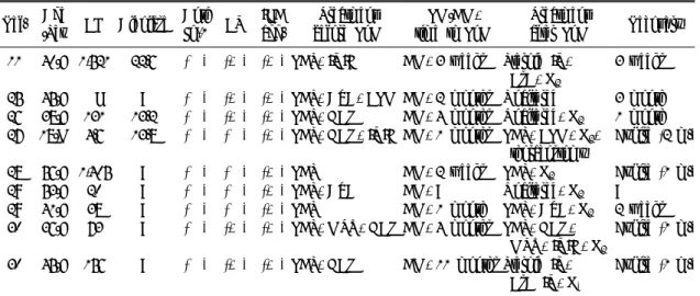
5명의 환자는 영상 자료가 남아있지 않아 제외하였 다. 78명은 각각 특발폐섬유증 14명(17.9%), 특발기 질화폐렴 30명(38.5%), 비특이간질폐렴 25명(32.1%), 급성 간질폐렴 1명(1.3%), 두 가지 이상이 혼합된 형 태의 간질폐질환 8명(10.3%)이었다(표 3). 두 가지 이 상이 혼합된 형태의 간질폐질환의 경우는 특발기질 화폐렴과 비특이간질폐렴이 혼재한 경우가 7명(9.0%), 특발폐섬유증과 비특이간질폐렴이 혼재한 경우가 1 명(1.3%)이었다. 간질폐질환은 51명(65.4%)에서 추적 관찰 기간 동안 큰 변화가 없었고, 17명(21.8%)에서 호전과 악화를 반복하여 서서히 악화되었으며, 10명 (12.8%)에서 호흡기 증상이 생긴 지 1달 이내에 매 우 빠르게 악화되었고, 이 경우에는 모두 사망하였 다(표 4).
4. 자발성 종격동기종 발생 환자들의 특징
추적 관찰 기간 동안 6명(3.7%)의 환자에서 7회의 자발 종격동기종이 발생하였다. 5명은 처음부터 피부 근육염을 진단받았으며 1명은 다발근육염 진단 시에 자발 종격동기종이 발생하였다가 9개월 뒤 피부 발
Table 5. Characteristics of 6 cases of spontaneous pneumomediastinum at dermatomyositis/polymyositis patients Age
/sex CK LDH Aldo. ANA Anti
Jo-1 CV ILD agg.
Treatment before PnM
PM/DM, time to PnM
Treatment
after PnM Recovery 59/F 24 142 18.4 (+)* (−) (−) (+) PDS DM, initial. Steroid pulse, CsA 5 month 51/F 32 305 5.4 (−) (−) (−) (+) PDS, CsA, HCQ DM, 1 month Steroid pulse,
CsA, O2
2 month
42/F 166 267 37.6 (+)† (−) (−) (+) PDS, CsA, HCQ DM, 2 years Steroid pulse, CsA, O2
2 weeks
43/F 125 448 12.9 (−) (−) (−) (+) PDS, CsA DM, 6 month IVIG, PDS, CsA, HCQ, O2,
5 month
44/F recur
49 290
251 346
3.7 7.1
(+)‡ (−) (−) (+) (+)
PDS, AZA PDS, CsA
PM, initial.
DM, 1 week
PDS, CsA, O2, Collar incision Unaltered, O2
2 month 1 month 54/F 26 122 7.1 (−) (−) (−) (+) PDS, CsA DM, 1 year IVIG, O2, PDS,
Tacrolimus
2 weeks
Aldo.: aldolase, CV: cutaneous vasculopathy, agg.: aggravation, PDS: prednisolone, CsA: cyclosporine A, HCQ: hydroxy- cloroquinolone, AZA: azathioprine, IVIG: intravascular immunoglobulin. *Speckled pattern 1:640, †Cytoplasmic pattern 1:320, ‡Cytoplasmic patthern 1:160
Table 4. Clinical course of interstitial lung disease among dermatomyositis/polymyositis patients
No interval
change
Slow progression
Rapid
progressionUnknown
Numbers Result
Alive (PnM) Expire
51
51
17
16 (6) 1
10
0 10
5
5 0 PnM: pneumomediastinum
진이 발생하면서 피부근육염으로 진단이 바뀌었고 이 때 종격동기종이 재발하였다. 평균 나이는 47.6세 (범위 42∼59세)이며 모두 여자 환자였다. 종격동기 종 발생 당시 5명의 환자에서 CK 수치는 정상 범위 였고, LDH 수치는 1명 외에는 모두 증가하였다. 항 Jo-1 항체는 모두 음성이었으며, 항핵항체는 3명의 환자에서 양성으로 각각 speckled pattern (1명), cyto- plasmic pattern (2명)이었다. 1명은 피부근육염 진단 3개월 후 간질폐질환이 발병하였고 나머지 환자들은 피부근육염 진단 당시에 간질폐질환을 동반하고 있 었다. 4명은 특발기질화폐렴, 1명은 비특이간질성폐 렴, 나머지 1명은 특발기질화폐렴과 비특이간질폐렴 이 혼재되었다. 자발 종격동기종은 모든 경우에서 간질폐질환이 악화되었을 때 발생하였는데 4명에서
목과 흉부에 피하기종을 동반하였다. 이들은 자발 종 격동기종이 발생하기 전에 스테로이드 및 면역 억제 제를 복용하고 있었다. 3명에서는 기침, 가래를 동반 하였으며, 6명 모두에서 입원 당시 또는 입원 기간 중에 갑작스럽게 악화한 호흡 곤란을 호소하여 단순 흉부X선 촬영을 하였다. 그 후 단순흉부X선 촬영 및 흉부 고해상단층촬영을 하여 자발 종격동기종의 경과를 추적하였다. 6명 모두에서 자발 종격동기종 발생 이후에는 고용량 스테로이드를 사용하였으며 이와 함께 사용하고 있던 면역억제제의 용량 조절과 함께 산소 치료를 병행하였고 2명의 환자에서 인 면 역글로불린 주사치료를 시행하였다. 목과 흉부에 광 범위한 피하기종을 동반한 환자 1명에서 칼라 절개 (collar incision)와 짜내기(squeezing)를 시행하여 호전 되었으며 이 환자는 9개월 후 재발하였으나 사용하
고 있는 스테로이드, フ 되었 Ē ꖎ傭u
Table 6. Comparison of clinical features between Interstitial lung disease patients with and without pneumomediastinum
Pneumomediastinum (+) Pneumomediastium (−)
Numbers of patients 6 (7.1%) 77 (92.9%)
Age 48.8 (42∼59) 45.4 (27∼75)
Male/Female 0/6 14/63
DM/PM 6/0 63/14
Follow up duration (months) 22 (2∼57) 30.3 (1∼155)
ANA (+) 3 (50%) 47 (61.0%)
CK/LDH (mean) 101.7/268.7 596.4/358.3
고 찰
피부근육염/다발근육염은 주로 근육과 피부를 침 범하는 아직까지 명확한 원인이 밝혀지지 않은 전신 적인 염증성 결체조직 질병이다. 피부근육염/다발근 육염에서 폐를 침범하는 경우에는 전반적인 폐포의 손상, 폐 혈관염, 감염성 폐렴, 폐부종과 종양 등을 일으킬 수 있다 (10). 간질폐질환은 피부근육염/다발 근육염 환자의 약 10∼43%에서 발견되는 가장 흔한 폐질환이다 (1-3). 피부근육염/다발근육염 환자에서 일반적으로 흔한 사망 원인은 종양, 감염과 심장 질 환으로 알려져 있으나 간질폐질환 역시 사망률에 영 향을 미치는 원인으로 알려져 있다. 강 등은 한국인 피부근육염/다발근육염에서의 간질폐질환 유병율은 40.3%였고, 이것이 나쁜 예후와 관련이 있다고 보고 하였다 (3). 또한 김 등은 염증성 근질환 환자의 생 존율에 대한 예후 인자 연구에서 고령, 치료 기간과 사망률간의 관계는 유의성이 없고, 간질폐렴과 악성 종양은 염증성 근질환과 관련하여 통계적으로 유의 하게 예후에 영향을 미친다고 보고하였다 (15). 본 연구에서도 간질폐질환의 유병율은 피부근육염/다발 근육염에서 각각 58.0%와 33.3%로서 기존의 보고와 큰 차이는 없었다. 사망의 원인에 있어서도 간질폐 질환이 높은 빈도를 보였다.
피부근육염/다발근육염에 동반한 자발 종격동기종 은 드물게 보고되어 있다. Schwarz 등이 1976년 처 음으로 피부근육염 환자에서 자발 종격동기종의 발 생을 보고하였고 (16), 그 이후로 추가적인 증례들이 보고되었으며 Kono 등은 피부근육염/다발근육염 환 자의 약 8.3%에서 종격동기종이 발생한다고 하였다
(17). 본 연구에 있어 종격동기종은 전체 환자의 3.7%에서 발생하였다. 그러나 모든 환자가 피부근육 염 환자였음을 감안하면 피부근육염 환자 중 5.0%
에서 발생하였다. 지금까지 보고된 몇몇 연구에서는 자발 종격동기종 발생 후 약 37.5∼52.5%의 사망률을 보인다고 알려져 있다 (8,18-20). Matsuda 등과 Jansen 등은 종격동기종을 동반한 피부근육염/다발근육염 환자 20명과 9명에서 각각 55%와 30%의 사망률을 보였다고 보고하였다 (8,21). 그렇지만 본 연구에서 사망한 증례가 없어 반드시 나쁜 예후를 보인다고 할 수는 없다.
피부근육염/다발근육염에서의 자발 종격동기종은 상 대적으로 드물지만 특징적인 합병증으로 아직까지는 그 기전이 명확히 밝혀지지 않았다. 발생 기전에는 3가지 가설이 있는데, 첫째는 간질폐질환에 의해 폐 가 섬유화되면서 폐포가 변성되고 폐막하 수포(sub- pleural cyst)를 형성하여 이 주머니가 터지면서 종격 동기종이 발생한다는 가설이다 (21). 두번째로는 Cicuttinit 등이 말하는 혈관염 이론이 있다 (22). 이 는 혈관병증에 의해 점막 경계(mucosal barrier)가 파 괴되면서 종격동기종이 발생한다는 이론으로 Kono 등과 Tong 등은 종격동기종이 발생한 환자 군에서 그렇지 않은 환자 군에서 보다 피부 혈관병증이 많 이 이환된다는 것을 보고하여 피부 혈관병증과 종격 동기종 간에 명확한 연관 관계가 있음을 보여주었다 (17,23). 마지막으로 간질폐질환에 스테로이드 사용 으로 폐간질이 약화됨으로 인해 종격동기종이 발생 한다는 가설이 있다 (9). Benoit Le Goff 등은 근육 약화가 없고 초기에 폐기능 검사에서 일산화탄소 확 산능력(DLCO) 또는 폐 용적(vital capacity) 감소가 심한 폐 침범이 있는 환자에서 고용량 스테로이드를
Table 7. Review of reported spontaneous pneumomediastinum cases with dermatomyositis/polymyositis in Korean Ref. Age
/Sex CK Aldolase Anti
Jo-1 CV ILD agg.
Treatment before PnM
PM/DM, time to PnM
Treatment
after PnM Recovery 11 40/F 1,521 22.6 (−) (+) (+) PDS, IVIG DM, 3 weeks Steroid IV,
CsA, O2
3 weeks
25 45/F NL ? (−) (+) (+) PDS, AZA, CPM DM, 2 months Unaltered 3 month 26 38/F 131 13.2 (−) (+) (+) PDS, HCQ DM, 4 months Unaltered, O2 1 month 27 18/M 4.6 13.8 (−) (−) (+) PDS, HCQ, IVIG DM, 1 months PDS, CPM, O2,
thoracostomy
Expire (2 m.)
28 56/F 1,405 ? (−) (−) (+) PDS DM, 2 weeks PDS, O2 Expire (1 m.) 29 53/F 20 ? (−) (−) (+) PDS, AZA DM, ? Unaltered, O2 ?
29 41/F 38 ? (−) (−) (+) PDS DM, 1 month PDS, AZA, O2 2 weeks 30 36/F 73 ? (−) (+) (+) PDS, MTX, HCQ DM, 4 months PDS, HCQ,
MTX, IVIG, O2
Expire (1 m.)
30 45/F 156 ? (−) (+) (+) PDS, HCQ DM, 11 months Steroid IV, CsA IV, O2
Expire (1 m.)
Ref.: reference, CV: cutaneous vasculopathy, agg.: aggravation, NL: normal, ?: unknown, PDS: prednisolone, CsA:
cyclosporine A, HCQ: hydroxycloroquinolone, AZA: azathioprine, IVIG: intravascular immunoglobulin, M: month
사용한 경우 종격동기종 발생 후 불량한 예후와 관 련이 있다고 보고하였다 (24). 또한 면역 억제제의 사용은 예후와 큰 관련이 없으나 50% 정도에서는 회복을 보였다고 보고하였다 (24).
저자들은 한국인 피부근육염/다발근육염 환자에서 종격동기종이 발생하여 국내와 외국에 증례 보고된 문헌들을 살펴보았다(표 7) (11,25-30). 한국에서 보 고된 종격동기종은 모두 9예로서 피부근육염 환자에 서 발생하였으며 다발근육염에서 보고된 증례는 없 었다. 평균 나이는 41.3세(범위 18∼56세)이고, 1명을 제외하고 모두 여자였다. 모든 증례에서 간질폐질환 이 동반되었고, 종격동기종이 발생하였을 때 간질폐 질환의 악화 소견을 보였다. 모든 환자에서 자발종 격동기종 발생 전에 스테로이드를 사용한 병력이 있 고, 간질폐질환을 동반하였던 것으로 보아 종격동기 종 발생에 간질폐질환의 악화와 스테로이드에 의한 폐간질의 악화 효과가 모두 작용을 했을 것이라고 추정된다. 그러나 발생 이후에 스테로이드 증량과 함께 면역억제제 치료, 산소 치료를 병행하여 호전 되었고 스테로이드 및 면역억제제에 반응이 불충분 하여 간질폐질환이 악화된 4명의 증례는 호흡부전으 로 사망한 것으로 보아 스테로이드가 종격동기종 발 생에도 영향이 있으나 고용량의 스테로이드 사용이 종격동기종 발생 후에 반드시 나쁜 예후와 관련이
있다고 말하기는 어렵다.
일반적으로 항 Jo-1 항체가 양성인 경우 간질폐질 환의 빈도가 높아 간질폐질환과 항 Jo-1 항체 사이 의 연관성이 있음이 밝혀진 바 있다 (31). 그러나 본 연구와 한국인 증례에서 간질폐질환 동반하고 있으 나 모두 항 Jo-1 항체 음성이었고, Yoshida 등이 고 찰한 문헌에서도 피부근육염 21명의 환자 중 16명의 환자에서 간질폐질환을 동반하였으나 항 Jo-1 항체 는 음성이었다 (12). 따라서 항 Jo-1 항체와 자발종 격동기종 발생은 큰 연관이 없는 것으로 생각된다.
앞서 말했듯이 피부 혈관병증이 발현한 피부근육 염 환자에서 자발 종격동기종이 호발한다고 보고되 고 있는데, 고찰한 문헌에서 피부 혈관병증이 동반 되었던 3명의 증례는 모두 호전된 것으로 보아 피부 혈관병증은 자발 종격동기종 발생 후의 예후에는 큰 영향을 미치지 않는 것으로 생각된다. Terao 등은 피부근육염/다발근육염 환자에서 간질폐질환의 정도 에 따라 자발 종격동기종을 두 그룹 분류하였는데, 피부 혈관병증 동반 여부와 상관없이 심한 간질폐질 환을 가진 군에서 경한 간질폐질환이나 폐질환이 없 는 군에 비해 예후가 나쁘다고 하였다 (19). 즉, 종 격동기종에서 피부 혈관병증 동반이 종격동기종의 발생에 영향을 미치기는 하나 예후와는 관련이 없다 고 생각된다.
피부근육염 환자에서 근육 효소 CK의 증가는 중 요한 검사 소견이고 Fudman 등은 CK 상승이 없는 경우 피부근육염 환자의 예후가 불량하다고 하였고 (32), Barvaux 등은 CK가 정상이거나 상승하더라도 미약한 경우의 환자에서 피부근염과 연관된 간질폐 질환을 동반하고 이 때 자발 종격동기종이 발생한 경우에는 예후가 매우 불량하다고 하였으나 (15), 한 국인 증례 15명 중 12명에서 자발 종격동기종 발생 당시 CK 수치는 정상 범위였고 이 중 3명은 사망을 하였다. CK 수치가 증가되어 있던 3명 중에서 1명 이 사망하여 자발 종격동기종이 발생한 환자에서 CK가 정상 혹은 상승이 미약하다고 하여 예후가 불 량하다고 단정적으로 말할 수는 없다.
현재까지 기존 문헌들에서 보고한 자발 종격동기 종의 증례는 대부분 피부근육염이었으며 다발근육염 에서 발생한 경우는 드물어 자발 종격동기종의 발병 은 간질폐질환을 동반한 피부근육염에서 호발한다고 할 수 있겠다. 또한 일부에서 종격동기종이 피부근 육염/다발근육염 환자에서 나쁜 예후를 나타내는 인 자로 생각되어 왔지만 본 연구에서 고찰한 바로는 한국인 피부근육염/다발근육염 환자에서 종격동기종 이 발생한 경우 스테로이드와 면역 억제제, 산소 치 료를 통해 대부분은 호전된 것으로 보아 종격동기종 이 피부근육염/다발근육염 환자에서 사망과 관련한 나쁜 예후 인자는 아닌 것으로 생각된다.
본 연구는 몇 가지 한계점을 가지고 있다. 첫째, 161명의 환자를 후향적으로 의무기록을 분석한 것으 로 추적관찰에서 소실된 환자들의 예후를 평가하지 않았다는 점이다. 이로 인해 악성종양, 간질폐질환의 발생, 감염 등의 질환 발생이 좀 더 많을 수 있고, 사망 환자 수와 사망 원인 질환의 비율이 달라질 수 있었다. 둘째, 간질폐질환의 분류에 있어서 조직 검 사가 이루어진 경우가 없어 표준 방법인 병리학적 분류가 아닌 방사선학적 분류를 하였기 때문에 실제 와의 결과가 상이할 수 있다. 셋째, 검사 수치에서 다른 환자들은 피부근육염/다발근육염 진단 당시의 CK/LDH를 조사하였으나, 자발 종격동기종이 발생한 환자는 종격동기종 발생시의 검사 수치와 비교를 한 한계점이 있다. 넷째, 자발 종격동기종이 발생하였던 본원 증례에서는 이로 인해 사망한 경우가 없어 자 발 종격동기종으로 사망한 환자와 그렇지 않은 환자
에서 종격동기종 발생 후 예후에 영향을 미치는 인 자에 대해 통계학적 분석을 하지 못하였다. 마지막 으로 고찰한 문헌들에서 추적 관찰 기간이 짧아 자 발 종격동기종 호전 이후의 경과는 알 수 없고, 한 국에서 보고되지 않은 자발 종격동기종이 있을 수 있어 발생 빈도는 실제로는 본 연구보다 높을 수 있 다. 앞으로 호전된 자발 종격동기종 환자들의 추후 임상 경과를 평가하여야 하며 종격동기종 이외의 다 른 원인에 의해 사망한 다른 피부근육염/다발근육염 환자들과 비교하여 예후에 미치는 영향에 대해 비교 하는 것이 필요할 것으로 생각된다. 또한 자발 종격 동기종 발생 후 피부근육염/다발근육염의 예후에 인 종 간에 차이가 있는 지, 자발 종격동기종이 발생한 다른 자가면역질환과 비교하여 예후에 미치는 영향 에 대해 연구해 보는 것도 필요할 것으로 생각된다.
결 론
결론적으로 본 연구에서 피부근육염/다발근육염 환자에서 자발 종격동기종의 발생률은 흔한 합병증 은 아니었다. 그러나 간질폐질환을 동반한 피부근육 염환자에서 종격동기종이 주로 발생하였다. 피부근 육염/다발근육염 환자에서 종격동기종이 예후에 나 쁜 영향을 미치는 인자라고 단정하기 어려우며 오히 려 적절한 치료를 시행하면 좋은 예후를 보일 수도 있다고 사료된다.
참고문헌
1) Dickey BF, Myers AR. Pulmonary disease in poly- myositis/dermatomyositis. Semin Arthritis Rheum 1984;14:60-76.
2) Marie I, Hachulla E, Cherin P, Dominique S, Hatron PY, Hellot MF, et al. Interstitial lung disease in polymyositis and dermatomyositis. Arthritis Rheum 2002;47:614-22.
3) Kang EH, Lee EB, Shin KC, Im CH, Chung DH, Han SK, et al. Interstitial lung disease in patients with polymyositis, dermatomyositis and amyopathic derma- tomyositis. Rheumatology (Oxford) 2005;44:1282-6.
4) Kim DH, Park JH, Chei CS, Hwang SW, Kim HY, Yoo BH. Spontaneous Pneumomediastinum: clinical investigation. Korean J Thorac Cardiovasc Surg 2006;
39:220-5.
5) Lee SY, Jo YM, Kim MS, Kim HI, Lee SW, Chung WT. A Case of Pneumomediastinum in the Course of Treatment in Lupus Myocarditis. J Korean Rheum Assoc 2008;15:322-7.
6) Patel A, Kesler B, Wise RA. Persistent pneumome- diastinum in interstitial fibrosis associated with rheu- matoid arthritis: treatment with high-concentration oxygen. Chest 2000;117:1809-13.
7) Kishi K, Kashiwabara K, Narushima K, Sarashina G, Kobayashi K, Yagyu H, et al. Mixed connective ti- ssue disease-associated interstitial pneumonia compli- cated by pneumomediastinum during prednisolone therapy. Nihon Kokyuki Gakkai Zasshi 2000;38:
480-4.
8) Matsuda Y, Tomii M, Kashiwazaki S. Fatal pneumo- mediastinum in dermatomyositis without creatine kinase elevation. Intern Med 1993;32:643-7.
9) Yamanishi Y, Maeda H, Konishi F, Hiyama K, Yamana S, Ishioka S, et al. Dermatomyositis asso- ciated with rapidly progressive fatal interstitial pneumonitis and pneumomediastinum. Scand J Rheu- matol 1999;28:58-61.
10) Kuroda T, Morikawa H, Satou T, Tanabe Y, Mura- kami S, Ito S, et al. A case of dermatomyositis complicated with pneumomediastinum successfully treated with cyclosporin A. Clin Rheumatol 2003;22:
45-8.
11) Rhee K, Jung C, Bae J, So M, Oh J, Kim S, et al.
A case of dermatomyositis complicated with Pneumo- mediastinum successfully treated with Cyclosporin A.
J Korean Rheum Assoc 2006;13:160-5.
12) Yoshida K, Kurosaka D, Kingetsu I, Hirai K, Yamada A. Pneumomediastinum in dermatomyositis itself is not a poor prognostic factor: report of a case and review of the literature. Rheumatol Int 2008;28:913-7.
13) Bohan A, Peter JB. Polymyositis and dermatomyositis (first of two parts) N Engl J Med 1975;292:344-7.
14) Lynch D, Travis W, Muller N, Galvin J, Hansell D, Grenier P, et al. Idiopathic Interstitial Pneumonias:
CT Features. Radiology 2005;236:10.
15) Kim HI, Baek HK, Jung JK, Jo YM, Lee SY, Lee SW, et al. Prognostic factors affecting survival rate in inflammatory myositis. J Korean Rheum Assoc 2009;
6:108-14.
16) Schwarz MI, Matthay RA, Sahn SA, Stanford RE, Marmorstein BL, Scheinhorn DJ. Interstitial lung disease in polymyositis and dermatomyositis: analysis of six cases and review of the literature. Medicine
(Baltimore) 1976;55:89-104.
17) Kono H, Inokuma S, Nakayama H, Suzuki M. Pneu- momediastinum in dermatomyositis: association with cutaneous vasculopathy. Ann Rheum Dis 2000;59:
372-6.
18) Barvaux V, Van Mullem X, Pieters T, Houssiau F.
Persistent pneumomediastinum and dermatomyositis:
a case report and review of the literature. Clin Rheu- matol 2001;20:359-61.
19) Terao M, Ozawa K, Inui S, Murota H, Yokomi A, Itami S. A case of dermatomyositis complicated with pneumomediastinum. Mod Rheumatol 2007;17:156-9.
20) de Souza Neves F, Shinjo S, Carvalho J, Levy-Neto M, Borges C. Spontaneous pneumomediastinum and dermatomyositis may be a not so rare association:
report of a case and review of the literature. Clin Rheumatol 2007;26:105-7.
21) Jansen T, Barrera P, Van Engelen B, Cox N, Laan R, Van de Putte L. Dermatomyositis with subclinical myositis and spontaneous pneumomediastinum with pneumothorax: case report and review of the litera- ture. Clin Exp Rheumatol 1998;16:733-5.
22) Cicuttini FM, Fraser KJ. Recurrent pneumomedia- stinum in adult dermatomyositis. J Rheumatol 1989;
16:384-6.
23) Tong S, Shi X, Su J, Zhao Y, Zhang F. Clinical analysis of pneumomediastinum complicated in poly- myositis and dermatomyositis. Zhonghua Yi Xue Za Zhi 2006;86:624.
24) Le Goff B, Cherin P, Cantagrel A, Gayraud M, Ha- chulla E, Laborde F, et al. Pneumomediastinum in interstitial lung disease associated with dermatomyo- sitis and polymyositis. Arthritis Care Res 2008;61:
108-18.
25) Park SH, Kum YS, Kim KC, Choe JY, Kim SK.
Pneumomediastinum and subcutaneous emphysema secondary to amyopathic dermatomyositis with crypto- genic organizing pneumonia in invasive breast cancer:
a case report and review of literature. Rheumatol Int 2009;29:1231-5.
26) Kim HJ, Hong YK, Yoo WH. Dermatomyositis, complicated with pneumomediastinum, successfully treated with cyclosporine A: a case report and review of literature. Rheumatol Int 2009;29:1101-4.
27) Oh JH, Ko JJ, Lee CK, Ko YH, Yun KS, Lim YS, et al. A case of spontaneous pneumomediastinum and subcutaneous emphysema in dermatomyositis: Inclu- ding analysis of cases with dermatomyositis and pneumomediastinum in the literature. Koreran J Med
1998;55:131-6.
28) Park JH, Park SH, Kim PS, Lee JW, Yoo WH, Joo YS, et al. Fatal pneumomediastinum and subcu- taneous emphysema in a patient with dermatomyo- sitis. J Korean Rheum Assoc 1999;6:287.
29) Song JS, Park YB, Lee JG, Kwon KH, Lee WK, Suh CH, et al. Two cases of spontaneous pneumome- diastinum in dermatomyositis. J Korean Rheum Assoc 1998;5:152-7.
30) Lee HY, Moon HI, Kang KY, Ju JH, Park SH, Kim
HY. Two cases of Spontaneous pneumomediastinum in amyopathic dermatomyositis. Korean J Med 2009;
77:479-83.
31) Hochberg M, Feldman D, Stevens M, Arnett F, Reichlin M. Antibody to Jo-1 in polymyositis/der- matomyositis: association with interstitial pulmonary disease. J Rheumatol 1984;11:663.
32) Fudman E, Schnitzer T. Dermatomyositis without creatine kinase elevation. A poor prognostic sign. Am J Med 1986;80:329-32.


