대 한 류 마 티 스 학 회 지
□ 증 례 □
Vol. 15, No. 4, December, 2008
― 328 ―
<접수일:2008년 9월 2일, 심사통과일:2008년 9월 26일>
※통신저자:배 상 철
서울시 성동구 행당동 17번지
한양대학교 류마티스병원 류마티스내과
Tel:02) 2290-9203, Fax:02) 2298-8231, E-mail:[email protected]
궤양대장염(ulcerative colitis)이 동반된 전신홍반루푸스 환자 1예
한양대학교 의과대학 내과학교실 류마티스병원 류마티스내과1, 소화기내과2, 조직병리과3
이경아
1ㆍ배상철
1ㆍ방소영
1ㆍ김원준
1ㆍ김혜영
1ㆍ손창남
1ㆍ이오영
2ㆍ백승삼
3= Abstract =
A Case of Systemic Lupus Erythematosus Patient with Ulcerative Colitis
Kyeong A Lee1, Sang-Cheol Bae1, So Young Bang1, Won Jun Kim1, Hye Young Kim1, Chang Nam Son1, Oh Young Lee2, Seung Sam Paik3
Department of Internal Medicine, Hanyang Univercity, Division of Rheumatology, Department of Internal Medicine, The Hospital for Rheumatic Diseases1, Division of
Gatroenterology2, Department of Internal Medicine, Department of Pathology3, Hanyang University College of Medicine, Seoul, Korea
Systemic lupus erythematosus (SLE) and ulcerative colitis (UC) are both considered as systemic diseases with an abnormal immune response that depends on interactions between susceptibility genes and environmental factors. Patients with UC may share common manifestations with SLE, for example peripheral arthritis, oral ulcer. Although many autoimmune disorders tend to coexist in one patient, these two diseases are rarely associated with each other.
We reported a case of a 36-year-old female where rectal bleeding was identified as ulcerative colitis. She was diagnosed as SLE prior to ulcerative colitis. There are no documented reports on SLE with ulcerative colitis in Korea.
Key Words: Systemic lupus erythematosus, Ulcerative colitis, Inflammatory bowel disease
서 론
전신홍반루푸스(systemic lupus erythematosus, 루푸
스)와 궤양대장염(ulcerative colitis)은 비정상적인 면 역반응을 갖는 전신질환이다. 루푸스는 위장관을 침 범하여 다양한 정도의 합병증을 유발할 수 있고, 궤 양대장염 역시 위장관 뿐만 아니라 관절염, 구강궤
― 이경아 외 : A Case of Systemic Lupus Erythematosus Patient with Ulcerative Colitis ―
― 329 ―
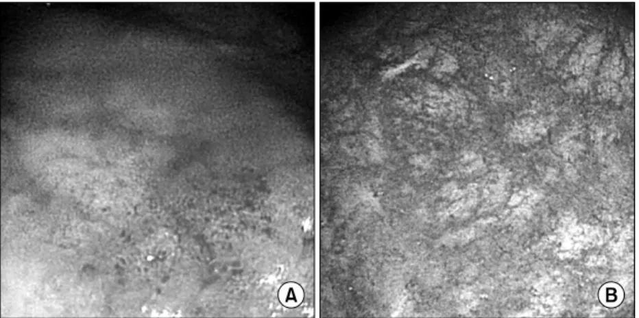
Fig. 1. Colonoscopic findings.
Lost of vascular mar- kings is observed and aphthous ulcers and friability are shown (A).
After 6 weeks of treatment, there has been a significant im- provement (B).
양등과 같은 루푸스와 유사한 소견을 나타내기도 한 다. 최근 저자들은 혈변을 주소로 내원한 루푸스환 자에서 병발한 궤양대장염 1예를 경험하였기에 보고 하는 바이다.
증 례
환 자: 38세 여자
주 소: 내원 3개월 전부터 빈번해지는 혈변 현병력: 6년 전 전신홍반루푸스를 진단받고 치료 중인 자로 내원 3개월 전부터 시작된 소량의 혈변이 있었고 최근 빈번해지는 다량의 혈변이 있어 입원함.
과거력: 6년 전 다발성관절염을 주소로 내원하여 항핵항체(ANA)양성, 항 ds-DNA항체양성, 백혈구와 림프구 감소증으로 전신홍반루푸스를 진단받았다.
그 후 임상소견과 질병의 활성도가 호전을 보여 현 재까지 항말라리아제제(hydroxychloroquine 400 mg)와 소량의 스테로이드(prednisolone 5 mg)로 치료 중이 었다
가족력: 특이사항 없음.
이학적 소견: 내원 시 체온 36.3oC, 맥박수 분당 60 회, 호흡수 분당 20회, 혈압은 100/60 mmHg였다. 장 음은 항진되지 않았고 배꼽 주변으로 경한 압통 외 에 간비종대나 촉지되는 림프절은 관찰되지 않았다.
검사실 소견: 말초 혈액검사에서 백혈구 5,600/mm3 (림프구 1,271/mm3), 혈색소 9.9 g/dL, 혈소판 208,000/ mm3이었다. 생화학 검사에서 나트륨 137 mEq/L, 칼륨 3.9 mEq/L, 총단백 5.8 g/dL, 알부민 4.0 g/dL, AST 22 IU/L, ALT 22 IU/L, BUN 11 mg/dL,
크레아티닌 0.8 mg/dL, 총빌리루빈 0.6 mg/dL, 직접 빌리루빈 0.3 mg/dL, 칼슘 8.5 mg/dL, 인 3.1 mg/dL, LDH 200 U/L, CPK 105 U/L로 정상범위였고, retic- ulocyte 1.4% (0.5∼2%), iron 33 μg/dL (50∼150 μ g/dL), TIBC 439 μg/dL (250∼400 μg/dL), ferrintin 40 ng/mL (30∼400 ng/dL)였다. 면역학적 검사에서 항핵 항체 양성(nucleolar with fine speckled level 4, 1:
2,560), 항ds-DNA 항체는 1년 전부터 음전된 상태가 지속되었다. 혈청 C3 90.3 mg/dL (90∼180 mg/dL), C4 14.2 mg/dL (10∼40 mg/dL), CH50 29.6 units/mL (23∼46 units/mL)로 정상범위 내에 있었다. 루푸스 항응고인자(lupus anticoagulant)와 ANCA는 음성이었 다. 요검사에서는 특이소견 없었다.
방사선학적 소견: 단순 복부 방사선 사진에서 특이 소견 없었고, 복부 전산화단층촬영에서 장벽의 비후 는 보이지 않았으나 우측 하복부의 창자사이막 (mesentery)에 반응성 림프절이 다수 관찰되었고 그 외 특이소견은 관찰되지 않았다.
대장내시경 소견: 항문고리(anal verge) 직상방의 직 장 점막에 혈관음영(vascular marking) 소실과 아프타 궤양들이 관찰되었다(그림 1).
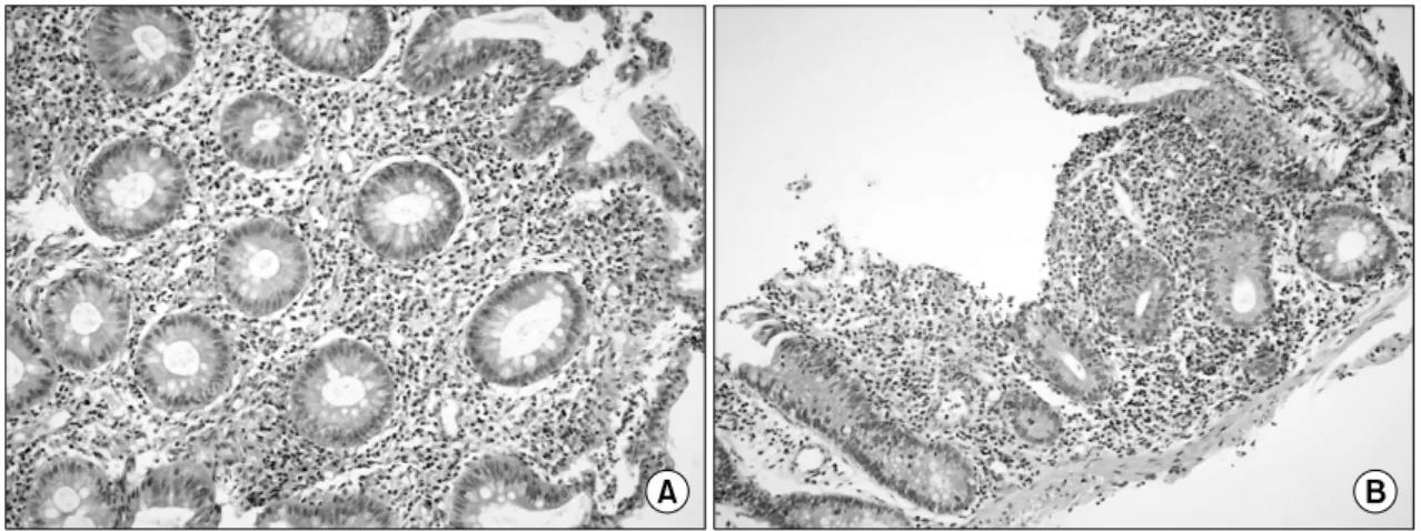
병리 소견: 직장 조직검사에서 전반적으로 만성염 증세포들이 미만성으로 점막 고유판을 침윤하고 있 었으며 점막 샘들은 전반적으로 위축되었고 술잔세 포들의 심한 소실이 관찰되었다. 일부에서는 작은 아프타성 궤양들이 동반되어 있었다(그림 2).
임상경과: 환자는 궤양대장염의 치료로 mesalazine 관제를 사용하였다. 이 후 환자의 증상과 추적 내시경 소견은 호전 되었고 현재 외래에서 추적 관찰 중이다.
― 대한류마티스학회지 제 15 권 제 4 호 2008 ―
― 330 ―
Fig. 2. Microscopic finding. The rectal mucosa shows heavy inflammatory infiltration in the lamina propria (A) and aphtous ulcer (B) with severe goblet cell depletion (H&E stain, ×200).
고 찰
전신홍반루푸스와 궤양대장염이 병발된 경우는 매 우 드물다. 국내에서 2005년 이 등에서 항인지질증 후군에서 크론병을 동반한 경우와 (1) 2006년 이 등에 서 루푸스 환자에서 동반된 크론병이 보고되었지만 (2) 루푸스 환자에서 병발한 궤양대장염은 아직 보 고가 없다. 국외에서도 1969년부터 2005년도 사이에 영문으로 보고된 루푸스와 궤양대장염이 병발한 증 례는 6예에 불과하다 (3).
루푸스와 궤양대장염의 발병원인은 잘 밝혀져 있 지 않지만, 감염은 루푸스의 병인에 대한 관련이 연 구되고 있고 이는 선천면역계통(innate immune sys- tem)을 비특이적으로 자극하여 T세포와 B세포의 활 성과 팽창을 촉진하는 것으로 여겨지고 있다. 바이 러스와 세균이 모두 발병에 관여할 수 있고 그 중에 서 Epstein-Barr virus (EBV)는 루푸스를 유발하는 초 기요인으로 고려될 수 있다는 가설이 있으며 (4), 궤 양대장염에서도 EBV 유도 활성표지자인 CD30의 혈 청수치가 증가되어있다는 보고가 있다 (5). 하지만 아직 궤양대장염과 루푸스의 발병원인이나 기전에 대한 연구는 아직은 부족한 상태이다. 그러나 궤양 대장염이 다른 자가면역 질환과 함께 나타나는 예가 종종 보고되고 있으며, 루푸스와 궤양대장염이 병발 한 국외의 증례들에서도 대부분에서 루푸스가 진단 된 이후에 궤양 대장염이 병발하였다 (3). 앞으로 루 푸스가 궤양대장염의 감수성을 증가시켰을 가능성에
대해 더 많은 연구가 필요하다.
루푸스 환자에서 위장관 증상이 발생한 경우 장혈 관염과 연관되어 나타나는 빈도가 드물지 않기 때문 에 이에 대한 감별이 필요하다. 루푸스 장염은 복통, 오심, 구토를 주 증상으로 하며 (6-8) 위장관 출혈은 5∼10%이고 (7,8) 대부분 공장과 회장을 가장 흔하 게 침범한다 (6). 루푸스 장염은 거의 항상 질병의 활성도와 연관이 있으며 (9) Buck 등은 SLEDAI가 8 점보다 큰 환자군에서만 루푸스 장염이 발생하였다 고 보고하였다 (10). 루푸스 장염 환자에서 대장내시 경 소견은 장점막의 부종과 궤양이 나타나며 혈관염 이 관찰된다 (6). 복부 전산화단층촬영소견은 다른 원인의 허혈성 장질환과 유사하게 나타나는데 장벽 의 비후 소견와 thumbprinting, double halo, target sign이 나타날 수 있다 (11). Sultan SM 등은 루푸스 장염의 조직소견은 소혈관의 동맥염(arteritis)과 세정 맥염(venulitis)이 관찰되며, 장점막에 염증세포가 급 성 및 만성적으로 침윤한다고 하였다 (9). 본 증례에 서는 입원 당시 SLEDAI가 0점이었고 병변의 위치가 직장에 국한되었으며 루푸스 장염을 의심할 만한 조 직소견과 방사선 소견이 관찰되지 않았기 때문에 루 푸스 장염을 배제 할 수 있었다.
직장염의 원인은 루푸스 장염이외에도 궤양 직장 염, 크론병, 창자침투성세균, 거대세포바이러스(cyto- megalovirus), 성매개질환에 의한 감염과 비소염성스 테로이드, 내시경전 장정결(bowel preparation)약제가 될 수 있다 (12). 궤양 직장염(ulcerative proctitis)의
― 이경아 외 : A Case of Systemic Lupus Erythematosus Patient with Ulcerative Colitis ―
― 331 ― 진단은 대개 내시경을 통해 내리는데 점막의 부종, 홍반(erythema), 연약성(friability), 혈관음영의 소실이 그 특징이며 조직소견은 점막의 구조가 파괴되어 있 고 염증세포들의 침윤과 점액고갈, 움고름집(crypt abscess)등이 관찰될 수 있다 (12). 반면 크론병은 직 장에 국한되어 나타나는 경우는 드물며, 내시경소견 은 충혈된 점막이 띄엄띄엄 보이고, 작고 깊은 궤양 이 종주형 또는 다각형 모양으로 나타나는 것이 특 징이다. 크론병의 조직소견은 술잔세포가 대개는 보 존되고 단핵세포의 점막침범이 있으며, 육아종을 형 성한다 (13). 본 증례에서 환자는 발열, 설사, 복통과 같은 증상은 없었으며 혈청 검사에서 특이소견이 없 었고, 조직검사에서 거대세포바이러스 감염에 합당 한 소견이 없었기 때문에 감염성 질환을 배제하였 다. 환자가 복용하던 약제 목록은 hydroxychloroquine 400 mg, prednisolone 5 mg였으며, 비소염성스테로이 드는 복용하지 않았기에 약제 관련성은 없었으며, 임상소견, 내시경 소견과 조직검사 소견이 궤양대장 염에 합당하여 진단하였다.
궤양대장염의 발병 위험은 장내 세균에 대해 방어 기전으로 작용하는 P-glycoprotein의 발현을 감소시키 는 다형태와 관련이 있다는 보고가 있으며 (5), 점막 염증의 유도와 조절에 점막내의 T세포의 역할과 장 내 세균과 선천 면역의 역할에 대한 연구가 이루어 지면서 IL-1 수용체 대항제의 다형태, Th2 세포와의 관련성, NF-κB 신호체계를 억제하는 전사인자인 PPAR-γ가 염증성 대장염에서 정상에서보다 낮게 발현된다는 사실이 보고 되면서 새로운 약제 개발에 대한 연구도 활발히 이루어 지고 있다 (14).
궤양직장염의 치료에서 국소 mesalazine이 국소 스 테로이드 보다 더 효과적이라는 다수의 연구결과 (12,15)에 따라 본 증례에서는 mesalazine 관제를 치 료제로 선택했으며 이후 환자의 증상과 내시경 소견 은 호전되었다.
요 약
결론적으로 전신홍반루푸스 환자에서 혈변이 발생 한 경우에는 루푸스 장염뿐 아니라 드물게 염증성 장질환인 경우도 있으므로, 이를 염두에 두어 내시 경 및 조직검사로 정확한 진단을 하여 적절한 치료 방법을 결정하는 것이 중요하다.
참고문헌
1) Lee JH, Lee JH, Kim SH, Kim JH, Hwang JU, Kim CK, et al. A case of antiphospholipid syndrome with Crohn's disease. Korean J Med 2005;69:222-6.
2) Lee BG, Jung PM, Kim HS, Kim IY, Cho MY, Lee CK, et al. A case of systemic lupus erythematosus patient with crohn's disease. J Korean Rheum Assoc 2006;13:321-9.
3) Nitzan O, Elias M, Saliba WR. Systemic lupus erythematous and inflammatory bowel disease. Eur J Intern Med 2006;17:313-8.
4) Riemekasten G, Hahn BH. Key autoantigen in SLE.
Rheumatology (Oxford) 2005;44:975-82.
5) Giacomelli R, Passacantando A, Parzanese I, Vernia P, Klidara N, Cucinelli F, et al. Serum levels of soluble CD30 are increased in ulcerative colitis (UC) but not in Crohn's disease (CD). Clin Exp Immunol 1998;111:532-5.
6) Kwok SK, Seo SH, Ju JH, Park KS, Yoon CH, Kim WU, et al. Lupus enteritis: clinical characteristics, risk factor for relapse and association with anti-endothelial cell antibody. Lupus 2007;16:803-9.
7) Hoffman BI, Katz WA. The gastrointestinal manifestations of systemic lupus erythematosus: a review of the literature. Semin Arthritis Rheum 1980;9:237-47.
8) Jovaisas A, Kraag G. Acute gastrointestinal mani- festations of systemic lupus erythematosus. Can J Surg 1987;30:185-7.
9) Sultan SM, Ioannou Y, Isenberg DA. A review of gastrointestinal manifestations of systemic lupus erythematosus. Rheumatology (Oxford) 1999;38:917-32.
10) Buck AC, Serebro LH, Quinet RJ. Subacute abdominal pain requiring hospitalization in a systemic lupus erythematosus patient: a retrospective analysis and review of the literature. Lupus 2001;10:491-5.
11) Thoeni RF, Cello JP. CT imaging of colitis.
Radiology 2006;240:623-38.
12) Miguel DR. Diagnosis and treatment of ulcerative proctitis. J Clin Gastroenterol 2004;38:733-40.
13) Nikolaus S, Schreiber S. Diagnostics of inflammatory bowel disease. Gastroenterology 2007;133:1670-89.
14) Anisur R, David AI. Systemic lupus erythematous. N Engl J Med 2008;358:929-39.
15) Daniel KP. Inflammatory bowel disease. N Engl J Med 2002;347:417-29.