Comparison of Contrast-Enhanced T2 FLAIR and 3D T1 Black-Blood Fast Spin-Echo for Detection of Leptomeningeal Metastases
INTRODUCTION
Early detection of leptomeningeal metastases is important to ensure appropriate therapy for preservation of neurologic function (1). Imaging plays a key role in the diagnosis and management of leptomeningeal metastases because CSF examination results are often false-negative and leptomeningeal metastases may be asymptomatic (2).
Previous studies established the value of contrast-enhanced fluid attenuated inversion recovery (FLAIR) sequence in imaging leptomeningeal metastases (3, 4), which may be attributed to three main factors. First, the FLAIR sequence does not allow
This is an Open Access article distributed under the terms of the Creative Commons Attribution Non-Commercial License (http://creativecommons.org/licenses/
by-nc/3.0/) which permits unrestricted non-commercial use, distribution, and reproduction in any medium, provided the original work is properly cited.
Received: March 5, 2018 Revised: April 11, 2018 Accepted: May 15, 2018 Correspondence to:
Yae Won Park, M.D.
Department of Radiology, Ewha Womans University Mokdong Hospital, 1071 Anyangchen-ro, Yangcheon-gu, Seoul 07985, Korea.
Tel. +82-2-2650-5114 Fax. +82-2-2650-5302 E-mail: [email protected]
Copyright © 2018 Korean Society of Magnetic Resonance in Medicine (KSMRM)
Original Article
Purpose: Imaging plays a significant role in diagnosing leptomeningeal metastases.However, the most appropriate sequence for the detection of leptomeningeal metastases has yet to be determined. This study compares the efficacies of contrast- enhanced T2 fluid attenuated inversion recovery (FLAIR) and contrast-enhanced 3D T1 black-blood fast spin echo (FSE) imaging for the detection of leptomeningeal metastases.
Materials and Methods: Tube phantoms containing varying concentrations of gadobutrol solution were scanned using T2 FLAIR and 3D T1 black-blood FSE.
Additionally, 30 patients with leptomeningeal metastases were retrospectively evaluated to compare conspicuous lesions and the extent of leptomeningeal metastases detected by T2 FLAIR and 3D T1 black-blood FSE.
Results: The signal intensities of low-concentration gadobutrol solutions (< 0.5 mmol/L) on T2 FLAIR images were higher than in 3D T1 black-blood FSE. The T2 FLAIR sequences exhibited significantly greater visual conspicuity scores than the 3D T1 black-blood sequence in leptomeningeal metastases of the pial membrane of cistern (P = 0.014). T2 FLAIR images exhibited a greater or equal extent (96.7%) of leptomeningeal metastases than 3D T1 black-blood FSE images.
Conclusion: Because of its high sensitivity even at low gadolinium concentrations, contrast-enhanced T2 FLAIR images delineated leptomeningeal metastases in a wider territory than 3D T1 black-blood FSE.
Keywords: Leptomeningeal metastases; Black-blood; Contrast-enhanced T2 FLAIR Yae Won Park1, Sung Jun Ahn2
1Department of Radiology, Ewha Womans University College of Medicine, Seoul, Korea
2Department of Radiology, Yonsei University, College of Medicine, Seoul, Korea Magnetic resonance imaging
contrast enhancement of vessels (5-8). Second, it reveals an observable T1 contrast with dark CSF signal (5). Third, T2 FLAIR with contrast is more sensitive than T1 weighted image (T1WI) in detecting low concentrations of gadolinium (5, 9).
Recently, three-dimensional T1 black-blood (3D T1 BB) fast spin-echo (FSE) imaging has been found effective for selective suppression of blood vessels and better detection of brain metastases (10-12). Two different techniques are used in black-blood imaging. The first technique is a velocity-selective preparation pulse (or motion-sensitized driven-equilibrium, MSDE), using a spin-echo-based preparative pulse with a velocity-encoding gradient that suppresses blood flow signal (13, 14). This technique utilizes a preparative sequence composed of three nonselective radiofrequency pulses with flip angles of 90°-180°-90°, with symmetric gradients around the 180° pulse. The flow signal is suppressed due to the phase rotation of the magnetized blood flow caused by the motion-probing gradient pulses. The MSDE implemented with 3D FSE imaging increases the contrast-to-noise ratio for brain metastases compared with the conventional gradient echo (GRE) sequence (15). The other technique entails variable flip angle modulation (16) of FSE. In case of small and variable refocusing flip angles, flow signal is suppressed.
Similar sequences using this technique are commercially available (CUBE, VISTA, and SPACE from GE Healthcare, Milwaukee, WI, USA; Philips Healthcare, Best, the Netherlands; and Siemens, Erlangen, Germany, respectively) (16-18). In variable flip angles, blood suppression varies from blood inflow, since blood suppression results from phase dispersion (10).
Although contrast-enhanced 3D T1 BB-FSE imaging presumably improves the diagnostic yield of leptomeningeal metastases, it is not established. In addition, comparison with contrast-enhanced T2 FLAIR in the detection of leptomeningeal metastases has yet to be performed.
Thus, the purpose of this study was to compare contrast- enhanced T2 FLAIR and contrast-enhanced 3D T1 BB-FSE imaging for the detection of leptomeningeal metastases.
MATERIALS AND METHODS
Phantom StudyIn order to determine the underlying mechanisms of contrast in contrast-enhanced FLAIR, and 3D T1 BB-FSE images, tube phantoms containing gadobutrol solutions
(Gadovist; Bayer Schering Pharma, Berlin, Germany) of varying concentrations (range, 0.0125 to 0.9 mmol/L) were scanned with a 3.0-MRI unit (Discovery MR750;
GE Healthcare, Milwaukee, WI, USA). A reference tube phantom containing 0.9% normal saline was also scanned simultaneously. The MSDE The pulse was applied before the 3D T1 CUBE sequence for 3D T1 BB FSE imaging.
Detailed MR parameters for T2 FLAIR and 3D T1 BB-FSE are summarized in Table 1.
Clinical Study Patients
We retrospectively screened consecutive patients diagnosed with clinically suspected brain metastasis at our hospital. Between May and December 2016, 372 patients were evaluated without the MRI protocol for brain metastasis, and identified 30 patients (6 males and 24 females; mean age, 60.6 years; age range 35-74 years) with leptomeningeal metastases. The MRI protocol was similar to the phantom study, and 3D T1 BB-FSE and T2 FLAIR images were acquired sequentially after injection of a gadolinium contrast agent (0.2 mmol/kg gadobutrol; Gadovist; Bayer HealthCare, Germany). The order of sequences was 3D T1 BB-FSE followed by T2 FLAIR in 20 patients, and the order of T2 FLAIR and 3D T1 BB-FSE was reversed for 10 patients.
Leptomeningeal metastases were diagnosed by the presence of one of the following positive findings on T2 FLAIR or 3D T1 BB-FSE images: 1) enhancement in the sulci of the
Table 1. Imaging Parameters for the T2 FLAIR and 3D T1 BB FSE Sequences
T2 FLAIR 3D BB T1 FSE
FOV (mm) 210 220
TR (ms) 12000 500
TE (ms) 144.46 24.511
Inversion time (ms) 2517
Matrix (RO/PE) 352 × 224 256 × 224
Slice thickness (mm) 4 1
ETL 30 24
Flip angle 111 Variable
Scan FOV (cm) 21 22
Scan time (min) 3 min 12 sec 4 min 54 sec 3D = three dimensional; BB = black blood; ETL = echo train length; FLAIR = fluid attenuated inversion recovery; FOV = field of view; FSE = fast spin echo; FSPGR = fast spoiled gradient-echo; PE = phase encoding; RO = readout; TE = echo time;
TR = repetition time
cerebral hemisphere or folia of the cerebellum, 2) cranial nerve enhancement, 3) subependymal enhancement, or 4) pial enhancement in the cistern (19-21). Focal enhancement restricted to a single gyrus suggested a sluggish flow. We also excluded patients exposed to supplemental oxygen, or carried a trauma history, or infarct symptoms, which may produce false positive interpretations (22-25). Our MRI protocol for leptomeningeal metastasis also includes routine T2WI and DWI. Primary malignancies among the 30 patients included lung cancer (n = 21), diffuse large B-cell lymphoma (n = 4), and breast (n = 2), liver (n = 1), cervical (n = 1), and rectal (n = 1) cancer. Our Institutional Review Board waived patient consent for this retrospective study.
Image Assessment
Image assessment was based on T2 FLAIR and 3D T1 BB FSE sequences. Sixty sequences (30 patients × 2 sequences) were saved as DICOM files and randomly sorted by the study coordinator (Y.W.P.). Two neuroradiologists with 6 and 11 years of experience in brain MRI, blinded to patients’
clinical history and diagnosis, independently reviewed these 60 sequences for evaluation of lesion conspicuity and the extent of leptomeningeal metastases. Cases presenting with prominent cerebrospinal fluid (CSF) flow artifacts on T2 FLAIR were excluded from the leptomeningeal metastases on pial membrane of cistern. Coronal and sagittal multiplanar reconstruction (MPR) images as well as axial images were included in the review of the 3D T1 BB FSE sequence.
Lesion conspicuity was assessed according to findings
of enhancement in the sulci of the cerebral hemisphere or folia of the cerebellum, cranial nerve enhancement, subependymal enhancement, and pial enhancement in the cistern. Each positive finding was scored 0-2 according to the degree of conspicuity: 0, negative; 1, suspicious of leptomeningeal metastases, but not clearly distinguished from adjacent structures; and 2, certain leptomeningeal metastases, clearly distinguished from adjacent structures.
In a second session, a week later, the reviewers compared T2FLAIR and 3D T1 BB FSE images to determine whether the extent of leptomeningeal seeding on T2 FLAIR images was larger, equal, or smaller compared to those on 3D T1 BB FSE images.
Statistical Analysis
Subjective assessments of the two reviewers were tabulated and summarized for each sequence. The visual conspicuity scores among the T2 FLAIR and 3D T1 BB FSE sequences were compared using Wilcoxon signed rank test.
The interobserver agreement for visual conspicuity was analyzed using weighted Cohen kappa coefficient. The κ values > 0.81, in the range of 0.61-0.80, and < 0.60 were considered to reflect excellent, good, and poor agreement, respectively. All P-values < 0.05 were statistically significant in the statistical analysis performed using SPSS Statistics 23.0 (IBM, Armonk, NY, USA) and MedCalc Statistical Software version 14.8.1 (MedCalc Software, Ostend, Belgium).
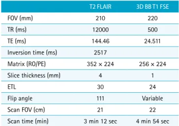
Fig. 1. Relative signal intensities of gadolinium solution based on gadolinium concentration at various pulse sequences: The signal intensities of low-concentration gadobutrol solutions (< 0.5 mmol/
L) relative to normal saline on T2 FLAIR images are higher than in 3D T1 BB FSE images. 3D = three dimensional; BB = black blood;
FLAIR = fluid attenuated inversion recovery; FSE = fast spin echo;
FSPGR = fast spoiled gradient echo
RESULTS
Phantom StudyThe results of the phantom study are shown in Figures 1 and 2. The signal intensity ratios of various concentrations of gadobutrol solution to normal saline on T2 FLAIR and 3D T1 BB-FSE images were plotted according to gadobutrol concentration. The results indicate that the signal intensities of low-concentration gadobutrol solutions (< 0.5 mmol/L) relative to normal saline on T2 FLAIR images were higher compared with the values in 3D T1 BB-FSE.
Clinical Study
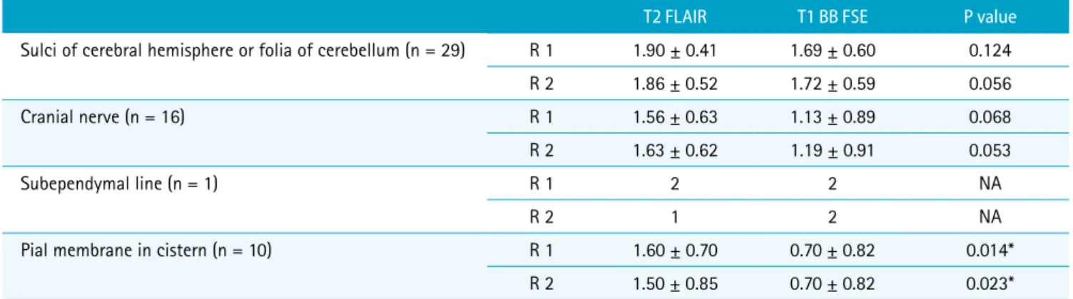
The visual conspicuity scores of the T2 FLAIR sequence for leptomeningeal metastases of the pial membrane in cistern were significantly greater than those of the 3D T1 BB-FSE sequence (P = 0.014 for reviewer 1 and P = 0.023 for reviewer 2). However, no significant differences were observed in visual conspicuity scores for leptomeningeal metastases of the cranial nerve, subependymal line, and pial membrane in the cistern among the three sequences (Table 2). The interobserver agreement for visual conspicuity scores was excellent (κ = 0.87). A representative case is illustrated in Figure 3.
In a majority of patients, T2 FLAIR images exhibited
Fig. 2. Images of phantom tubes containing low concentrations of gadobutrol solution (0.0125-0.9 mmol/L) acquired by T2 FLAIR and 3D T1 BB FSE MRI. 3D = three- dimensional; BB = black blood;
FLAIR = fluid attenuated inversion recovery; FSE = fast spin echo;
FSPGR = fast spoiled gradient echo Table 2. Visual Conspicuity Scores of Each Sequence for Detection of Leptomeningeal Metastases
T2 FLAIR T1 BB FSE P value
Sulci of cerebral hemisphere or folia of cerebellum (n = 29) R 1 1.90 ± 0.41 1.69 ± 0.60 0.124
R 2 1.86 ± 0.52 1.72 ± 0.59 0.056
Cranial nerve (n = 16) R 1 1.56 ± 0.63 1.13 ± 0.89 0.068
R 2 1.63 ± 0.62 1.19 ± 0.91 0.053
Subependymal line (n = 1) R 1 2 2 NA
R 2 1 2 NA
Pial membrane in cistern (n = 10) R 1 1.60 ± 0.70 0.70 ± 0.82 0.014*
R 2 1.50 ± 0.85 0.70 ± 0.82 0.023*
Visual conspicuity scores for leptomeningeal metastases: 0, negative; 1, suspicious of leptomeningeal metastases, but not clearly distinguished from adjacent structures;
and 2, certain leptomeningeal metastases, clearly distinguished from adjacent structures.
*P < 0.05
BB = black blood; FLAIR = fluid attenuated inversion recovery; FSE = fast spin echo; NA = not applicable; R1 = Reviewer 1; R2 = Reviewer 2
greater (43.3% and 33.3% for reviewers 1 and 2, respectively) or equal (53.4% and 63.4% for reviewers 1
and 2, respectively) extent for leptomeningeal metastases compared with 3D T1 BB FSE images (Table 3; Fig. 4).
DISCUSSION
In this study, leptomeningeal metastases of the pial membrane in cistern were more conspicuous in contrast- enhanced T2 FLAIR images than in contrast-enhanced 3D T1-weighted BB-FSE. In addition, the T2 FLAIR sequence demonstrated leptomeningeal metastases across wider or equal regions than the 3D T1 BB-FSE sequence.
a b c
Fig. 3. Images of a 73-year-old female patient with primary lung cancer acquired using the contrast-enhanced T2 FLAIR (a-c) and T1 BB-FSE (d-f) sequences. In T2 FLAIR images, leptomeningeal enhancement is clearly visualized in the pial membrane of the cistern and the cranial nerve (arrows; visual conspicuity score: 2). In 3D T1 BB-FSE images, leptomeningeal enhancement is not clearly demarcated in the pial membrane of the cistern (arrow; visual conspicuity score: 0) but suspected in the cranial nerve (conspicuity score: 1). 3D = three-dimensional; BB = black blood; FLAIR = fluid attenuated inversion recovery; FSE = fast spin echo
d e f
Table 3. Extent of Leptomeningeal Seeding
Reviewer 1 Reviewer 2 T2 FLAIR > 3D T1 BB FSE 13/30 (43.3) 10/30 (33.3) T2 FLAIR = 3D T1 BB FSE 16/30 (53.3) 19/30 (63.4) T2 FLAIR < 3D T1 BB FSE 1/30 (3.3) 1/30 (3.3) Data are presented as numbers of patients/total patients (%).
BB = black blood; FLAIR = fluid attenuated inversion recovery; FSE = fast spin echo
These results are clinically significant. Imaging with the T2 FLAIR sequence after gadolinium injection during MRI for brain metastases might be disputed given that the 3D T1 BB-FSE sequence might serve as an alternative to MRI for detection of leptomeningeal metastases. However, considering our results, T2 FLAIR imaging after gadolinium injection may still be of value.
Previous studies reported a higher signal intensity of T2 FLAIR compared with variants of T1-weighted images at lower concentrations of gadolinium (5, 7). In the previous studies, the signal intensity of gadolinium on T2 FLAIR was higher than that of 2D T1-weighted images (repetition time [TR]/echo time [TE] = 650 ms/25 ms) at lower gadolinium concentrations with magnetization transfer pulse (< 1 mmol/L) and without magnetization transfer pulse (< 0.5 mmol/L). Further, in a study comparing 3D T2 FLAIR with 3D magnetization prepared rapid acquisition gradient echo (MPRAGE) (TR/TE/inversion time [TI] = 1900 ms/4.7 ms/900 ms), the signal intensity of 3D T2 FLAIR was higher than that of 3D MPRAGE at lower gadolinium concentrations (< 0.5 mmol/L) (7). Our study suggested that the addition of preparation pulse to 3D T1 FSE may not affect the superiority of T2 FLAIR over T1-weighted image in demonstrating low gadolinium concentration (<
0.5 mmol/L). This phenomenon is explained by the unique T1 weighting of the T2 FLAIR sequence. Because of the mild T1 weighting induced by the long inversion time and T1 shortening caused by gadolinium, the T2 FLAIR sequence is more sensitive than conventional contrast- enhanced T1 sequence as well as black-blood T1 sequence in demonstrating lower concentrations of gadolinium (5, 9,
24).
In our in vivo study, the contrast-enhanced T2 FLAIR sequence revealed leptomeningeal metastases more clearly than the 3D T1 BB-FSE sequence in pial membrane of cistern. Moreover, although statistically not significant, both reviewers graded the T2 FLAIR highly even in the leptomeningeal metastases of cerebral hemisphere or folia of the cerebellum and cranial nerves. In addition, visualization of leptomeningeal metastases was wider on contrast-enhanced T2 FLAIR that on 3D T1 BB. In leptomeningeal metastases, gadolinium leaks into the adjacent CSF through damaged vessels and is diluted, which corresponds to the low-concentration setting used in the phantom study (26). Therefore, the T2 FLAIR sequence might be superior to 3D T1 BB-FSE in terms of visualization of leptomeningeal metastases (5, 9).
Different acquisition time, different MR parameters, and different types of gadolinium-based contrast agents may affect the conspicuity of leptomeningeal seeding. A delay in imaging time may be effective in increasing the contrast intensity, because it prolongs the perfusion of contrast agent by the aberrant and leaky neovasculature within the metastases (27). However, an optimal MR acquisition time has yet to be determined for evaluation of leptomeningeal metastases. We presume that the optimal acquisition time may vary from that of brain parenchymal metastases, because extravasated gadolinium diluted by CSF affected the leptomeningeal metastasis. In our protocol, 2D FLAIR yielded a thicker slice (4 mm) than 3D sequences (1 mm), and the partial volume effects may have affected the assessment of the images. However, even at a lower Fig. 4. Images of a 50-year-old female patient acquired using the T2 FLAIR (a) and 3D T1 BB- FSE (b) sequences. The extent of leptomeningeal seeding in the T2 FLAIR image is greater than in the 3D T1 BB-FSE image (white arrows). 3D = three-dimensional;
BB = black blood; FLAIR = fluid attenuated inversion recovery; FSE
= fast spin echo
a b
spatial resolution along the z-axis, T2 FLAIR identified leptomeningeal metastases wider than the 3D T1 BB FSE.
Further, the use of a different contrast agent may have affected the conspicuity of different types of sequences due to varying R1 and R2 relaxation times (28). However, a previous study did not reveal a significant difference in efficacy of evaluation based on differences in contrast agents used in central nervous system applications (29).
The present study has several limitations. First, our hospital does not perform CSF analysis routinely for the diagnosis of leptomeningeal metastases. We did not perform CSF analysis due to the invasive procedure involved (30) and the low sensitivity (20, 31, 32). We assumed that appropriate neuroimaging abnormalities, along with clinical features, are adequate for the diagnosis of leptomeningeal metastases. Second, we did not assess the diagnostic accuracies of both sequences for detection of leptomeningeal seeding, in the absence of a gold standard sequence for diagnosis. However, all of our cases showed leptomeningeal enhancement in both sequences, without significant variation in diagnostic accuracy. Third, the extent of leptomeningeal metastases may be exaggerated due to the contrast leakage. Fourth, we could not directly measure the gadolinium concentration in vivo. Fifth, we used a double-dose contrast media solution, which may have improved the lesion detection. However, we speculate that a similar trend may occur in a single-dose study.
In conclusion, the contrast-enhanced T2 FLAIR sequence delineates leptomeningeal metastases of the pial membrane in cistern more clearly than the contrast-enhanced 3D T1 BB-FSE sequence. In addition, because of its higher sensitivity even at low gadolinium concentrations, the contrast-enhanced T2 FLAIR sequence demonstrates leptomeningeal metastases in a wider region than the contrast-enhanced 3D T1 BB-FSE sequence.
REFERENCES
1. Grossman SA, Krabak MJ. Leptomeningeal carcinomatosis.
Cancer Treat Rev 1999;25:103-119
2. Wasserstrom WR, Glass JP, Posner JB. Diagnosis and treatment of leptomeningeal metastases from solid tumors:
experience with 90 patients. Cancer 1982;49:759-772 3. Kremer S, Abu Eid M, Bierry G, et al. Accuracy of delayed
post-contrast FLAIR MR imaging for the diagnosis of leptomeningeal infectious or tumoral diseases. J Neuroradiol 2006;33:285-291
4. Misaki K, Nakada M, Hayashi Y, et al. Contrast-enhanced fluid-attenuated inversion recovery MRI is useful to detect the CSF dissemination of glioblastoma. J Comput Assist Tomogr 2001;25:953-956
5. Mathews VP, Caldemeyer KS, Lowe MJ, Greenspan SL, Weber DM, Ulmer JL. Brain: gadolinium-enhanced fast fluid-attenuated inversion-recovery MR imaging. Radiology 1999;211:257-263
6. Terae S, Yoshida D, Kudo K, Tha KK, Fujino M, Miyasaka K.
Contrast-enhanced FLAIR imaging in combination with pre- and postcontrast magnetization transfer T1-weighted imaging: usefulness in the evaluation of brain metastases.
J Magn Reson Imaging 2007;25:479-487
7. Fukuoka H, Hirai T, Okuda T, et al. Comparison of the added value of contrast-enhanced 3D fluid-attenuated inversion recovery and magnetization-prepared rapid acquisition of gradient echo sequences in relation to conventional postcontrast T1-weighted images for the evaluation of leptomeningeal diseases at 3T. AJNR Am J Neuroradiol 2010;31:868-873
8. Tomura N, Narita K, Takahashi S, et al. Contrast-enhanced multi-shot echo-planar FLAIR in the depiction of metastatic tumors of the brain: comparison with contrast- enhanced spin-echo T1-weighted imaging. Acta Radiol 2007;48:1032-1037
9. Jackson EF, Hayman LA. Meningeal enhancement on fast FLAIR images. Radiology 2000;215:922-924
10. Park J, Kim J, Yoo E, Lee H, Chang JH, Kim EY. Detection of small metastatic brain tumors: comparison of 3D contrast- enhanced whole-brain black-blood imaging and MP-RAGE imaging. Invest Radiol 2012;47:136-141
11. Kammer NN, Coppenrath E, Treitl KM, Kooijman H, Dietrich O, Saam T. Comparison of contrast-enhanced modified T1- weighted 3D TSE black-blood and 3D MP-RAGE sequences for the detection of cerebral metastases and brain tumours.
Eur Radiol 2016;26:1818-1825
12. Park J, Kim EY. Contrast-enhanced, three-dimensional, whole-brain, black-blood imaging: application to small brain metastases. Magn Reson Med 2010;63:553-561 13. Korosec FR, Grist TM, Polzin JA, Weber DM, Mistretta CA.
MR angiography using velocity-selective preparation pulses and segmented gradient-echo acquisition. Magn Reson Med 1993;30:704-714
14. Norris DG, Schwarzbauer C. Velocity selective radio- frequency pulse trains. J Magn Reson 1999;137:231-236 15. Nagao E, Yoshiura T, Hiwatashi A, et al. 3D turbo spin-
echo sequence with motion-sensitized driven-equilibrium preparation for detection of brain metastases on 3T MR imaging. AJNR Am J Neuroradiol 2011;32:664-670
16. Busse RF, Brau AC, Vu A, et al. Effects of refocusing flip
angle modulation and view ordering in 3D fast spin echo.
Magn Reson Med 2008;60:640-649
17. Takemoto K, Takano K, Abe H, et al. The new MRI modalities
"BPAS and VISTA" for the diagnosis of VA dissection. Acta Neurochir Suppl 2011;112:59-65
18. Kato Y, Higano S, Tamura H, et al. Usefulness of contrast- enhanced T1-weighted sampling perfection with application-optimized contrasts by using different flip angle evolutions in detection of small brain metastasis at 3T MR imaging: comparison with magnetization-prepared rapid acquisition of gradient echo imaging. AJNR Am J Neuroradiol 2009;30:923-929
19. Collie DA, Brush JP, Lammie GA, et al. Imaging features of leptomeningeal metastases. Clin Radiol 1999;54:765-771 20. Freilich RJ, Krol G, DeAngelis LM. Neuroimaging
and cerebrospinal fluid cytology in the diagnosis of leptomeningeal metastasis. Ann Neurol 1995;38:51-57 21. Straathof CS, de Bruin HG, Dippel DW, Vecht CJ. The
diagnostic accuracy of magnetic resonance imaging and cerebrospinal fluid cytology in leptomeningeal metastasis.
J Neurol 1999;246:810-814
22. Ahn SJ, Suh SH, Lee KY, Kim JH, Seo KD, Lee S. Hyperintense vessels on T2-PROPELLER-FLAIR in patients with acute MCA stroke: prediction of arterial stenosis and perfusion abnormality. AJNR Am J Neuroradiol 2015;36:2042-2047 23. Ahn SJ, Lee KY, Ahn SS, Suh H, Kim BS, Lee SK. Can FLAIR
hyperintense vessel (FHV) signs be influenced by varying MR parameters and flow velocities? A flow phantom analysis. Acta Radiol 2016;57:580-586
24. Ahn SJ, Chung TS, Chang JH, Lee SK. The added value of double dose gadolinium enhanced 3D T2 fluid-attenuated inversion recovery for evaluating small brain metastases.
Yonsei Med J 2014;55:1231-1237
25. Jeong HK, Oh SW, Kim J, Lee SK, Ahn SJ. Reduction of oxygen-induced CSF hyperintensity on FLAIR MR images in sedated children: usefulness of magnetization-prepared FLAIR imaging. AJNR Am J Neuroradiol 2016;37:1549- 1555
26. Siegal T, Sandbank U, Gabizon A, et al. Alteration of blood-brain-CSF barrier in experimental meningeal carcinomatosis. A morphologic and adriamycin-penetration study. J Neurooncol 1987;4:233-242
27. Ludemann L, Grieger W, Wurm R, Wust P, Zimmer C.
Quantitative measurement of leakage volume and permeability in gliomas, meningiomas and brain metastases with dynamic contrast-enhanced MRI. Magn Reson Imaging 2005;23:833-841
28. Pintaske J, Martirosian P, Graf H, et al. Relaxivity of Gadopentetate Dimeglumine (Magnevist), Gadobutrol (Gadovist), and Gadobenate Dimeglumine (MultiHance) in human blood plasma at 0.2, 1.5, and 3 Tesla. Invest Radiol 2006;41:213-221
29. van der Molen AJ, Bellin MF. Extracellular gadolinium- based contrast media: differences in diagnostic efficacy.
Eur J Radiol 2008;66:168-174
30. Ruff RL, Dougherty JH Jr. Complications of lumbar puncture followed by anticoagulation. Stroke 1981;12:879-881 31. Clarke JL, Perez HR, Jacks LM, Panageas KS, Deangelis LM.
Leptomeningeal metastases in the MRI era. Neurology 2010;74:1449-1454
32. Glass JP, Melamed M, Chernik NL, Posner JB. Malignant cells in cerebrospinal fluid (CSF): the meaning of a positive CSF cytology. Neurology 1979;29:1369-1375