Korean Journal of Urology
ⒸThe Korean Urological Association, 2013 884 Korean J Urol 2013;54:884-887
www.kjurology.org
http://dx.doi.org/10.4111/kju.2013.54.12.884
Case Report
Spermatocytic Seminoma With Sarcoma: An Indication for Adjuvant Chemotherapy in Localized Disease
David Wetherell, Nathan Lawrentschuk, Dennis Gyomber
Department of Urology, University of Melbourne, Austin Hospital, Heidelberg, Australia
Spermatocytic seminoma (SCS) with sarcoma is an extremely rare testicular tumor with only 11 cases previously described in the literature. We present the 12th case of SCS with sarcoma in a 29-year-old male. SCS itself is an uncommon germ cell tumor with a relatively indolent clinical course that mostly affects males around the fifth dec- ade of life. Sarcomatous differentiation of SCS occurs in 5% to 6% of cases and correlates with a higher possibility of metastatic disease and a poor prognosis. Clinically, this tu- mor manifests as a slow-growing testicular mass often with an accelerated period of secondary growth. After a concise review of the literature, we conclude that SCS with sarcoma should be treated by radical inguinal orchidectomy with strong consideration given to adjuvant chemotherapy.
Keywords: Orchiectomy; Sarcoma; Seminoma; Testicular neoplasms; Testis
This is an Open Access article distributed under the terms of the Creative Commons Attribution Non-Commercial License (http://creativecommons.org/licenses/by-nc/3.0) which permits unrestricted non-commercial use, distribution, and reproduction in any medium, provided the original work is properly cited.
Article History:
received 5 March, 2012 accepted 17 April, 2012
Corresponding Author:
David Wetherell Department of Urology, University of Melbourne, Austin Hospital, 145 Studley Road, Heidelberg, Victoria 3084, Australia
TEL: +61-3-9496-5000 FAX: +61-3-9496-3617
E-mail: drwetherell@doctors.org.uk
FIG. 1. Gray-scale ultrasound scan demonstrating a large cystic mass in the right testis.
INTRODUCTION
Spermatocytic seminoma (SCS) is an uncommon germ cell tumor (GCT). It accounts for 4% to 7% of all cases of tes- ticular seminoma [1]. The mean age of diagnosis of SCS is 54 years [2]. Clinically, SCS is an indolent neoplasm with rare potential to metastasize and therefore is associated with a good prognosis [3]. Sarcomatous transformation of GCTs is associated with a significant propensity for ag- gressive and malignant tumor behavior [4]. Specifically, the development of a sarcomatous component in testicular GCT is described as an uncommon phenomenon [4]. The de- velopment of a sarcomatous component in SCS is ex- tremely rare, and only a handful of cases have been de- scribed in the literature (Table 1). We present an exception- ally rare case of SCS with undifferentiated sarcomatous transformation in a young male. We believe this to be only the 12th report to date of SCS with sarcomatous differentiation.
CASE REPORT
A 29-year-old male of Syrian origin presented with a pain- less right-sided testicular swelling of 12 months’ duration.
Clinical examination revealed a nontender enlarged right testis. The tumor markers alpha-fetoprotein (α-FP) and be- ta human chorionic gonadotropin (β-HCG) were negative.
An ultrasound scan of the scrotum revealed a 7-cm hypo- echoic, hypervascular and cystic mass replacing the right
Korean J Urol 2013;54:884-887
Spermatocytic Seminoma With Sarcoma 885
FIG. 2 Microscopic chondroid-type change demonstrating sarco-
ma differentiation (×200). FIG. 3 Microscopic demonstration of the interface between the sarcoma (top right) and seminoma (bottom left) elements (×40).
testis (Fig. 1). The patient underwent a right radical in- guinal orchidectomy. A staging computed tomography scan was negative for lymphatic or metastatic disease.
Macroscopically, a lobulated, circumscribed, focally ne- crotic, soft tumor (60 mm×55 mm×50 mm) was found re- placing most of the right testis. The tumor appeared to be confined within the testis. The majority of the tumor was polymorphous and was composed of small, intermediate, and large epithelioid cells arranged in sheets with broad areas of necrosis. Microscopic features of these cells were consistent with SCS. Additionally, there were ill-defined areas of undifferentiated sarcoma (Figs. 2, 3) that were composed of highly pleomorphic, epithelioid, and spin- dle-shaped cells with high mitotic activity invested in fi- brous and fibromyxoid stroma. The tumor did not extend beyond the tunica albuginea. The spermatic cord and epi- didymis were uninvolved. No lymphovascular invasion was identified. Immunohistochemical analysis revealed focal staining of a small proportion of the SCS cells with an- tibody to CD117 (c-KIT). Immunohistochemical stains of antibodies to PLAP, HCG, AFP, CD30, and CD68 and the myeloid markers CD34 and CD117 were all negative. The histological diagnosis was SCS with undifferentiated sar- coma confined to the testis. Further oncological treatment with adjuvant chemotherapy was strongly advised but the patient refused and moved to a different locality.
DISCUSSION
Testicular seminoma accounts for 40% to 50% of all GCTs of the testis [5], with SCS accounting for less than 1% of all testicular tumors [6]. SCS occurs purely in the testis and in an older age group (mean age, 54 years; range, 25 to 87 years) compared with classic seminoma (mean age, 41 years; range, 30 to 40 years) [1,2,7]. Diagnosis of SCS is based on histopathological features including the size spec- trum of cells, lack of cytoplasmic glycogen, and lymphocytic
infiltrate [8]. Absence of granulomas and spherical, dark nuclei help to distinguish these tumors from classic semi- nomas [1,8]. Importantly, SCSs are not associated with concurrent germ cell components, unlike a third of classic seminomas with teratomatous-type association [1]. The clinical manifestation of SCS is classically described as a painlessly enlarging testicular mass [8]. Metastasis of SCS is extremely rare and has only been seen in 1 or 2 cases [8], leading to a favorable prognosis after orchidectomy, which is considered the gold standard treatment.
SCS with sarcoma is an extremely rare diagnosis with on- ly 11 cases previously reported in the literature (Table 1) and with this article illustrating the 12th case. This sarcom- atous differentiation occurs in approximately 5% to 6% of SCS [2,8] and is associated with aggressive behavior and poor prognosis [8]. In the few reported cases of SCS with a sarcomatous component, the mean patient age was 51 years (range, 40 to 66 years), which is similar to the age associated with pure SCS. In most of the cases outlined in Table 1, there was a dual component associated with the clinical history.
First, the slow progression of a painless unilateral scrotal mass (mean, 4 years from 7 cases) is followed by a secondary accelerated period of growth, which is often painful (mean, 2 months from 6 cases). The second component of rapid growth has been suggested to be the latent sarcomatous dif- ferentiation of a pure SCS [1]. The most common sarcoma- tous differentiation was rhabdomyosarcoma, followed by undifferentiated and spindle cell sarcoma (Table 1). No ter- atomatous component was identified in any of the cases [10]. In most cases, serum tumor markers such as α-FP, β-HCG, and lactate dehydrogenase; were normal. Average tumor size was 14 cm, with most tumors being treated by radical orchidectomy and adjuvant chemotherapy. Meta- static disease at the time of presentation or histological di- agnosis was present in six cases, with two cases developing metastatic disease months or years after orchidectomy and two cases without any evidence of metastatic disease at any
Korean J Urol 2013;54:884-887
886 Wetherell et al
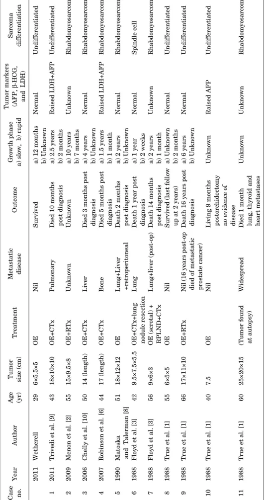
TABLE 1. Summary of the published case reports of spermatocytic seminoma with sarcoma Case no.YearAuthorAge (yr)Tumor size (cm)TreatmentMetastatic disease OutcomeGrowth phase a) slow, b) rapid Tumor markers (AFP, β-HCG, and LDH)
Sarcoma differentiation 1 2 3 4 5 6 7 8 9 10 11
2011 2011 2009 2006 2007 1990 1988 1988 1988 1988 1988 1988
Wetherell Trivedi et al. [9] Menon et al. [2] Chelly et al. [10] Robinson et al. [6] Matoska and Talerman [8] Floyd et al. [3] Floyd et al. [3] True et al. [1] True et al. [1] True et al. [1] True et al. [1]
29 43 55 50 44 51 42 56 55 66 40 60
6×5.5×5 18×10×10 15×9.5×8 14 (length) 17 (length) 18×12×12 9.5×7.5×5.5 9×6×3 6×5×5 17×11×10 7.5 25×20×15
OE OE+CTx OE+RTx OE+CTx OE+CTx OE OE+CTx+lung nodule resection OE (scrotal) + RPLND+CTx OE OE+RTx OE (Tumor found at autopsy)
Nil Pulmonary Unknown Liver Bone Lung+Liver +retroperitoneal Lung Lung+liver (post-op) Nil Nil (16 years post-op died of metastatic prostate cancer) Nil Widespread
Survived Died 10 months post diagnosis Unknown Died 3 months post diagnosis Died 5 months post diagnosis Death 2 months post diagnosis Death 1 year post diagnosis Death 14 months post diagnosis Survived (last follow up at 2 years) Death 16 years post diagnosis Living 9 months postorchidectomy no evidence of disease Died 1 month lung, thyroid and heart metastases
a) 12 months b) Unknown a) 2.5 years b) 2 months a) 10 years b) 7 months a) 4 years b) Unknown a) 1.5 years b) 1 month a) 2 years b) Unknown a) 1 year b) 2 weeks a) 2 years b) 1 month a) Unknown b) 2 months a) 6 years b) Unknown Unknown Unknown
Normal Raised LDH+AFP Unknown Normal Raised LDH+AFP Normal Normal Unknown Normal Normal Raised AFP Unknown
Undifferentiated Undifferentiated Rhabdomyosarcoma Rhabdomyosarcoma Rhabdomyosarcoma Rhabdomyosarcoma Spindle cell Rhabdomyosarcoma Undifferentiated Undifferentiated Undifferentiated Rhabdomyosarcoma AFP, alpha-fetoprotein; β-HCG, beta-human chorionic gonadotropin; LDH, lactate dehydrogenase; OE, radical inguinal orchidectomy; CTx, chemotherapy; RTx, radiotherapy; RPLND, retroperitoneal lymph node dissection; Post-op, postoperation.
Korean J Urol 2013;54:884-887
Spermatocytic Seminoma With Sarcoma 887
stage. One case described by Menon et al. [2] lacked details on any metastatic disease but mentioned further care at an oncology unit. Poor prognosis of these cases is evident by mortality in seven cases with a mean survival of 7 months.
In one case reported by True et al. [1], the patient developed metastases 16 years after the diagnosis, which was as- sumed to be a result of metastatic prostate carcinoma.
The diagnosis of SCS is rare below the age of 40 years [9]
and our case of SCS with undifferentiated sarcoma in a 29-year-old man is the only case below this age reported in the literature. Our case classically reported a long duration (12 months) of painless testicular swelling. No period of painful accelerated growth was reported. The size of the tu- mor in this case was considerably smaller at 6 cm compared with the average 14 cm calculated from previous cases.
Although SCS with sarcoma is associated with a poor prog- nosis and metastasis at diagnosis is seen in half of cases, our case had no evidence of extratesticular spread.
Previous articles have estimated up to 16 case reports in the literature [10]. Our literature review revealed 11 defi- nite cases of SCS with sarcomatous components. We be- lieve that overestimation of the number of cases has arisen because of the duplication of some reported cases (case 7 in Table 1) as explained by Matoska and Talerman [8]. Case number 11 (Table 1), one of the five first cases described by True et al. [1], was found to have extensive metastasis at presentation and histological diagnosis was made at autopsy. The highly aggressive nature of sarcomatous dif- ferentiation together with the assumed late presentation (patient age, 60 years; tumor size, 25 cm) accounts for the absence of surgical or oncological management in this case.
In case 7, the method of orchidectomy was via a scrotal ap- proach rather than the conventional, and now standard, in- guinal approach. This variation in surgical technique was questioned by Floyd et al. [3] and a hemiscrotectomy and retroperitoneal lymph node dissection was performed.
Rhabdomyosarcoma appears to be associated with meta- static disease in every case: 1, 2, 3, 7, and 9 [10]. Owing to the limited number of cases, no definitive conclusions can be drawn about the association of specific differentiation of sarcomatous elements and the likelihood of metastatic disease. The general consensus is that SCS with sarcoma, of any differentiation or undifferentiated, is associated with a high probability of early metastases. The mecha- nism of transformation is unknown but one hypothesis is anaplastic conversion or transformation of SCS [9]. The rapid acceleration growth phase demonstrated clinically is
likely to represent this period of transition and histological differentiation into sarcoma [9]. Most articles suggest that in the presence of sarcomatous differentiation in SCS, ad- juvant chemotherapy following orchidectomy is the choice of treatment for these highly malignant tumors [2,7,9].
In conclusion, SCS with sarcomatous differentiation is an exceptionally rare tumor with only 12 cases reported in the literature. The development of this sarcomatous ele- ment transforms the prognosis of the relatively indolent SCS tumor to a highly aggressive malignant neoplasm.
These tumors often present with concurrent metastatic disease and thus treatment with adjuvant chemotherapy following radical inguinal orchidectomy should be consid- ered in all cases.
CONFLICTS OF INTEREST The authors have nothing to disclose.
REFERENCES
1. True LD, Otis CN, Delprado W, Scully RE, Rosai J. Spermatocytic seminoma of testis with sarcomatous transformation: a report of five cases. Am J Surg Pathol 1988;12:75-82.
2. Menon S, Karpate A, Desai S. Spermatocytic seminoma with rhabdomyosarcomatous differentiation: a case report with a re- view of the literature. J Cancer Res Ther 2009;5:213-5.
3. Floyd C, Ayala AG, Logothetis CJ, Silva EG. Spermatocytic semi- noma with associated sarcoma of the testis. Cancer 1988;61:
409-14.
4. Malagon HD, Valdez AM, Moran CA, Suster S. Germ cell tumors with sarcomatous components: a clinicopathologic and im- munohistochemical study of 46 cases. Am J Surg Pathol 2007;31:
1356-62.
5. Henley JD, Ulbright TM. Seminoma: a review with emphasis on morphologic patterns and differential diagnosis. Pathol Case Rev 2005;10:167-75.
6. Robinson A, Bainbridge T, Kollmannsberger C. A spermatocytic seminoma with rhabdomyosarcoma transformation and ex- tensive metastases. Am J Clin Oncol 2007;30:440-1.
7. Eble JN. Spermatocytic seminoma. Pathol Case Rev 2005;10:
176-80.
8. Matoska J, Talerman A. Spermatocytic seminoma associated with rhabdomyosarcoma. Am J Clin Pathol 1990;94:89-95.
9. Trivedi P, Pasricha S, Gupta A. Spermatocytic seminoma asso- ciated with undifferentiated sarcoma: a rare case report. Indian J Pathol Microbiol 2011;54:138-40.
10. Chelly I, Mekni A, Gargouri MM, Bellil K, Zitouna M, Horchani A, et al. Spermatocytic seminoma with rhabdomyosarcomatous contingent. Prog Urol 2006;16:218-20.