INTRODUCTION
The global human genome project was launched in the 1990’s and researchers expect to understand information of the whole human genome, especially genes which encode functional proteins. However, they recognized that the human genome contains only about 20000 protein-coding genes [1,2].
It is not surprising that the numbers of protein-coding genes are pretty similar in number to those of Caenorhabditis elegans.
Because the protein-coding genes could not properly explain the human gene expression complexity in view of physiologi- cal and evolutional aspects, investigators have suspected that the potential non-protein coding but transcriptionally active genes (>98% of transcribed genes) may be fully accountable for gene expression complexity of human [3-5].
Recently, investigators categorized the non-coding RNAs (ncRNAs) as short ncRNA, mid-size ncRNA, and long non-
Roles of Long Non-Coding RNAs on Tumorigenesis and Glioma Development
Ju Young Park1*, Jeong Eun Lee1*, Jong Bae Park2,3, Heon Yoo2,3, Seung-Hoon Lee2,3, Jong Heon Kim1,3
1Cancer Cell and Molecular Biology Branch, 2Specific Organs Cancer Branch, Research Institute, National Cancer Center, Goyang, Korea
3Department of System Cancer Science, Graduate School of Cancer Science and Policy, National Cancer Center, Goyang, Korea
Received March 19, 2014 Revised March 27, 2014 Accepted March 31, 2014 Correspondence Jong Heon Kim
Cancer Cell and Molecular Biology Branch, Research Institute, National Cancer Center, 323 Ilsan-ro, Ilsandong-gu, Goyang 410-769, Korea Tel: +82-31-920-2204
Fax: +82-31-920-2006 E-mail: [email protected]
*These two authors contributed equally to this work.
More than 98% of eukaryotic transcriptomes are composed of non-coding RNAs with no functional protein-coding capacity. Those transcripts also include tens of thousands of long non-coding RNAs (lncRNAs) which are emerging as key elements of cellular homeostasis, essentially tumorigenesis steps. However, we are only beginning to understand the nature and extent of the involvement of ln- cRNAs on tumorigeneis. Here, we highlight recent progresses that have identified a myriad of molecu- lar functions on tumorigenesis for several lncRNAs including metastasis-associated lung adenocarci- noma transcript 1 (MALAT1), prostate cancer associated non-coding RNA 1 (PRNCR1), prostate cancer gene expression marker 1 (PCGEM1), H19, and homeobox transcript antisense intergenic RNA (HO- TAIR), and several new lncRNAs for glioma development. Potential therapeutic approaches for the ln- cRNAs in various human diseases are also discussed.
Key Words Non-coding RNA; lncRNA; Tumorigenesis; Glioma.
coding RNA (lncRNA) by their lengths, and sometimes ncRNAs are subdivided by function, loci, and post-transcrip- tional modification. Among these, most onco-molecular biol- ogists are deeply interested in the short ncRNAs [e.g., microR- NAs (miRNAs)] [6] and very recently lncRNAs for their research projects as an aspect of gene expression regulation [7-11]. This review, we essentially highlight and introduce our recent understanding of roles of lncRNAs in tumorigenesis, including glioma development.
LNCRNAS: MODE OF ACTION
Recent reports showed that lncRNAs have multifunctional roles on modulating embryonic pluripotency, differentiation, development, and various diseases, essentially in cancers [7-11].
Therefore, dysregulation of lncRNAs has been shown to be as- sociated with a broad range of defects on those physiological phenomena. lncRNAs may be classified according to their mode of action and functions in cells such as, 1) mediators on signaling pathway, 2) serving as molecular decoys, 3) work as molecular guides for the ribonucleoprotein complexes to cer-
This is an Open Access article distributed under the terms of the Creative Commons Attribution Non-Commercial License (http://creativecommons.org/licenses/by-nc/3.0) which permits unrestricted non-commercial use, distribution, and reproduction in any medium, provided the original work is properly cited.
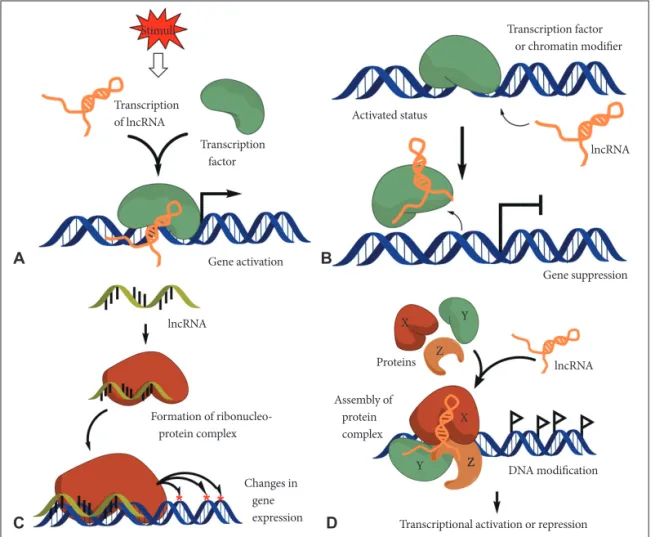
tain specific chromatin site, and also have 4) scaffold function for the proper complex formation (Fig. 1) [11].
lncRNAs: mediators on signaling pathway
In most cases, transcription of lncRNAs is temporally reg- ulated and the expression has tissue specificity. Moreover, their expression is modulated in response to the internal and exter- nal stimuli [12]. Therefore, lncRNAs may serve as molecular signaling mediators which modulate a certain set of gene ex- pression temporally and even spatially (Fig. 1A).
lncRNAs: molecular decoys
lncRNAs may serve as molecular decoys which take pro- teins or RNAs away from a specific location. Sometimes decoy lncRNAs can serve as “sponge” to proteins (e.g., transcription factors and chromatin modifiers) and small ncRNAs (e.g., miRNAs) (Fig. 1B) [13]. This event can lead to overall tran- scriptome change of cells.
lncRNAs: work as molecular guides
lncRNAs can serve as the molecular guides by locating cer- tain ribonucleoprotein complexes to a specific target site on the chromatin (Fig. 1C) [14]. The gene expression can be al- tered either in neighboring genes (in cis) or distantly located ones (in trans).
lncRNAs: scaffold function
lncRNAs can support the assembly of protein complexes which link the factors together to generate brand new func- tions (Fig. 1D). Some lncRNAs possess distinct protein-bind- ing domains that combine each molecule together. This event may have impact on action of transcription or repression.
LNCRNAS ON TUMORIGENESIS
Generally the lncRNAs designate ncRNAs which is more than 200 nt long [7-11]. 1) Usually those modulate DNA meth- ylation which is closely related with genome imprinting (e.g.,
Fig. 1. Schematic diagram of lncRNA action mechanisms. A: Mediators on signaling pathway: lncRNAs can serve as molecular signaling mediators which modulate certain set of gene expression in conjunction with specific transcription factors or chromatin modifiers. B: Molec- ular decoys: lncRNAs can serve as the molecular decoy which takes away proteins or RNAs from the specific location. C: Work as molecu- lar guides: lncRNAs can serve as the molecular guides by locating certain ribonucleoprotein complexes to specific target site on chromatin.
D: Scaffold function: lncRNAs can support the assembly of protein complexes which link the factors together to generate brand new func- tions. IncRNA: long non-coding RNA.
Stimuli
Transcription of lncRNA
Transcription factor
Activated status
Gene activation
Gene suppression
lncRNA
lncRNA lncRNA
Proteins X
X Y
Y ZZ
Z
Formation of ribonucleo- protein complex
Transcription factor or chromatin modifier
Changes in gene expression
Assembly of protein complex
DNA modification
Transcriptional activation or repression
A
C
B
D
X-chromosome inactivation by lncRNA XIST) [15]. 2) Several large intergenic non-coding RNAs (lincRNAs) which locate intergenic (gene to gene) region induced by tumor suppressor p53 in response to DNA damage (e.g., lincRNA-p21) [16]. 3) The ultra-conserved region (UCR) is extremely conserved DNA sequences among species which is more than 200 nt long [17,18]. From this region the lncRNA T-UCR is tran- scribed actively. Although the exact physiological roles of T- UCR have not been reported yet, however investigators as- sumed that T-UCRs have existed from early evolution stage, and have special roles for interacting with miRNA or different distribution in tissues.
In most cases lncRNAs are involved in transcriptional regu- lation [19]. The lncRNAs can be classified by their action mechanisms, 1) acting in cis; the lncRNA transcription affects the surrounding coding gene expression, 2) modulating his- tone modification [14,20], and 3) acting in trans; XIST induces X-chromosome inactivation of female [15].
Recent reports showed that the dysfunction of lncRNAs is closely associated with tumor formation, proliferation, inva- sion, and metastasis [3,8,9,11]. In this review, we highlight several essential lncRNAs which are closely related with tu-
morigeneis. As the lncRNAs 1) metastasis-associated lung ad- enocarcinoma transcript 1 (MALAT1) and 2) prostate cancer associated non-coding RNA 1 (PRNCR1), prostate cancer gene expression marker 1 (PCGEM1), and as the lincRNA 3) H19 and 4) homeobox transcript antisense intergenic RNA (HOTAIR) are discussed.
lncRNA MALAT1
The lncRNA MALAT1 is a 7-kb long, spliced non-coding RNA, also known as non-coding nuclear-enriched abundant transcript 2 (NEAT2), which is highly conserved amongst mammals and dominantly expressed in the nucleus [21]. In- terestingly, a conserved tRNA-like sequence at the 3ʹ end is cleaved off and processed to generate a short tRNA-like ncRNA, MALAT1-associated small cytoplasmic RNA (mas- cRNA) [22]. Moreover, MALAT1 modulates the speckle as- sociation of a subset of pre-mRNA splicing factors [23-25].
MALAT1 RNA is usually overexpressed in cancer tissues, and overexpression of MALAT1 is associated with cell hyper- proliferation and metastasis [21,26]. Some reports showed that MALAT1 regulates the expression of metastasis-associated genes and MALAT1 regulates expression of cell cycle genes
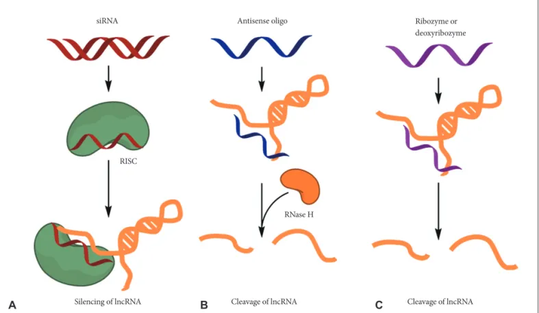
Fig. 2. Potential therapeutic approaches for targeting lncRNAs. Several methods, including small interfering RNAs (siRNAs), antisense oli- gonucleotides (ASOs) and ribozymes or deoxyribozymes, can be used to block the function of lncRNAs. A: Synthetic double-stranded short RNA can be delivered to cells and the antisense strand of the siRNA duplex loads on to the RNA-induced silencing complex (RISC) and degrades the targeted lncRNA. B: ASOs are single-stranded, chemically modified DNA oligomers (less than 25 nt in length) that are de- signed to be complementary to a target lncRNA. ASOs form a heteroduplex with the target lncRNA, and RNase H recognizes the lncRNA- DNA heteroduplex and cleaves the RNA strand. C: However, ribozymes or deoxyribozymes do not dependent on the RISC, which mediates siRNA-induced degradation, or on RNase H. IncRNA: long non-coding RNA.
siRNA
RISC
RNase H
Silencing of lncRNA Cleavage of lncRNA Cleavage of lncRNA
Antisense oligo Ribozyme or
deoxyribozyme
B C
A
and performs key roles in G1/S and mitotic progression [27].
It also known that MALAT1 controls cell cycle progression by modulating the oncogenic transcription factor B-MYB (Mybl2) [28]. Tripathi et al. [27] showed that specific deletion of MALAT1 in cells leads to activation of p53 and its target genes. MALAT1-depleted cells show cell cycle defects which are sensitive to the p53 levels, indicating that p53 is a main downstream mediator for the MALAT1 activity. In MALAT1- depleted cells, replication of the S-phase is reduced, and rather cell population of G1 and G2/M increased. Also, expression of genes associated with cell cycle arrest (e.g., tumor suppressor p53, cdk inhibitor p21) also increases. In addition, reduced ex- pression of oncogenic transcription factor B-MYB which is re- lated with G2/M progression, was observed in MALAT1-de- pleted cells [27].
lncRNAs PRNCR1 and PCGEM1
Two lncRNAs, PRNCR1 (also known as PCAT8) and PC- GEM1, are usually overexpressed in aggressive prostate can- cers and have a close relationship with castration resistance and proliferation of cancer cells [29-31]. PRNCR1 and PC- GEM1, specifically interact with androgen receptor (AR) and strongly enhance androgen receptor-mediated gene activation in both ligand-dependent and -independent manner [14].
After the interaction between PRNCR1 and AR, acetylation occurs at the C-terminal of AR protein, which accelerates the association of DOT1L (disruptor of telomeric silencing 1 like histone H3 methyltransferase) to the PRNCR1-AR complex.
Consequently, DOT1L mediates N-terminal acetylation of AR protein which enhances recruitment of lncRNA PCGEM1. In the castration-resistant prostate cancer cells, expression for short hairpin RNA targeting these two lncRNAs strongly sup- pressed proliferation of cancer cells and xenograft tumor growth in mice [14]. Taken together, the data indicate that ln- cRNAs are potentially essential for the castration resistance in prostate cancers. However, the specific mechanistic roles of these two lncRNAs in castration resistance during prostate cancer development are still under investigation.
lincRNA H19
The lincRNA H19 gene encodes around 2.3-kb long tran- script which does not contain any known open reading frames [32-34]. It is normally transcribed by RNA polymerase II like- wise general mRNA transcripts and transported to the cyto- plasm after sequential capping-polyadenylation-splicing steps.
Furthermore, H19 lincRNA is one of the most highly ex- pressed in the placenta and evolutionarily conserved at the nu- cleotide level between human and rodent [34]. Interestingly, H19 is the imprinted gene which is only transcribed from the maternally inherited allele; the paternal H19 allele dose not ex-
pressed. H19 gene is located on downstream of growth-pro- moting insulin-like growth factor 2 (igf2) and H19 and igf2 share common imprinting mechanism. Recent reports show that H19 is closely associated with tumorigenesis and fetal growth syndrome. However, the exact physiological functions of H19 are largely unknown yet [35-40].
Intriguingly, recent reports showed that H19 serves as miR- NA precursor. miR-675 is located in the first exon of H19, and expressed through the excision from full H19 lincRNA tran- script in the placenta of gestational time point which shows to stop normal growth [41]. The overexpression of miR-675 in embryonic cell and extra-embryonic cell caused decrease of cell proliferation. The target gene of miR-675 (ex. lgf1r) is de- repressed in H19 null placenta. Furthermore, processing of miR-675 from H19 is known to be regulated by stress-response RNA-binding protein HuR. HuR inhibits miR-675 processing by binding to H19 transcript. Release of miR-675 from H19 transcript inhibits cell proliferation rapidly in response to cel- lular stress or oncogenic signals [34]. miR-675 processing mechanism from H19 seems to be associated with molecular pathology in fetal growth and tumorigenesis.
lincRNA HOTAIR
HOTAIR usually shows overexpressed patterns in early and metastatic breast cancer cells [20]. HOTAIR regulates the gene expression by interacting with polycomb repressive complex 2 (PRC2) and lysine-specific demethylase 1A. Together with these two enzymes, HOTAIR can control methylation and de- methylation status of histones [20]. PRC2 seems to be more important related with cancer because cancer cells which con- tain lysine-27 methylated histone H3 showed similar gene ex- pression of embryonic fibroblasts. Generally, PRC2 expression induces metastasis of cancer cells. However metastasis can be suppressed when PRC2 is too overexpressed.
LNCRNAS ON GLIOMA DEVELOPMENT
A glioma is a type of tumor that arises from glial cells mostly in the human brain. High-grade gliomas usually have a ten- dency to infiltrate into the extracellular matrix of the brain and this trait makes it difficult to perform surgery and radio-thera- py [42]. Therefore the understanding of the molecular mecha- nism of infiltrative phenotype of gliomas and identification of key regulator(s) for invasion are essential for the efficient treat- ment of this hardly curable disease [43,44].
Recent reports showed that several lncRNAs have close rela- tionship with glioma development. Some lncRNAs that may contribute to brain development and certain specific differen- tially expressed lncRNAs may play an important role in the pathogenesis of glioblastoma multiforme [45]. The most high-
ly upregulated lncRNA is Colorectal Neoplasia Differentially Expressed (CRNDE) [46]. The importance of CRNDE and its roles in specialized processes such as brain function have not been addressed yet.
Another lncRNA, maternally expressed gene 3 (MEG3), has been found that it markedly decreased in glioma tissues com- pared with adjacent normal tissues [47]. Moreover, overex- pression of the lncRNA MEG3 in human glioma cell lines in- hibits cell proliferation and promotes cell apoptosis. Therefore MEG3 might have inhibitory role in glioma development and can serve as potential drug in anti-glioma therapy.
The lincRNA H19 and its derivative miR-675 were positively correlated with glioma grades. Moreover, H19-derived miR- 675 regulated cadherin 13 which is the directly target of miR- 675, thereby modulating glioma cell invasion [48]. The poten- tial oncogenic role of lincRNA H19/miR-675 may serve as development of anti-glioma therapy.
THERAPEUTIC APPROACHES FOR TARGETING LNCRNAS
Various therapeutic approaches for the lncRNAs have been developed and several pharmaceutical companies are also ac- tively developing lncRNA-targeting therapeutics [49,50]. First, the small interfering RNAs (siRNAs) against the specific ln- cRNA can be used for the strategies to regulate lncRNA func- tion (Fig. 2A). In most cases, predominant localization of ln- cRNAs is in the nucleus, and thus siRNAs may be less accessible to lncRNAs than mRNAs. However, successful knockdown of lncRNAs have been reported by many re- searchers irrespective of their subcellular localization.
Other strategies [e.g., antisense oligonucleotides (ASOs) as well as ribozymes or deoxyribozymes] can be adapted to di- rectly target lncRNAs when an overall secondary structure or the nucleotide sequence is not favorable for the optimized de- sign of siRNAs (Fig. 2B). Antisense oligonucleotides have ad- vantages over siRNAs including independence of RNA-in- duced silencing complex (RISC) machinery, high specificity, and low off-target effects. Recent reports showed that MALAT1 function on lung cancer cells in mouse successfully inhibited by using ASOs [22,23,27].
Ribozymes or deoxyribozymes (e.g., hammerhead ribo- zyme) bind to a target sequence complementarily and pro- mote the cleavage of the flanking RNA region (Fig. 2C). These may be useful for the targeting of lncRNAs that are not favor- able for optimal siRNA design [49,50].
CONCLUSION
Recent global analysis showed that cancer transcriptome is
more complex than previously expected. Dysregulated expres- sion of lncRNAs, including protein-coding genes and miRNAs have potential pervasive roles as the driver of human cancers and development and progression of the cancers [3,8,11]. The epigenomic reprogramming by lncRNAs can be applicable to many other human diseases characterized by aberrant lncRNA expression [51]. Therefore, in the context of cancer cells, ecto- pic expression or specific knockdown of lncRNAs such as PRNCR1, PCGEM1, and HOTAIR seems to re-impose that chromatin state, thereby enabling gene expressions are more favorable or unfavorable to the mobilization and matrix inva- sion of cancer cells.
As the non-epigenomic regulation, such as regulation of al- ternative splicing (e.g., MALAT1) [23] and generation of miR- NA precursor (e.g., lincRNA H19) [41] lncRNAs can modu- late gene expression more favorable to tumor development.
Therefore, understanding the precise molecular mecha- nisms of lncRNAs to the various biological processes will be a critical step in exploring new strategies in future cancer ther- apy.
Conflicts of Interest
The authors have no financial conflicts of interest.
Acknowledgments
This research was supported by Basic Science Research Program through the National Research Foundation of Korea (NRF) funded by the Ministry of Education (NRF-2011-0024692) and a grant from the National Cancer Center, Republic of Korea (1410080).
REFERENCES
1. Carninci P, Kasukawa T, Katayama S, et al. The transcriptional land- scape of the mammalian genome. Science 2005;309:1559-63.
2. International Human Genome Sequencing Consortium. Finishing the euchromatic sequence of the human genome. Nature 2004;431:931-45.
3. Esteller M. Non-coding RNAs in human disease. Nat Rev Genet 2011;
12:861-74.
4. Kapranov P, Cheng J, Dike S, et al. RNA maps reveal new RNA classes and a possible function for pervasive transcription. Science 2007;316:
1484-8.
5. Kapranov P, Willingham AT, Gingeras TR. Genome-wide transcrip- tion and the implications for genomic organization. Nat Rev Genet 2007;8:413-23.
6. Esquela-Kerscher A, Slack FJ. Oncomirs - microRNAs with a role in cancer. Nat Rev Cancer 2006;6:259-69.
7. Bartolomei MS, Zemel S, Tilghman SM. Parental imprinting of the mouse H19 gene. Nature 1991;351:153-5.
8. Qiu MT, Hu JW, Yin R, Xu L. Long noncoding RNA: an emerging paradigm of cancer research. Tumour Biol 2013;34:613-20.
9. Gibb EA, Vucic EA, Enfield KS, et al. Human cancer long non-coding RNA transcriptomes. PLoS One 2011;6:e25915.
10. Hung T, Chang HY. Long noncoding RNA in genome regulation:
prospects and mechanisms. RNA Biol 2010;7:582-5.
11. Shi X, Sun M, Liu H, Yao Y, Song Y. Long non-coding RNAs: a new frontier in the study of human diseases. Cancer Lett 2013;339:159-66.
12. Wang KC, Chang HY. Molecular mechanisms of long noncoding RNAs. Mol Cell 2011;43:904-14.
13. Kallen AN, Zhou XB, Xu J, et al. The imprinted H19 lncRNA antago-
nizes let-7 microRNAs. Mol Cell 2013;52:101-12.
14. Yang L, Lin C, Jin C, et al. lncRNA-dependent mechanisms of andro- gen-receptor-regulated gene activation programs. Nature 2013;500:
598-602.
15. Plath K, Fang J, Mlynarczyk-Evans SK, et al. Role of histone H3 lysine 27 methylation in X inactivation. Science 2003;300:131-5.
16. Huarte M, Guttman M, Feldser D, et al. A large intergenic noncoding RNA induced by p53 mediates global gene repression in the p53 re- sponse. Cell 2010;142:409-19.
17. Bejerano G, Pheasant M, Makunin I, et al. Ultraconserved elements in the human genome. Science 2004;304:1321-5.
18. Lujambio A, Portela A, Liz J, et al. CpG island hypermethylation-asso- ciated silencing of non-coding RNAs transcribed from ultraconserved regions in human cancer. Oncogene 2010;29:6390-401.
19. Rinn JL, Chang HY. Genome regulation by long noncoding RNAs.
Annu Rev Biochem 2012;81:145-66.
20. Gupta RA, Shah N, Wang KC, et al. Long non-coding RNA HOTAIR reprograms chromatin state to promote cancer metastasis. Nature 2010;464:1071-6.
21. Ji P, Diederichs S, Wang W, et al. MALAT-1, a novel noncoding RNA, and thymosin beta4 predict metastasis and survival in early-stage non- small cell lung cancer. Oncogene 2003;22:8031-41.
22. Wilusz JE, Freier SM, Spector DL. 3’ end processing of a long nuclear- retained noncoding RNA yields a tRNA-like cytoplasmic RNA. Cell 2008;135:919-32.
23. Tripathi V, Ellis JD, Shen Z, et al. The nuclear-retained noncoding RNA MALAT1 regulates alternative splicing by modulating SR splic- ing factor phosphorylation. Mol Cell 2010;39:925-38.
24. Jurica MS, Moore MJ. Pre-mRNA splicing: awash in a sea of proteins.
Mol Cell 2003;12:5-14.
25. Manley JL, Krainer AR. A rational nomenclature for serine/arginine- rich protein splicing factors (SR proteins). Genes Dev 2010;24:1073-4.
26. Gutschner T, Hämmerle M, Eissmann M, et al. The noncoding RNA MALAT1 is a critical regulator of the metastasis phenotype of lung cancer cells. Cancer Res 2013;73:1180-9.
27. Tripathi V, Shen Z, Chakraborty A, et al. Long noncoding RNA MALAT1 controls cell cycle progression by regulating the expression of oncogenic transcription factor B-MYB. PLoS Genet 2013;9:e1003368.
28. Joaquin M, Watson RJ. Cell cycle regulation by the B-Myb transcrip- tion factor. Cell Mol Life Sci 2003;60:2389-401.
29. Petrovics G, Zhang W, Makarem M, et al. Elevated expression of PC- GEM1, a prostate-specific gene with cell growth-promoting function, is associated with high-risk prostate cancer patients. Oncogene 2004;
23:605-11.
30. Chung S, Nakagawa H, Uemura M, et al. Association of a novel long non-coding RNA in 8q24 with prostate cancer susceptibility. Cancer Sci 2011;102:245-52.
31. Komiya A, Yasuda K, Watanabe A, Fujiuchi Y, Tsuzuki T, Fuse H. The prognostic significance of loss of the androgen receptor and neuroen- docrine differentiation in prostate biopsy specimens among castration- resistant prostate cancer patients. Mol Clin Oncol 2013;1:257-62.
32. Brannan CI, Dees EC, Ingram RS, Tilghman SM. The product of the H19 gene may function as an RNA. Mol Cell Biol 1990;10:28-36.
33. Rachmilewitz J, Goshen R, Ariel I, Schneider T, de Groot N, Hochberg
A. Parental imprinting of the human H19 gene. FEBS Lett 1992;309:
25-8.
34. Keniry A, Oxley D, Monnier P, et al. The H19 lincRNA is a develop- mental reservoir of miR-675 that suppresses growth and Igf1r. Nat Cell Biol 2012;14:659-65.
35. Luo M, Li Z, Wang W, Zeng Y, Liu Z, Qiu J. Long non-coding RNA H19 increases bladder cancer metastasis by associating with EZH2 and inhibiting E-cadherin expression. Cancer Lett 2013;333:213-21.
36. Luo M, Li Z, Wang W, Zeng Y, Liu Z, Qiu J. Upregulated H19 contrib- utes to bladder cancer cell proliferation by regulating ID2 expression.
FEBS J 2013;280:1709-16.
37. Matouk IJ, DeGroot N, Mezan S, et al. The H19 non-coding RNA is essential for human tumor growth. PLoS One 2007;2:e845.
38. Matouk IJ, Mezan S, Mizrahi A, et al. The oncofetal H19 RNA connec- tion: hypoxia, p53 and cancer. Biochim Biophys Acta 2010;1803:443- 39. Tsang WP, Ng EK, Ng SS, et al. Oncofetal H19-derived miR-675 regu-51.
lates tumor suppressor RB in human colorectal cancer. Carcinogenesis 2010;31:350-8.
40. Zhang L, Yang F, Yuan JH, et al. Epigenetic activation of the MiR-200 family contributes to H19-mediated metastasis suppression in hepato- cellular carcinoma. Carcinogenesis 2013;34:577-86.
41. Cai X, Cullen BR. The imprinted H19 noncoding RNA is a primary microRNA precursor. RNA 2007;13:313-6.
42. Gwak HS, Kim TH, Jo GH, et al. Silencing of microRNA-21 confers radio-sensitivity through inhibition of the PI3K/AKT pathway and enhancing autophagy in malignant glioma cell lines. PLoS One 2012;
7:e47449.
43. Kwak HJ, Kim YJ, Chun KR, et al. Downregulation of Spry2 by miR- 21 triggers malignancy in human gliomas. Oncogene 2011;30:2433- 44. Kim YJ, Park SJ, Choi EY, et al. PTEN modulates miR-21 processing 42.
via RNA-regulatory protein RNH1. PLoS One 2011;6:e28308.
45. Ellis BC, Molloy PL, Graham LD. CRNDE: A Long Non-Coding RNA Involved in CanceR, Neurobiology, and DEvelopment. Front Genet 2012;3:270.
46. Graham LD, Pedersen SK, Brown GS, et al. Colorectal Neoplasia Dif- ferentially Expressed (CRNDE), a Novel Gene with Elevated Expres- sion in Colorectal Adenomas and Adenocarcinomas. Genes Cancer 2011;2:829-40.
47. Wang P, Ren Z, Sun P. Overexpression of the long non-coding RNA MEG3 impairs in vitro glioma cell proliferation. J Cell Biochem 2012;
113:1868-74.
48. Shi Y, Wang Y, Luan W, et al. Long Non-Coding RNA H19 Promotes Glioma Cell Invasion by Deriving miR-675. PLoS One 2014;9:e86295.
49. Li CH, Chen Y. Targeting long non-coding RNAs in cancers: progress and prospects. Int J Biochem Cell Biol 2013;45:1895-910.
50. Ling H, Fabbri M, Calin GA. MicroRNAs and other non-coding RNAs as targets for anticancer drug development. Nat Rev Drug Discov 2013;12:847-65.
51. Guttman M, Amit I, Garber M, et al. Chromatin signature reveals over a thousand highly conserved large non-coding RNAs in mammals.
Nature 2009;458:223-7.