818
서 론
후방십자인대 손상에 대한 진단 및 치료는 전방십자인 대에 비하여 연구된 바가 적으나 최근 교통사고와 스포츠 활동의 증가로 인하여 슬관절 손상의 빈도가 높아지고 자 기공명영상과 관절경적 진단도 발달하여 후방십자인대 손상의 진단 및 치료에 대한 관심이 높아지고 있는 추세 이다8,11,12).
후방십자인대의 손상에 대한 치료는 보존적 방법보다
는 수술적 방법이 점차 인정을 받고 있으며 이에 따라 시 술건수도 늘어가고 있다. 수술적 방법에 있어서도 기존의 tibial tunnel법을 이용한 방법과 이식건이 마모되는 현 상을 줄일 수 있고 수술 후 초기에 강력한 고정을 얻을 수 있는 tibial inlay법을 이용한 방법이 있다2,23,25,26,33)
. 후방십자인대 손상에 의한 후방 불안정성을 치료하기 위한 두 가지 후방십자인대 재건술에는 서로 장단점이 있 고 어느 것이 더 좋은 방법인가에 대해서는 논란이 있다.
이에 본원에서 시행한 tibial tunnel법과 tibial inlay법을
Tibial Inlay법과 Tibial Tunnel법을 이용한 관절경적 후방 십자 인대 재건술의 평가
서정탁․천상진․김정일․이춘기․박원로
부산대학교 의과대학 정형외과학교실
Arthroscopic Reconstruction of the Posterior Cruciate Ligament:
Comparison of Tibial Inlay and Tibial Tunnel Techniques
Jeung Tak Suh, M.D., Sang Jin Cheon, M.D., Jeung Il Kim, M.D., Choon Key Lee, M.D., and Won Ro Park, M.D.
Department of Orthopaedic Surgery, College of Medicine, Pusan National University, Busan, Korea
P u rp o s e : To compare the results of posterior cruciate ligament reconstructions by tibial inlay and tibial tunnel techniques.
M a te ria ls a n d M e th o d s : Despite of conservative treatment, all patients (31 cases) had pain and grade 2 or m ore posterior instability. Posterior drawer test and posterior drawer stress radiography were perform ed. Clinically, Lysholm knee score and Tegner activity score w ere evaluated.
R e s u lts : In the tibial tunnel group, posterior drawer test demonstrated grade 1 instability in 7 cases, grade 2 in 4 cases, and grade 3 in 1 case at the last follow -up. In the tibial inlay group, there w as grade 1 instability in 14 cases and grade 2 in 5 cases. O n posterior drawer stress radiography, the m ean side-to-side difference in m easurem ent of the tibial tunnel group im proved from 12.4 m m preoperatively to 4.0 m m at follow -up, and that of the tibial inlay group im proved from 11.8 m m to 2.9 m m . Lysholm knee score and Tegner activity score im proved to 86.8 points and 5.83 points, respectively, in the tibial tunnel group, and to 88.2 points and 5.84 points, in the tibial inlay group.
C o n c lu s io n : PCL reconstruction with the tibial inlay technique tends to maintain better posterior stability, but there is no statistically significant difference between the two techniques. Further study m ay be required.
K e y W o rd s : PCL reconstruction, Tibial inlay technique, Tibial tunnel technique
통신저자:서 정 탁
부산광역시 서구 아미동 1-10 부산대학교 의과대학 정형외과학교실
TEL: 051-240-7248․FAX: 051-247-8395 E-mail: [email protected]
Address reprint requests to Jeung Tak Suh, M.D.
Department of Orthopaedic Surgery, Pusan National University Hospital, 1-10, Ami-dong, Seo-gu, Busan 602-739, Korea
Tel: +82.51-240-7248, Fax: +82.51-247-8395 E-mail: [email protected]
이용한 후방십자인대 재건술의 결과를 비교, 분석하였다.
대상 및 방법
1999년 2월부터 2003년 12월까지 후방십자인대 손상 후 본원에 내원한 환자를 대상으로 하였으며 재건술의 적 응증은 6주간의 보존적 치료에도 불구하고 지속적인 동 통을 호소하면서 이학적 검사 상 등급 2 이상의 후방 전 위를 보이는 환자로 하였다. 이 중 후외방 불안정 또는 후방십자인대 외의 다른 구조물의 심각한 동반 손상이 있 거나 2년 이상 추시가 불가능하였던 환자는 대상에서 제 외하였다.
본 연구의 대상이 된 환자는 tibial tunnel법을 이용한 군이 12명(남자 9명, 여자 3명), tibial inlay법을 이용한 군이 19명(남자 14명, 여자 5명)으로 평균 연령은 각각 34.6세, 35.1세였다. 수상 기전에 있어서도 두 군 간의 의미 있는 차이는 없었으며 후방십자인대 재건술의 방법 을 결정하는 데 있어서도 특별한 기준은 없었다.
두 군 간의 결과 비교는 후방 전위 검사, 후방 전위 스 트레스 방사선 사진, Lysholm 슬관절 점수와 Tegner 활 동도 점수를 이용하였다. 후방 전위 검사는 수술 전, 수술 직후, 최종 추시 시에 시행하였고, 등급 1은 5 mm미만의 전위, 등급 2는 5 mm 이상, 10 mm 미만의 전위, 등급 3은 10 mm 이상의 전위로 정의하였다. 후방 전위 스트 레스 방사선 사진은 수술 전, 최종 추시 시 시행하였고, 슬관절을 90도 굴곡 시킨 상태에서 Telos 기구(Telos stress device; Austin & Associates, Fallston, MD, USA)를 이용하여 20 lb의 부하를 주고 촬영하였다. 후방 전위의 정도는 대퇴골에서 내측과 또는 외측과의 후연, 경골에서 내측 고평부 또는 외측 고평부의 후연을 기준으 로 경골의 후방 피질골에 평행하도록 접선을 그어 측정하 였으며 건측과의 차이를 구하였다29,38).
Lysholm 슬관절 점수와 Tegner 활동도 점수는 수술 전과 최종 추시 시에 측정하여 비교하였으며, 특히 Lysholm 슬관절 점수에서는 불안정성과 동통이 차지하 는 50점을 별도로 계산하여 비교하였다27,39).
대상이 된 환자들의 평균 추시 기간은 수술 후 24개월 에서 68개월로 평균 32.4개월이었으며 최종 추시로 간주 하고 검사를 시행한 시기는 수술 후 24개월에서 36개월 사이로 평균 28.9개월이었다.
통계적 분석은 SPSS (SPSS for Windows Release
13.0; SPSS, Chicago, IL, USA)를 이용하였으며 Mann- Whitney test로 검증하였다.
1. 수술 방법
Tibial tunnel법에 의한 재건술 시 대부분의 경우 전신 마취 하에서 수술을 시행하였으며 지혈대를 사용하였다.
먼저 전외측 및 전내측 도달법으로 기본적인 진단적 관절 경을 시행하였고, 전외측에서 과간 절흔을 통해 후내측 구획으로 관절경을 삽입하고 슬관절을 90o 굴곡 상태로 고정하였다. 이때 투과되는 광원을 이용하여 후내측 입구 (portal)를 만들고 후외측 입구도 유사한 방법으로 만들 었다. 경격막 입구(trans-septal portal)을 만들기 위해 서 관절경을 후내방에서 삽입하고 후외방에서 변환 막대 (switching stick)을 삽입하여 격막을 내측으로 밀고 후 방십자인대 후방에서 격막의 중앙에 작은 구멍을 만들었 다. 전동형 쉐이버를 후외측 입구로 삽입하고 이 구멍을 통과하여 인대의 잔존 부위를 손상시키지 않으면서 충분 한 시야를 확보하기 위해 후격막을 제거하였다. 후방십자 인대의 경골부착부를 확인하고 후관절낭을 충분히 하방 으로 박리한 후, 후방십자인대 도자(PCL guide)의 고리 (hook)를 전내측 입구를 통하여 후방십자인대 경골부착 부로 삽입하였으며 관절선에 대하여 약 1 cm 가량 하방 에서 후방십자인대 경골부착부의 하외방에 위치시켰다.
45도에서 50도로 맞추어진 후방십자인대 도자를 이용하 여 영상증폭기 하에서 도자핀의 삽입을 시행하였으며, 그 방향은 후방 근위 경골 피질골의 방향과 평행하도록 하였 고 후방십자인대 경골부착부의 중앙 혹은 약간 외측을 통 과하도록 하였다. 도자핀의 위치를 영상증폭기를 통해 확 인한 후 7 mm의 확공기로 터널을 만들었으며 이식편의 크기에 따라 9 mm까지 확공하였다.
다음으로 대퇴골 터널을 뚫기 위해 내측 광근(vastus medialis)의 내측 연을 따라 슬개골의 상단 높이에서 3∼
4 cm 가량 피부 절개를 가하고 대퇴골 내과 외측면에서 관절 연골경계의 8 mm 후방을 목표로 하여 원위 대퇴골 내측에서 관절 내로 우측은 1시 30분, 좌측은 10시 30분 방향으로 만들었으며, 가급적이면 파열된 후방십자인대 의 잔여부분과 활액막을 보존하고자 노력하였다. 아킬레 스 동종 이식건은 슬관절 내의 통과가 쉽도록 하기 위해 끝을 얇게 처리한 후 Ethibond (Ethicon, Somerville, NJ, USA) 5 호사로 양면을 맞물리게 고리형 봉합(in-
terlocking loop suture)하여 처리하였고 반대측 끝은 종골 골편을 대퇴 터널에 견고하게 고정되도록 깔대기 모 양으로 만들었다. Wire loop를 경골 터널에서 관절 내를 거쳐 대퇴골 터널로 통과시켜두고 이식편을 연결하여 대 퇴골로 들어가고 경골에서 빠지도록 통과시켰다. 대퇴골 부위에서는 금속 간섭나사를 이용하여 고정하였고 경골 부위에서는 생분해성 나사로 고정한 뒤 6.5 mm 해면골 나사 또는 staple로 고정을 보강하였다.
Tibial inlay법을 이용한 재건술에서도 대퇴골에 대해 서는 tibial tunnel법에서 시행한 방법과 거의 동일한 술 기를 이용하였다. 경골부착부에 대해서는, 환자를 횡와 위에서 복와위로 한 후 슬관절을 신전, 고관절을 외전 및 외회전하여 비복근의 내측두의 근위부와 슬와부의 피부 선을 따라 피부 절개를 가한 뒤 비복근 내측두의 내측연 과 반막양근의 외측연을 노출시켰다. 비복근과 슬와동 맥, 정맥 및 경골신경을 포함하여 외측으로 견인하여 관 절낭을 노출시킨 뒤 후방십자인대 경골부착 부위를 촉지 하여 관절낭에 절개를 가하였다. Burr를 이용하여 경골 부착부에 폭 1.5 cm, 깊이 1 cm, 길이 2.5 cm의 홈을 만든 후, 대퇴골 터널에서 전방 십자 인대와 후방십자인 대의 잔존 부위 사이로 통과시켜 두었던 wire loop를 이 용하여 이식편을 통과시키고 2개의 4.5 mm 유관 나사로 이식골편의 경골부를 고정하였으며 적당한 장력을 준 뒤 대퇴골에서 생분해성 간섭나사와 staple로 고정하였다.
2. 수술 후 재활
재건술 후 약 2∼3주까지는 장하지 석고 붕대로 슬관절 을 신전 상태로 유지한 채로 고정하였고, 이후 보조기를 착용한 채로 서서히 슬관절 운동 범위를 증가시켜 재건술 후 6주까지 90o 굴곡을 얻을 수 있도록 지시하였다. 보조 기는 후방 함몰(posterior sagging)을 방지하기 위해서 재건술 후 3개월까지 착용하도록 하였고 이 기간 동안 목
발도 사용하도록 지시하였다.
결 과
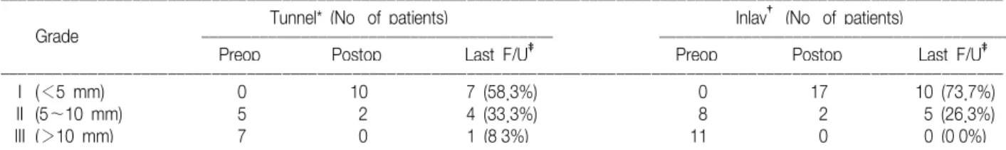
후방 전위의 정도는 tibial tunnel법을 이용한 군에서 수술 전 등급 2가 5명(41.7%), 등급 3이 7명(58.3%)이었 으며, tibial inlay법을 이용한 군에서는 등급 2가 8명 (42.1%), 등급 3이 11명(57.9%)이었다. 수술 직후 시행 한 후방 전위의 정도는 tibial tunnel법을 이용한 군에서 는 등급 1이 10명(83.3%), 등급 2가 2명(16.7%), tibial inlay법을 이용한 군에서는 등급 1이 17명(89.5%), 등급 2가 2명(10.5%)로 향상되었으며 두 군간의 차이는 뚜렷 한 차이는 없었다. 한편 최종 추시 시에는 tibial tunnel 법을 이용한 군에서는 등급 1이 7명(58.3%), 등급 2가 4명(33.3%), 등급 3이 1명(8.3%)이었으며, tibial inlay 법을 이용한 군에서는 등급 1이 14명(73.7%), 등급 2가 5명(26.3%)이었으며 등급 3을 보이는 예는 없었다 (Table 1). 수술 직후와 최종 추시 시의 후방 전위의 정도 가 1개 등급 저하된 예가 tibial tunnel법을 이용한 군에 서는 5명(41.7%), tibial inlay법을 이용한 군에서는 3명 (15.8%)이었으며, 특히 tibial tunnel법을 이용한 군에서 최종 추시 시 등급 3을 보인 1명은 이식건의 파열이 의심 되었으나 환자의 거부로 더 이상의 검사 및 추시가 불가 능하였다(Fig. 1). 두 군 간의 최종 추시 시의 후방 전위 정도는 tibial inlay법을 이용한 군에서 다소 잘 유지되는 경향을 보였으나 통계적 유의성은 찾아 볼 수 없었다 (p=0.236)(Fig. 2).
후방 전위 스트레스 방사선 검사의 결과는 tibial tun- nel 법을 이용한 군에서 수술 전 환측에서 평균 15.7 mm 의 전위를 보였으며 건측에서는 평균 3.3 mm를 보였다.
최종 추시 시에는 환측이 평균 7.3 mm의 전위를 보여 건측과의 차이는 수술 전 평균 12.4 mm에서 최종 추시 시 평균 4.0 mm로 향상되었다. 한편 tibial inlay 법을
Table 1. Results of the Posterior Drawer Test in the Tibial Tunnel Group and the Tibial Inlay Group
ꠏꠏꠏꠏꠏꠏꠏꠏꠏꠏꠏꠏꠏꠏꠏꠏꠏꠏꠏꠏꠏꠏꠏꠏꠏꠏꠏꠏꠏꠏꠏꠏꠏꠏꠏꠏꠏꠏꠏꠏꠏꠏꠏꠏꠏꠏꠏꠏꠏꠏꠏꠏꠏꠏꠏꠏꠏꠏꠏꠏꠏꠏꠏꠏꠏꠏꠏꠏꠏꠏꠏꠏꠏꠏꠏꠏꠏꠏꠏꠏꠏꠏꠏꠏꠏꠏꠏꠏꠏꠏꠏꠏꠏꠏꠏꠏꠏꠏꠏꠏꠏꠏꠏꠏꠏꠏꠏꠏꠏꠏꠏꠏꠏꠏ Tunnel* (No. of patients) Inlay† (No. of patients)
Grade ꠏꠏꠏꠏꠏꠏꠏꠏꠏꠏꠏꠏꠏꠏꠏꠏꠏꠏꠏꠏꠏꠏꠏꠏꠏꠏꠏꠏꠏꠏꠏꠏꠏꠏꠏꠏꠏꠏꠏꠏꠏꠏ ꠏꠏꠏꠏꠏꠏꠏꠏꠏꠏꠏꠏꠏꠏꠏꠏꠏꠏꠏꠏꠏꠏꠏꠏꠏꠏꠏꠏꠏꠏꠏꠏꠏꠏꠏꠏꠏꠏꠏꠏꠏꠏ
Preop. Postop. Last F/U‡ Preop. Postop. Last F/U‡
ꠏꠏꠏꠏꠏꠏꠏꠏꠏꠏꠏꠏꠏꠏꠏꠏꠏꠏꠏꠏꠏꠏꠏꠏꠏꠏꠏꠏꠏꠏꠏꠏꠏꠏꠏꠏꠏꠏꠏꠏꠏꠏꠏꠏꠏꠏꠏꠏꠏꠏꠏꠏꠏꠏꠏꠏꠏꠏꠏꠏꠏꠏꠏꠏꠏꠏꠏꠏꠏꠏꠏꠏꠏꠏꠏꠏꠏꠏꠏꠏꠏꠏꠏꠏꠏꠏꠏꠏꠏꠏꠏꠏꠏꠏꠏꠏꠏꠏꠏꠏꠏꠏꠏꠏꠏꠏꠏꠏꠏꠏꠏꠏꠏꠏ
I (<5 mm) 0 10 7 (58.3%) 0 17 10 (73.7%)
II (5∼10 mm) 5 2 4 (33.3%) 8 2 5 (26.3%)
III (>10 mm) 7 0 1 (8.3%) 11 0 0 (0.0%)
ꠏꠏꠏꠏꠏꠏꠏꠏꠏꠏꠏꠏꠏꠏꠏꠏꠏꠏꠏꠏꠏꠏꠏꠏꠏꠏꠏꠏꠏꠏꠏꠏꠏꠏꠏꠏꠏꠏꠏꠏꠏꠏꠏꠏꠏꠏꠏꠏꠏꠏꠏꠏꠏꠏꠏꠏꠏꠏꠏꠏꠏꠏꠏꠏꠏꠏꠏꠏꠏꠏꠏꠏꠏꠏꠏꠏꠏꠏꠏꠏꠏꠏꠏꠏꠏꠏꠏꠏꠏꠏꠏꠏꠏꠏꠏꠏꠏꠏꠏꠏꠏꠏꠏꠏꠏꠏꠏꠏꠏꠏꠏꠏꠏꠏ
*Tunnel, Group reconstructed by the tibial tunnel technique; †Inlay, Group reconstructed by the tibial inlay technique; F/U‡, Follow-up.
이용한 군에서는 수술 전 환측에서 평균 14.4 mm의 전 위를 보였으며 건측에서는 평균 2.6 mm, 최종 추시 시 환측에서 평균 5.4 mm의 전위를 보여 환측과의 차이가 수술 전 평균 11.8 mm에서 최종 추시 시 평균 2.9 mm 로 향상되었다(Table 2). 방사선학적 검사에서도 단순한 평균값 비교에서 tibial inlay 법을 이용한 군이 다소 양 호한 결과를 보였으나 통계적 의의는 찾아 볼 수 없었다 (p=0.101).
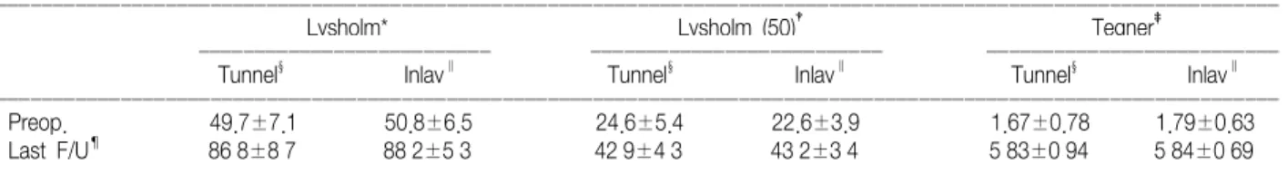
Lysholm knee socre는 tibial tunnel법을 이용한 군 에서 수술 전 평균 49.7점에서 최종 추시 시 86.8점으로, tibial inlay법을 이용한 군에서도 수술 전 평균 50.8점에 서 최종 추시 시 88.2점으로 향상되었다. 또한 Ly- sholm 슬관절 점수의 총 점수 100점 중 불안정성과 동통 만을 별도로 분리하여 50점 만점으로 계산한 점수에서도 tibial tunnel법을 이용한 군에서 수술 전 평균 24.6점, 최종 추시 시 평균 42.9점, tibial inlay법을 이용한 군에 서 수술 전 평균 22.6점, 최종 추시 시 평균 43.2점으로 뚜렷한 차이를 보이지 않았다. 한편 Tegner 활동도 점수 도 수술 전 tibial tunnel법을 이용한 군에서 1.67점, tibial inlay법을 이용한 군에서 1.79점, 최종 추시 시 각 각 5.83점, 5.84점으로 두 군 간에 큰 차이를 볼 수가 없었다(Table 3).
Fig. 2. The radiographs (A) before reconstruction, (B) in the nor- mal knee, (C) and after reconstruction demonstrate posterior drawer stress views of a patient who underwent reconstruction with the tibial inlay technique. The results of the posterior drawer test improved from grade 3 preoperatively to grade 1 at the last follow up.
A B C
Table 3. Comparison of Clinical Scores between the Tibial Tunnel Group and the Tibial Inlay Group (mean±SD)
ꠏꠏꠏꠏꠏꠏꠏꠏꠏꠏꠏꠏꠏꠏꠏꠏꠏꠏꠏꠏꠏꠏꠏꠏꠏꠏꠏꠏꠏꠏꠏꠏꠏꠏꠏꠏꠏꠏꠏꠏꠏꠏꠏꠏꠏꠏꠏꠏꠏꠏꠏꠏꠏꠏꠏꠏꠏꠏꠏꠏꠏꠏꠏꠏꠏꠏꠏꠏꠏꠏꠏꠏꠏꠏꠏꠏꠏꠏꠏꠏꠏꠏꠏꠏꠏꠏꠏꠏꠏꠏꠏꠏꠏꠏꠏꠏꠏꠏꠏꠏꠏꠏꠏꠏꠏꠏꠏꠏꠏꠏꠏꠏꠏꠏ
Lysholm* Lysholm (50)† Tegner‡
ꠏꠏꠏꠏꠏꠏꠏꠏꠏꠏꠏꠏꠏꠏꠏꠏꠏꠏꠏꠏꠏꠏꠏꠏꠏꠏ ꠏꠏꠏꠏꠏꠏꠏꠏꠏꠏꠏꠏꠏꠏꠏꠏꠏꠏꠏꠏꠏꠏꠏꠏꠏꠏ ꠏꠏꠏꠏꠏꠏꠏꠏꠏꠏꠏꠏꠏꠏꠏꠏꠏꠏꠏꠏꠏꠏꠏꠏꠏꠏ
Tunnel§ Inlay∥ Tunnel§ Inlay∥ Tunnel§ Inlay∥
ꠏꠏꠏꠏꠏꠏꠏꠏꠏꠏꠏꠏꠏꠏꠏꠏꠏꠏꠏꠏꠏꠏꠏꠏꠏꠏꠏꠏꠏꠏꠏꠏꠏꠏꠏꠏꠏꠏꠏꠏꠏꠏꠏꠏꠏꠏꠏꠏꠏꠏꠏꠏꠏꠏꠏꠏꠏꠏꠏꠏꠏꠏꠏꠏꠏꠏꠏꠏꠏꠏꠏꠏꠏꠏꠏꠏꠏꠏꠏꠏꠏꠏꠏꠏꠏꠏꠏꠏꠏꠏꠏꠏꠏꠏꠏꠏꠏꠏꠏꠏꠏꠏꠏꠏꠏꠏꠏꠏꠏꠏꠏꠏꠏꠏ
Preop. 49.7±7.1 50.8±6.5 24.6±5.4 22.6±3.9 1.67±0.78 1.79±0.63
Last F/U¶ 86.8±8.7 88.2±5.3 42.9±4.3 43.2±3.4 5.83±0.94 5.84±0.69
ꠏꠏꠏꠏꠏꠏꠏꠏꠏꠏꠏꠏꠏꠏꠏꠏꠏꠏꠏꠏꠏꠏꠏꠏꠏꠏꠏꠏꠏꠏꠏꠏꠏꠏꠏꠏꠏꠏꠏꠏꠏꠏꠏꠏꠏꠏꠏꠏꠏꠏꠏꠏꠏꠏꠏꠏꠏꠏꠏꠏꠏꠏꠏꠏꠏꠏꠏꠏꠏꠏꠏꠏꠏꠏꠏꠏꠏꠏꠏꠏꠏꠏꠏꠏꠏꠏꠏꠏꠏꠏꠏꠏꠏꠏꠏꠏꠏꠏꠏꠏꠏꠏꠏꠏꠏꠏꠏꠏꠏꠏꠏꠏꠏꠏ
*Lysholm, Lysholm knee score; †Lysholm (50), Consisted of only instability and pain scores; ‡Tegner, Tegner activity score; §Tunnel, Group reconstructed by the tibial tunnel technique; ∥Inlay, Group reconstructed by the tibial inlay technique; ¶F/U, Follow-up.
Fig. 1. The radiographs (A) before reconstruction, (B) in the nor- mal knee, and (C) after reconstruction demonstrate posterior drawer stress views of a patient who underwent reconstruction with the tibial tunnel technique. It shows grade 3 posterior instability at both the preoperative examination and the last follow up. The double-headed arrows indicate the distance from the most posterior contours of the femoral condyle and tibial plateau.
A B C
Table 2. Results of Posterior Drawer Stress Views in the Tibial Tunnel Group and the Tibial Inlay Group (Side-to-Side Differences) ꠏꠏꠏꠏꠏꠏꠏꠏꠏꠏꠏꠏꠏꠏꠏꠏꠏꠏꠏꠏꠏꠏꠏꠏꠏꠏꠏꠏꠏꠏꠏꠏꠏꠏꠏꠏꠏꠏꠏꠏꠏꠏꠏꠏꠏꠏꠏꠏꠏꠏꠏꠏꠏꠏ
Tunnel* Inlay†
(mm, mean±SD) (mm, mean±SD) ꠏꠏꠏꠏꠏꠏꠏꠏꠏꠏꠏꠏꠏꠏꠏꠏꠏꠏꠏꠏꠏꠏꠏꠏꠏꠏꠏꠏꠏꠏꠏꠏꠏꠏꠏꠏꠏꠏꠏꠏꠏꠏꠏꠏꠏꠏꠏꠏꠏꠏꠏꠏꠏꠏ
Preop. 12.4±2.9 11.8±2.8
Last F/U‡ 4.0±2.3 2.9±0.9
Difference 8.4±3.7 8.9±2.7
ꠏꠏꠏꠏꠏꠏꠏꠏꠏꠏꠏꠏꠏꠏꠏꠏꠏꠏꠏꠏꠏꠏꠏꠏꠏꠏꠏꠏꠏꠏꠏꠏꠏꠏꠏꠏꠏꠏꠏꠏꠏꠏꠏꠏꠏꠏꠏꠏꠏꠏꠏꠏꠏꠏ
*Tunnel, Group reconstructed by the tibial tunnel technique; †Inlay, Group reconstructed by the tibial inlay technique; ‡F/U, Follow-up.
수술 후 발생한 합병증으로는 tibial tunnel법을 이용한 군에서 심한 슬관절 강직을 보인 1예가 있었으나 외래 추 시 상 슬관절 운동범위가 향상되어 마지막 추시에서 약 100도의 굴곡이 가능하였고, tibial inlay법을 이용한 군 에서 이식건 골편의 삽입부위에 심한 부종을 보이는 1예 가 있었으나 감염은 아니었고 동종골에 대한 반응성 염증 으로 판단되었다.
고 찰
후방십자인대는 내측 대퇴과의 외측면 후방에서 기시 하여 경골 과간의 후면에 부착한다. 후방십자인대는 거의 수직으로 주행하고 있으며 경골 부착점은 경골 과간 후방 의 관절면에서 1 cm 가량 아래쪽에 위치하고 있다13,21,32,36)
. 후방십자인대의 길이는 약 38 mm, 폭은 약 13 mm이며 단면의 대부분을 차지하고 생역학적으로 중요한 역할을 하는 전외측 다발은 슬관절이 굴곡할 때 긴장되고, 후내 측 다발은 신전할 때 긴장된다. 후방십자인대의 등장점에 대한 연구는 재건술 시의 중요성 때문에 많은 연구가 있 었으나 아직 논란의 여지가 있고 가장 등장성이 있다고 주장되는 위치보다는 전외측 다발을 재건하는 것이 임상 적으로 결과가 좋다고 알려져 있다10,14,15,18,19)
. 후방십자인대 손상 시 치료 방법에 대해서는 보존적 치 료와 수술적 치료 사이에서 지금까지 많은 논란이 있었으 나 점차 수술적 치료에 비중이 높아지는 추세이고 특히 젊고 활동적인 환자에 있어서는 더욱 그러하다는 의견이 지배적이다4,5,7,8,11,12,19,20,22,37)
.
후방십자인대의 수술적 치료 방법은 1983년 Clancy7,8) 에 의해 경골과 대퇴골에 터널을 뚫고 자가 슬개건을 이 용한 전방도달법으로 시작되어 널리 사용되었다. 전방도 달법은 대부분의 관절경 술식과 인공관절 치환술 등에 익 숙한 많은 술자들에게 친숙하다는 장점이 있지만 전방도 달법의 경우 경골 터널과 이식건이 예각을 이룸으로써 급 격한 방향 전환이 마찰을 일으켜서 이식건의 마모를 초래 할 수 있어(the killer turn) 초기에 이식건의 긴장도를 조절하는데 문제점이 있다고 하였다2,31,33). Burks와 Schaffer6)는 비복근의 내측부를 외측으로 견인하여 슬 와부의 신경, 혈관에 손상을 주지 않고 후방십자인대의 경골 부착점에 도달할 수 있는 후방도달법을 보고하였고 이에 1995년 Berg2)는 자가골-슬개건-골을 이용하여 tibial inlay법을 이용한 후방십자인대 재건술을 소개하
였으며 이는 전방에서 관절경적 술식을 이용하고 후방에 서 관혈적인 방법으로 후방십자인대의 부착부위를 고정 하여 이식건이 마찰되는 문제를 해결하여 좋은 결과를 보 였다고 보고하였다6,25,33).
Tibial inlay법을 이용한 후방십자인대 재건술은 수술 중 환자의 자세를 바꿔야하고 후방에 추가적인 피부절개 가 필요하기 때문에 수술 시간이 길어지고 감염의 발생이 높아질 수 있다는 점과 기술적인 요구도가 높다는 점이 단점으로 여겨지나, tibial tunnel법을 이용한 방법에 비 해 killer turn을 근본적으로 해결하여 이식건의 마모를 막을 수 있고 초기에 경골 부착점에 견고한 고정을 얻을 수 있으며 크기가 큰 이식건을 사용할 수 있고 이식건의 길이 조절이 용이하다는 장점이 있다5,6,9,16,17,23,25,31,33,34,40)
. 사체 실험을 통한 연구에서 tibial inlay법과 tibial tunnel법을 이용한 재건술 후 후방 안정성을 비교한 실 험에서 양자 간 큰 차이가 없다는 보고가 있는 반면 tibial inlay법에서 이식편의 변성이 적고 후방 전위 정도가 적 다는 보고도 있다3,30,35). 또한 실제 임상을 통한 연구의 결과를 보면 tibial tunnel법에 의한 재건술의 경우, Becker 등1)의 보고에 의하면 후방 전위 스트레스 방사선 검사에서 수술 전 평균 9.5 mm에서 수술 후 4.1 mm로 향상되었다고 하고 Kim과 Oh28)는 수술 전 평균 18.2 mm에서 수술 후 5.0 mm로 향상되었다고 보고하였다.
한편 tibial inlay법을 이용한 재건술의 경우, Jung 등24) 은 추시 시 건측에 비해 평균 3.4 mm의 후방 전위를 보 였다고 보고하였고 Berg2)의 연구에 의하면 후방 전위가 수술 후 평균 2.0 mm로 향상되었다고 한다. 이는 최종 추시 시 tibial tunnel법을 이용한 군에서 4.0 mm, tibial inlay법을 이용한 군에서 평균 2.9 mm의 후방 전위를 보인 본 연구의 결과와 큰 차이가 없어 보이지만 여러 가 지 연구 방법의 차이로 직접적인 비교는 불가능할 것으로 판단된다.
문헌고찰을 통한 다양한 치료 방법의 결과는, 보존적 치료에 비해 수술적 치료가 우세한 결과를 보인다는 주장 에는 대체로 이견이 없지만, 수술적 치료에 있어서 세부 적인 술식, 평가 방법, 추시 기간 등이 표준화된 것이 없 어 다양한 재건술 사이의 객관적인 결과 비교가 어려운 것이 사실이다.
본 연구의 대상이 된 후방십자인대 재건술은 tibial inlay법과 tibial tunnel법을 이용한 두 가지 군으로 나뉘
었고 모든 재건술은 경골 부착부에 대한 고정방법 외에는 대체로 동일한 술식으로 동일한 술자에 의해 비슷한 시기 에 이루어 졌다. 두 가지 군의 재건술 후 결과는 통계적으 로 유의성이 있는 뚜렷한 차이점을 보이지 않았지만, 임 상적 결과의 차이보다 방사선학적 검사 및 이학적 검사에 서 tibial inlay법을 이용한 군에서 다소 우세한 결과를 보이는 경향이 있었으며 이는 더 많은 증례로 더 긴 추시 기간을 가지고 비교, 분석해 보아야 할 문제라고 생각된 다. 아울러 이차 관절경 소견을 추가로 연구해 보는 것도 가치가 있을 것이라고 판단된다.
결 론
본 연구에서는 tibial inlay법을 이용한 재건술이 후방 안정성의 유지에 있어서 다소 우세한 결과를 보였으나 통 계적 유의성은 없었으며 더 많은 증례와 더 긴 추시 기간 을 통한 연구가 필요하리라 생각된다.
참고문헌
1. Becker R, Ropke M, Nebelung W: Clinical outcome of arthroscopic posterior cruciate ligament-plasty. Unfallchirurg, 102: 354-358, 1999.
2. Berg EE: Posterior cruciate ligament tibial inlay recons- truction. Arthroscopy, 11: 69-76, 1995.
3. Bergfeld JA, McAllister DR, Parker RD, Valdevit AD, Kambic HE: A biomechanical comparison of posterior cruciate ligament reconstruction techniques. Am J Sports Med, 29:
129-136, 2001.
4. Bianchi M: Acute tears of the posterior cruciate ligament:
Clinical study and results of operative treatment in 27 cases.
Am J Sports Med, 11: 308-314, 1983.
5. Burger RS, Larson RJ: The knee. 1st ed, Philadelphia, WE Saunders Co: 513-598, 1993.
6. Burks RT, Schaffer JJ: A simplified approach to the tibial attachment of the posterior cruciate ligament. Clin Orthop Relat Res, 254: 216-219, 1990.
7. Clancy WG Jr.: Repair and reconstruction of the posterior cruciate ligament. In: Chapman MW ed. Operative orthopaedics. 1st ed. Philadelphia, Lippincott: 1651-1665, 1988.
8. Clancy WG Jr, Shelbourne KD, Zoellner GB, Keene JS, Reider B, Rosenberg TD: Treatment of knee joint instability
secondary to rupture of the posterior cruciate ligament. Report of a new procedure. J Bone Joint Surg Am, 65: 310-322, 1983.
9. Cooper DE, Warren RF, Warner JJP: The posterior cruciate ligament and posterolateral structures of the knee: anatomy, function and patterns of injury. In: Tullos HS, ed.
Instructional Course Lectures, Vol 40. Rosemont, American Academy of Orthopaedic Surgeons: 249-270, 1991.
10. Covey CD, Sapega AA: Injury of the Posterior cruciate ligament. J Bone Joint Surg Am, 75: 1376-1386, 1993.
11. Dandy DJ, Pusey RJ: The long-term results of unrepaired tears of the posterior cruciate ligament. J Bone Joint Surg Br, 64: 92-94, 1982.
12. Fanelli GC, Giannotti BF, Edson CJ: The posterior cruciate ligament arthroscopic evaluation and treatment. Arthroscopy, 10: 673-688, 1994.
13. Feagin JA Jr: The crucial ligaments. 1st ed. New York, Churchill Livingstone: 71-106, 1988.
14. Girgis FG, Marshall JL, Monajem A: The cruciate ligaments of the knee joint. Anatomical, functional and experimental analysis. Clin Orthop, 106: 216-231, 1975.
15. Gollehon DL, Torzilli PA, Warren RF: The role of the posterolateral and cruciate ligaments in the stability of the human knee. A biomechanical study. J Bone Joint Surg Am, 69: 233-242, 1987.
16. Good L, Tarlow SD, Odensten M, Gillquist J: Load tolerance, security and failure modes of fixation devices for synthetic knee ligaments. Clin Orthop Relat Res, 253: 190-196, 1990.
17. Grood ES, Hefzy MS, Lindenfield TN: Factors affecting the region of most isometric femoral attachments. Part I: The posterior cruciate ligament. Am J Sports Med, 17: 197-207, 1989.
18. Hughston JC: The posterior cruciate ligament in knee joint stability. In proceedings of the American Academy of ortho- paedic surgery. J Bone Joint Surg Am, 51: 1045-1046, 1969.
19. Hughston JC, Bowden JA, Andrews JR, Norwood LA:
Acute tears of the posterior cruciate ligament. J Bone Joint Surg Am, 62: 438-450, 1980.
20. Hughston JC, Degenhardt TC: Reconstruction of the pos- terior cruciate ligament. Clin Orthop Relat Res, 164: 59-77,
1982.
21. Harner CD, Xerogeanes JW, Livesay GA, et al: The human posterior cruciate ligament complex: an interdisciplinary study.
Ligament morphology and biomechanical evaluation. Am J Sports Med, 23: 736-45, 1995.
22. Insall JN: Surgery of the knee. 2nd ed. New York, Churchill Livingstone: 384-387, 1984.
23. Jung YB, Chang EC, Yum JK: Second look findings after arthroscopic posterior cruciate ligament reconstruction. J Kore- an Knee Soc, 9: 35-42, 1997.
24. Jung YB, Tae SK, Jung HJ, Lee KH: Replacement of the torn posterior cruciate ligament with a mid-third patellar tendon graft with use of a modified tibial inlay method. J Bone Joint Surg Am, 86: 1878-1883, 2004.
25. Jung YB, Tae SK, Yum JK, Koo BH: Arthroscopic posterior cruciate ligament reconstruction with two graft tendons by combined femoral dual tunnel and modified tibial inlay method.
J Korean Knee Soc, 10: 119-124, 1998.
26. Jung YB, Tae SK, Yum JK, Koo BH: The results of posterior cruciate ligament reconstruction: Transtibial two tunnel technique vs. modified tibial inlay technique. J Korean Arthro- scopy Society, 2: 135-140, 1998.
27. Kettelkamp DB, Thompson C: Development of a knee scoring scale. Clin Orthop Relat Res, 107: 93-99, 1975.
28. Kim MK: The reconstruction of the posterior cruciate ligament using autogenous bone-patellar tendon-bone under arthroscopy.
J Korean Orthop Assoc, 33: 280-288, 1998.
29. Margheritini F, Mancini L, Mauro CS, Mariani PP: Stress radiography for quantifying posterior cruciate ligament deficiency. Arthroscopy, 19: 706-711, 2003
30. Margheritini F, Mauro CS, Rihn JA, Stabile KJ, Woo SL, Harner CD: Biomechanical comparison of tibial inlay versus transtibial techniques for posterior cruciate ligament recon- struction: analysis of knee kinematics and graft in situ forces.
Am J Sports Med, 32: 587-593, 2004.
31. Miller MD, Bergfeld JA, Fowler PJ, Harner CD, Noyes FR:
The posterior cruciate ligament injured knee: principles of evaluation and treatment. In: Zuckerman JD, ed. Instructional Course Lectures, Vol 48. Rosemont, American Academy of Orthopaedic Surgeons: 199-207, 1999.
32. Miller MD, Harner CD: The anatomic and surgical considera- tions for posterior cruciate ligament reconstruction. In: Jackson DW, ed. Instructional Course Lectures, Vol 44. Rosemont, American Academy of Orthopaedic Surgeons: 431-440, 1995.
33. Miller MD, Olszewski AD: Posterior cruciate ligament injuries: New treatment options. Am J Knee Surg, 8: 145-154, 1995.
34. Noyes FR, Butler DL, Grood ES, Zernicke RF, Hefzy MS:
Biomechanical analysis of human ligament grafts used in knee-ligament repairs and reconstruction. J Bone Joint Surg Am, 66: 344-352, 1984.
35. Oakes DA, Markolf KL, McWilliams J, Young CR, McAllister DR: Biomechanical comparison of tibial inlay and tibial tunnel techniques for reconstruction of the posterior cruciate ligament. Analysis of graft forces. J Bone Joint Surg Am, 84: 938-944, 2002.
36. Race A, Amis AA: The mechanical properties of the two bundles of the human posterior cruciate ligament. J Biomech, 27: 13-24, 1994.
37. Southmayd WW, Rubin BD: Reconstruction of the posterior cruciate ligament using the semimembranosus tendon. Clin Orthop Relat Res, 150: 196-197, 1980.
38. Staubli HU, Jakob RP: Posterior instability of the knee near extension. A clinical and stress radiographic analysis of acute injuries of the posterior cruciate ligament. J Bone Joint Surg Br, 72: 225-30, 1990.
39. Tegner Y, Lysholm J: Rating systems in the evaluation of knee ligament injuries. Clin Orthop Relat Res, 198: 43-49, 1985.
40. Trickey EL: Rupture of the posterior cruciate ligament of the knee. J Bone Joint Surg Br, 50: 334-341, 1968.
= 국문초록 =
목적: 후방 불안정성의 치료로 이용되고 있는 tibial tunnel법과 tibial inlay법을 이용한 후방 십자 인대 재건술의 치료 결과를 비교 및 분석하고자 하였다.
대상 및 방법: 보존적 치료에도 불구하고 통증과 불안정성을 호소하고 등급 2 이상의 후방 전위를 보인 31명의 환자들을 대상으로 하였으며 평가는 수술 전 및 최종 추시 관찰 시의 후방 전위 검사, 후방 전위 스트레스 방사선 사진 촬영, Lysholm knee score와 Tegner activity score로 하였다.
결 과: 후방 전위 검사에서 최종 추시 시 tibial tunnel 군은 등급 1이 7예(58.3%), 등급 2가 4예(33.3%), 등급 3이 1예(8.3%)였고, tibial inlay 군은 등급 1이 14예(73.7%), 등급 2가 5예(26.3%)였다. 후방 전위 스트레스 방사선 사진상 건측과의 차이는 tibial tunnel 군에서는 수술 전 평균 12.4 mm, tibial inlay 군에서는 평균 11.8 mm였고, 최종 추시 시에는 각각 평균 4.0 mm 및 2.9 mm로 향상되었다. 최종 추시 시 Lysholm knee score는 tibial tunnel 군에서 86.8점으로, tibial inlay 군에서 88.2점으로 향상되었으며, Tegner activity score 는 각각 5.83점, 5.84점으로 향상되었다.
결 론: 최종 수시 시 후방 안정성은 tibial inlay법에 의한 후방십자인대 재건술이 비교적 잘 유지되는 경향을 보였으나 통계적 유의성은 없었으며 더 많은 증례와 더 긴 추시 기간을 통한 연구가 필요하리라 생각된다.
색인 단어: 후방 십자 인대 재건술, Tibial inlay법, Tibial tunnel법