Prognostic value of COL6A3 in pancreatic adenocarcinoma
Christos Svoronos1, Georgios Tsoulfas2, Maria Souvatzi3, and Efthimios Chatzitheoklitos4
1Department of Surgery, AMEOS Klinikum, Halberstadt, Germany, 2First Department of Surgery, Aristotle University of Thessaloniki, Thessaloniki, Greece, 3St Mary’s Hospital Imperial College Healthcare NSH Trust, London, UK,
4Unit of Pancreatic Surgery, Interbalkan Center, Thessaloniki, Greece
Backgrounds/Aims: Pancreatic cancer is one of the most fatal human malignancies with poor prognosis, despite ad- vances in therapy. Here, we evaluated the potential role of collagen type VI 3 chain (COL6A3) as a non-invasive biomarker for pancreatic adenocarcinoma. Methods: In this study, we investigated immunohistochemically the ex- pression of COL6A3 in 30 patients with resectable pancreatic adenocarcinoma by immunohistochemistry in a tissue sample of the cancer and a tissue sample of normal pancreas for each patient. Also, we looked for associations be- tween COL6A3 and other prognostic factors of pancreatic cancer. Results: All of the pancreatic cancer tissue samples revealed in different ranges of intensity from weak (+1) in 16.67%, moderate (+2) in 50%, to strongly positive (+3) in 33.33% staining for COL6A3. We found no moderate or strongly positive staining in normal pancreatic tissue. There was only weak positive staining in 23 samples (76.67%) and 7 (23.30%) were negative. Also, there was significant correlation between COL6A3 moderate and strongly expression and negative prognostic factors for pancreatic cancer.
Conclusions: The greatest density of COL6A3 was observed in pancreatic cancer tissues and was correlated with negative prognostic factors for pancreatic cancer. Therefore, we suggest that COL6A3 could be used as prognostic factor in pancreatic cancer, but more studies need to prove its value. (Ann Hepatobiliary Pancreat Surg 2020;24:52-56) Key Words: Pancreatic adenocarcinoma; COL6A3; Prognosis
Received: October 2, 2019; Revised: November 29, 2019; Accepted: December 2, 2019 Corresponding author: Christos Svoronos
Department of Surgery, AMEOS Klinikum, Richard Wagner Strasse 65, Halbertstadt 38820, Germany Tel: +49-15203877197, Fax: +49-3941645370, E-mail: [email protected]
Copyright Ⓒ 2020 by The Korean Association of Hepato-Biliary-Pancreatic Surgery
This is an Open Access article distributed under the terms of the Creative Commons Attribution Non-Commercial License (http://creativecommons.org/
licenses/by-nc/4.0) which permits unrestricted non-commercial use, distribution, and reproduction in any medium, provided the original work is properly cited.
Annals of Hepato-Biliary-Pancreatic Surgery ∙ pISSN: 2508-5778ㆍeISSN: 2508-5859
INTRODUCTION
Pancreatic cancer is the third leading cause of can- cer-related death and by 2030, it will be the second. The five-year survival rate for pancreatic cancer is reported at 8%, which is the lowest among many other common types of cancer.1 However, pancreatic cancer is difficult to diag- nose early and more than 80% of pancreatic cancers are locally advanced or metastatic at the time of diagnosis.2 Identification of prognostic factors may improve the pre- diction of survival and selection of therapy.
The tumor microenvironment influences tumor genesis and metastasis of cancer cells. The dense stroma sur- rounding malignant epithelial cells is the histological hall- mark of pancreatic adenocarcinoma.3 The stroma consists of numerous cellular, as well as acellular elements. The cellular components include fibroblasts, stellate cells, im- mune cells, endothelial cells, and nerve cells. The acel-
lular compartment is comprised of extracellular matrix, which consists of collagen, fibrinogen, hyaluronan, and fibrin.4 The interactions between cancer cells and the mi- croenvironment are complicated, as the stroma elements can provide either a support or a barrier for tumor growth or metastasis. Collagen is by far the most abundant and well-characterized component of the extracellular matrix in pancreatic cancer.5 Currently, 28 different types of col- lagen have been described in the extracellular matrix in the pancreas. Collagen type VI is abundantly present in pancreas and it is the predominant constituent subtype, immediately surrounding islets in the pancreas.6 Type VI collagen is structured as a trimer composed of three dif- ferent alpha chains: alpha-1(VI), alpha- 2(VI), and al- pha-3(VI). It is now clear that type VI collagen has a vital role and can suppress apoptosis and oxidative damage, and enhance cell growth.7,8 Many recent studies have shown the overexpression of COL6A3 in many types of
cancer, which is also associated with poor prognosis in many of them, such as colon, prostate and lung cancer.9-11 However, there are few studies in the literature that have investigated the association of COL6A3 with pancreatic cancer.
The goal of this study was to investigate the expression of COL6A3 in pancreatic cancer adenocarcinoma and to compare it with the expression in the adjacent sections of pancreas where there were no cancer lesions. Furthermore, the prognostic value of COL6A3 was evaluated by com- paring the expression of COL6A3 with clinicopathological prognostic factors for pancreatic cancer in these patients.
MATERIALS AND METHODS
Sample collections and patients’ characteristics Human pancreatic adenocarcinoma samples were ob- tained from 30 patients, who underwent a pancreaticoduo- denectomy for pancreatic cancer in the Agios Dimitrios General Hospital surgical department. Also, from each pa- tient one sample of normal pancreatic tissue was obtained without any cancer lesions. For each patient, the following information was collected: demographic data including age and sex, tumor staging, tumor characteristics, including histological type, differentiation, and infiltration of the lymph nodes, perineural, and vascular structures. Tumor staging was determined based on the AJCC Cancer Staging Manual, 7th Edition (2010).12 We also evaluated the lymph node ratio (LNR) by dividing the number of the positive lymph nodes with the number of all lymph nodes that were examined, the modified Glasgow prognostic score (mGPS) which incorporates the C-reactive protein and al- bumin values, the neutrophil/lymphocyte ratio (NLR), the platelet/lymphocyte ratio (PLR), and the lymphocyte/
monocyte ratio (LMR) for each patient.
Immunochemistry
Immunostaining for COL6A3 was performed on in- dividual formalin-fixed, paraffin-embedded pancreatic ad- enocarcinoma and healthy pancreatic tissue sections using mouse polyclonal antibody raised against the COL6A3 (11a), (H3-2 ή sc-81766, Santa Cruz Biotechnology, Santa Cruz, CA, USA). All specimens were embedded in paraf- fin and cut onto glass slides as 5 m sections for immuno- chemistry. Slides were twice de-paraffinized using xylene
solution, for 5 minutes each time. Then tissues were dehy- drated by passing through descending concentrations (100%, 96%, 80%, 75%, 70%) of alcohol and washed off with distilled water. To remove the endogenous perox- idase activity, sections were incubated with 3% hydrogen peroxide at room temperature for 30 min. Antigen un- masking was performed in 0.01 M sodium citrate buffer pH 6.1, for 15 min (min) at high power, according to the manufacturer’s instructions. Non-specific antibody binding was blocked using Sniper, a specific blocking reagent for mouse and rabbit primary antibodies (Sniper, Biocare Medical, Concord, California, USA) for 5 min. The sec- tions were incubated for 1 h (h), at room temperature, with the primary antibodies against COL6A3 diluted 1:200 in phosphate buffered saline (PBS) according to the manufacturer’s instructions. The resultant immune perox- idase activity was developed using a DAB substrate kit (Vector Laboratories, California, USA) for 10 min. Sections were counterstained with Harris’ hematoxylin and mount- ed in Entellan (Merck, Darmstadt, Germany). Negative control sections, where the primary or secondary anti- bodies were omitted were also prepared.
Immunohistochemistry analysis
Immunohistochemical analyses were scored by two in- dependent pathologists. The observers were blinded to the clinicopathological data for each specimen. Protein bands were visualized and the intensity of COL6A3 staining was scored as follows: – (negative staining), + (weak positive staining), ++ (moderate positive staining), and +++ (strong positive staining).
Statistical analysis
Statistical analysis was performed using SPSS software (SPSS standard version 16.0). Clinical characteristics were analyzed using Pearson’s chi-square or Fisher’s exact test for categorical and dichotomous variables, and Student’s t-test for continuous variables. Statistical significance was assumed for a two-tailed p-value of <0.05.
RESULTS
Study patients’ characteristics
Samples were collected from 30 patients with pancre- atic adenocarcinoma. The median age was 68.2 (44-86)
Table 1. Clinicopatholigical characteristics of the cohort pa- tients (n=30)
Age (y) 68.2 (44-86)
Sex (male/female) 18 (60%)/12 (40%) AJCC stage
Ia 1 (3.33%)
Ib 2 (6.67%)
IIa 6 (20%)
IIb 21 (70%)
Tumor differentiation
G1 0
G2 11 (36.67%)
G3 19 (63.33%)
N Status
NO 9 (30%)
N1 21 (70%)
Perineural invasion
Positive 19 (63.33%)
Negative 11 (36.67%)
Microvascular invasion
Positive 21 (70%)
Negative 9 (30%)
Fig. 1. Immunochistochemical expression of COL6A3 in pan- creatic adenocarcinoma and pancreatic healthy tissue.
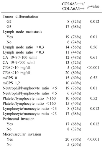
Table 2. Strong or moderate positive expression of COL6A3 and prognostic factor of pancreatic cancer
COL6A3+++/
COLA6A3++ p-value Tumor differentiation
G2 8 (32%) 0.012
G3 17 (68%)
Lymph node metastasis
Yes 19 (76%) 0.01
No 6 (24%)
Lymph node ratio >0.3 14 (56%) 0.56 Lymph node ratio <0.3 11 (44%)
CA 19-9>100 u/ml 12 (48%) 0.61
CA 19-9<00 u/ml 13 (52%)
CEA>10 mg/dl 5 (20%) <0.001
CEA<10 mg/dl 20 (80%)
mGPS 0 15 (60%) 0.52
mGPS 1,2 10 (40%)
Neutrophil/lymphocyte ratio >5 19 (76%) 0.01 Neutrophil/lymphocyte ratio <5 6 (24%)
Platelet/lymphocyte ratio >160 10 (60%) 0.52 Platelet/lymphocyte ratio <160 15 (40%)
Lymphocyte/monocyte ratio <3 8 (32%) 0.012 Lymphocyte/monocyte ratio <3 17 (68%)
Perineural invasion
Yes 17 (68%) 0.012
No 8 (32%)
Microvascular invasion
Yes 20 (80%) <0.001
No 5 (20%)
years. All the patients had resectable pancreatic tumors and underwent a pancreaticoduodenectomy. The pathologic evaluation of the surgical specimens revealed an R0 re- section in all the patients of the study. Most of the pa- tients had AJCC stage IIb (70%) and IIa (20%). The pan- creatic adenocarcinoma was moderately- and poorly-dif- ferentiated in 11 (36.67%) and 19 (63.33%) patients, re- spectively. Perineural invasion was identified in 19 (63.33%) patients, whereas vascular invasion was found in 21 (70%) patients. The clinicopathologic characteristics of the pa- tients in this study are summarized in Table 1.
Expression of COL6A3 protein
According to the protein band intensity in the stroma regions, the expression of COL6A3 in the stroma of pan- creatic adenocarcinoma was strong positive, moderate pos- itive and weak positive in 10 (33.33%), 15 (50%), and 5 (16.67%) samples, respectively. Among the normal pan- creatic tissue samples the expression of COL6A3 was negative and weak positive in 23 (76.67%) and 7 (23.33%) samples, respectively. There was no strong or moderate positive expression of COL6A3 observed in normal pan- creatic tissue (Fig. 1).
COL6A3 expression is associated with prognostic factors of pancreatic cancer
In order to evaluate the impact of COL6A3 expression on prognosis of pancreatic cancer, we evaluated the asso- ciation between the positive expressions of COL6A3 and other prognostic factors of pancreatic cancer. Among the prognostic factors, which were evaluated in this study,
lymph node ratio >0.3 (p=0.56), CA 19-9>100 u/ml (p=
0.61), CEA>10 mg/dl, mGPS, and platelet/lymphocyte ratio (p=0.52) had no statistically significant association with the strong positive or moderate positive expression of COL6A3. In contrast, tumor differentiation (p=0.012), lymph node metastasis (p=0.01), neutrophil/lymphocyte ratio >5 (p=0.01), lymphocyte/monocyte ratio <3 (p=0.012), perineural invasion (p=0.012), and microvascular invasion (p<0.001) were associated with strong or moderate pos- itive expression of COL6A3 (Table 2).
DISCUSSION
Pancreatic adenocarcinoma remains a lethal disease with very poor prognosis. Although surgery is the only curative treatment option, the 5-year survival, even after curative resection, is still only 15-25%.13,14 Multiple previous stud- ies have demonstrated that clinicopathologic factors such as tumor stage, histological differentiation, nodal involve- ment, perineural invasion and microvascular invasion are statistically significant prognostic variables.15,16 Also, nu- merous studies have shown the prognostic value of LNR in pancreatic cancer.17,18 Furthermore, various inflammatory markers, as mGPS, NLR, PLR, and LMR have been shown to be significant prognostic factors of the disease.19-22 Nevertheless, the number of patients alive at 5 years makes these prognostic variables only marginally influential.
Therefore, it is important to understand the intrinsic prop- erties of the development and the progression of the pan- creatic cancer and to identify more accurate prognostic factors for more effective therapies.
The COL6A3 is an extracellular matrix protein, which is usually found in most connective tissues, including muscle, skin, tendon, and vessels. Recently, many studies revealed the important role of COL6A3 in the diagnosis and prognosis of colorectal, lung and prostate cancer.
First, Arafat et al.23 in 2011, compared ,using Western blot analysis and immunochemistry, 8 specimens of pan- creatic adenocarcinoma with premalignant lesions and normal pancreas, and showed a high expression of COL6A3 in pancreatic adenocarcinoma and that a dynam- ic process of alternative splicing of the COL6A3 gene is exclusively associated with malignancy. Another study, from Kang et al.24 at 2014, comparing the serum levels of COL6A3 in patients with pancreatic cancer and pa-
tients with benign lesions and healthy people, revealed that COL6A3 protein levels were significantly elevated in patients with pancreatic cancer and demonstrated the po- tential clinical significance of circulating COL6A3 in the diagnosis of pancreatic cancer.
In the present study, we investigated the expression of COL6A3 in pancreatic adenocarcinoma and compared it with the expression of COL6A3 in normal pancreas of the same patients. Our immunohistochemical analysis revealed that COL6A3 was predominantly expressed in the pancre- atic cancer stroma. This difference highlights the important role of COL6A3 in the microenvironment during the de- velopment and progression of pancreatic cancer. Also, we found that high expression of COL6A3 was associated with most of the negative prognostic factors for pancreatic cancer. Specifically, most of the patients with strong or moderate positive expression of COL6A3 had poor differ- entiated tumors, lymph node metastasis by the diagnosis, NLR>5, LMR<3 (p=0.012), and perineural and micro- vascular invasion. These results reflect that overexpression of COL6A3 is associated with poor prognosis of pancre- atic cancer and suggest that COL6A3 could be used as a prognostic factor in pancreatic adenocarcinoma.
In summary, our results demonstrate that COL6A3 is overexpressed in pancreatic cancer stroma, suggesting that COL6A3 is involved in the progress of pancreatic cancer.
Also, COL6A3 was associated with negative prognostic factors of the disease, indicating that it could be a useful prognostic factor for pancreatic adenocarcinoma. In order to verify these results, a larger sample size and more in-depth analyses will be needed.
ORCID
Christos Svoronos: https://orcid.org/0000-0002-6375-6134 Georgios Tsoulfas: https://orcid.org/0000-0001-5043-7962 Maria Souvatzi: https://orcid.org/0000-0002-5269-5671 Efthimios Chatzitheoklitos: https://orcid.org/0000-0002- 9912-5190
AUTHOR CONTRIBUTIONS
Conceptualization: Christos Svoronos, Georgios Tsoulfas.
Data curation: Christos Svoronos, Georgios Tsoulfas, Efthimios Chatzitheoklitos. Formal analysis: Christos Svoronos, Georgios
Tsoulfas, Efthimios Chatzitheoklitos, Maria Souvatzi.
Methodology: Christos Svoronos, Georgios Tsoulfas. Project administration: Christos Svoronos, Georgios Tsoulfas, Efthimios Chatzitheoklitos. Visualization: Christos Svoronos, Georgios Tsoulfas. Writing - original draft: Christos Svoronos. Writing - review & editing: Christos Svoronos, Georgios Tsoulfas, Efthimios Chatzitheoklitos, Maria Souvatzi.
REFERENCES
1. Siegel RL, Miller KD, Jemal A. Cancer Statistics, 2017. CA Cancer J Clin 2017;67:7-30.
2. Jemal A, Bray F, Center MM, Ferlay J, Ward E, Forman D.
Global cancer statistics. CA Cancer J Clin 2011;61:69-90.
3. Rucki AA, Zheng L. Pancreatic cancer stroma: understanding bi- ology leads to new therapeutic strategies. World J Gastroenterol 2014;20:2237-2246.
4. Neesse A, Bauer CA, Öhlund D, Lauth M, Buchholz M, Michl P, et al. Stromal biology and therapy in pancreatic cancer: ready for clinical translation? Gut 2019;68:159-171.
5. Weniger M, Honselmann KC, Liss AS. The extracellular matrix and pancreatic cancer: a complex relationship. Cancers (Basel) 2018;10:E316.
6. Llacua LA, Hoek A, de Haan BJ, de Vos P. Collagen type VI interaction improves human islet survival in immunoisolating microcapsules for treatment of diabetes. Islets 2018;10:60-68.
7. Zazuli Z, Barliana MI, Mulyani UA, Perwitasari DA, Ng H, Abdulah R. Polymorphism of PXR gene associated with the in- creased risk of drug-induced liver injury in Indonesian pulmo- nary tuberculosis patients. J Clin Pharm Ther 2015;40:680-684.
8. Moon JY, Chang BC, Lee KE, Bang JS, Gwak HS. Effects of pregnane X receptor genetic polymorphisms on stable warfarin doses. J Cardiovasc Pharmacol Ther 2015;20:532-538.
9. Liu W, Li L, Ye H, Tao H, He H. Role of COL6A3 in colorectal cancer. Oncol Rep 2018;39:2527-2536.
10. Duan Y, Liu G, Sun Y, Wu J, Xiong Z, Jin T, et al. COL6A3 polymorphisms were associated with lung cancer risk in a Chinese population. Respir Res 2019;20:143.
11. Xie X, Liu X, Zhang Q, Yu J. Overexpression of collagen VI
3 in gastric cancer. Oncol Lett 2014;7:1537-1543.
12. American Cancer Society. American Joint Committee on Cancer (AJCC) TNM staging system. New York: American Cancer Society;
2016 [cited 2017 Jan 4]. Available from: http://www.cancer.org/
cancer/pancreaticcancer/detailedguide/pancreatic-cancer-staging.
13. Lin R, Han CQ, Wang WJ, Liu J, Qian W, Ding Z, et al.
Analysis on survival and prognostic factors in patients with re- sectable pancreatic adenocarcinoma. J Huazhong Univ Sci Technolog Med Sci 2017;37:612-620.
14. Yamamoto T, Yagi S, Kinoshita H, Sakamoto Y, Okada K, Uryuhara K, et al. Long-term survival after resection of pancre- atic cancer: a single-center retrospective analysis. World J Gastroenterol 2015;21:262-268.
15. Vernerey D, Huguet F, Vienot A, Goldstein D, Paget-Bailly S, Van Laethem JL, et al. Prognostic nomogram and score to pre- dict overall survival in locally advanced untreated pancreatic cancer (PROLAP). Br J Cancer 2016;115:281-289.
16. Wang XD, Qian JJ, Bai DS, Li ZN, Jiang GQ, Yao J. Marital status independently predicts pancreatic cancer survival in pa- tients treated with surgical resection: an analysis of the SEER database. Oncotarget 2016;7:24880-24887.
17. Butturini G, Stocken DD, Wente MN, Jeekel H, Klinkenbijl JH, Bakkevold KE, et al.; Pancreatic Cancer Meta-Analysis Group.
Influence of resection margins and treatment on survival in pa- tients with pancreatic cancer: meta-analysis of randomized con- trolled trials. Arch Surg 2008;143:75-83; discussion 83.
18. Pawlik TM, Gleisner AL, Cameron JL, Winter JM, Assumpcao L, Lillemoe KD, et al. Prognostic relevance of lymph node ratio following pancreaticoduodenectomy for pancreatic cancer. Surgery 2007;141:610-618.
19. Asari S, Matsumoto I, Toyama H, Shinzeki M, Goto T, Ishida J, et al. Preoperative independent prognostic factors in patients with borderline resectable pancreatic ductal adenocarcinoma fol- lowing curative resection: the neutrophil-lymphocyte and plate- let-lymphocyte ratios. Surg Today 2016;46:583-592.
20. Yu J, Ding Z, Yang Y, Liu S. Increased platelet-to-lymphocytes ratio is associated with poor long-term prognosis in patients with pancreatic cancer after surgery. Medicine (Baltimore) 2018;97:
e11002.
21. Li W, Tao L, Zhang L, Xiu D. Prognostic role of lymphocyte to monocyte ratio for patients with pancreatic cancer: a system- atic review and meta-analysis. Onco Targets Ther 2017;10:3391- 3397.
22. La Torre M, Nigri G, Cavallini M, Mercantini P, Ziparo V, Ramacciato G. The glasgow prognostic score as a predictor of survival in patients with potentially resectable pancreatic adeno- carcinoma. Ann Surg Oncol 2012;19:2917-2923.
23. Arafat H, Lazar M, Salem K, Chipitsyna G, Gong Q, Pan TC, et al. Tumor-specific expression and alternative splicing of the COL6A3 gene in pancreatic cancer. Surgery 2011;150:306-315.
24. Kang CY, Wang J, Axell-House D, Soni P, Chu ML, Chipitsyna G, et al. Clinical significance of serum COL6A3 in pancreatic ductal adenocarcinoma. J Gastrointest Surg 2014;18:7-15.