INTRODUCTION
The material of choice for dental implants is commercially pure titanium because of its high biocompatibility and suitability of tooling. This biocompatible material1enables direct bone anchorage called osseointegration.2In an attempt to achieve stable bonding between the titanium implants and bone, the surfaces of titanium dental implants have been modified by additive methods (titanium plasma spray) or by subtractive methods (acid etching, sandblasting) to increase the surface area and promote cell attachment .3,4
Healing around implants can be influenced by (1) physicochemical properties of the implant material, (2) mechanical properties of the implant, (3) surface topography of the material ; macrotopography and microtopography, (4) overall shape and design of the implant.5The surface topography of implant material can influence adherence, attachment, spreading of cells and modify and control the osseointegration process. Recent studies on the effect of various surface topographies on cell
adhesion and proliferation have already been reported or are in progress.6-9
One drawback of the titanium as implants material from an esthetic point of view is that the dark color of titanium can shine through the thin bone and mucosa.10 Also, soft tissue shrinkage, gingival recession, and peri-implant lesions may leave the implant fixture top or titanium abutment visible. One possible solution to these problems with titanium would be to make implants and transgingival abutments from tooth-colored materials such as zirconia.
Zirconia (ZrO2) has adequate mechanical properties for use in medical and dental purposes. Its mechanical properties are similar to those of stainless steel and its ivory color which is similar to that of natural teeth, makes it useful in the esthetically important area of the oral cavity.11 Its ability to transmit light renders it a suitable material in esthetic restorations. Zirconia exists in three phases (monoclinic, tetragonal and cubic) depending on the temperature. By mixing ZrO2with other metallic oxides, such as MgO, CaO, or Y2O3, greater molecular stability can be obtained.11Yttrium-stabilized zirconia, also known as
CELLULAR ATTACHMENT AND GENE EXPRESSION OF OSTEOBLAST-LIKE CELLS ON
ZIRCONIA CERAMIC SURFACES
Ahran Pae1*, DMD, MSD, PhD, Heesu Lee2, DMD, MSD, PhD, Hyeong-Seob Kim3, DMD, MSD, PhD, Jin Baik4, DMD, MSD, PhD, Yi-Hyung Woo5, DMD, MSD, PhD
1Assistant Professor, Department of Dentistry, School of Medicine, Ewha Womans University
2Assistant Professor, Department of Oral Anatomy, School of Dentistry, Kangnung National University
3Associate Professor, Department of Prosthodontics, School of Dentistry, Kyung-Hee University
4Assistant Professor, Department of Prosthodontics, School of Dentistry, Kyung-Hee University
5Professor, Director, Department of Prosthodontics, School of Dentistry, Kyung-Hee University
Corresponding Author: Ahran Pae
Department of Dentistry, School of Medicine, Ewha Womans University
911-1 Mokdong, Yangcheon-Ku, Seoul, 158-710, Korea +82 2 2650 2797: e-mail, [email protected] Received January 21, 2008 Last Revison February 4, 2008 Accepted June 20, 2008.
※ This work was supported by the Ewha Womans University Research Grant of 2007.
tetragonal zirconia polycrystal (TZP), is the combination with the best mechanical properties and is presently commercially available. Every transition between the different crystalline phases is due to stress on the zirconia surface, and this produces a volumetric change in the crystal where the compressive force is applied. When stress occurs on zirconia surface, cracking energy creates T-M (tetragonal-monoclinic) transition. This crystalline modification is followed by a 4 % volumetric expansion that seals the crack.12,13
Biomaterial properties of zirconia compared to titanium proved to have more advantages. Bacterial adhesion, which is an important aspect in order to maintain zirconia restorations without periodontal alterations, proved to be satisfactorily slight.14,15Scarano et al. reported a degree of coverage by bacteria of 12.1 % on zirconia as compared to 19.3 % on titanium.16 Rimondini et al. confirmed these results with an in vivo study, in which Y-TZP accumulated fewer bacteria than titanium in terms of the total number of bacteria and presence of potential putative pathogens as rods.17Inflammatory infiltrate, microvessel density, and vascular endothelial growth factor expression were found to be higher around the titanium caps than around the ZrO2 ones.18Zirconium oxide is also able to modulate immunity and cell cycle regulation.19 Additionally, allergic reactions and sensitivities to titanium have been reported.20,21
Zirconia is a biocompatible material and has the highest mechanical properties among oxide ceramics. Its biocompatibility as dental implant material has been demonstrated in several animal investigations.22-27Also, biological response of osteoblast-like cells between zirconia/alumina ceramic and pure titanium has been proved to be comparable.28
In this study, we discuss zirconia surfaces provided with micrometer-sized grooves. Such microgrooves influence cell behavior: the cells align themselves, and migrate guided by the surface grooves. This phenomenon is known as
“contact guidance”.29The microgrooves create a pattern of mechanical stress, which influences cell spreading and causes cell alignment. Matsuzaka et al. confirmed that the
‘contact guidance’behavior of cells on microgrooved surfaces, i.e. on smooth surfaces cell position is at random, whereas on any type of grooves cells will align themselves towards the groove direction.30
Anselme reported that cellular proliferation decreased as surface roughness increased,31while Mustafa and colleagues demonstrated that proliferation and differentiation were enhanced by surface roughness.32,33Surface of material not only regulates bone growth but also osteoblast differentiation by modulating the expression of key osteoblast genes in osteogenesis. These studies indicate that zirconium oxide can be suitable for implant materials.
This study was performed to define attachment and growth behavior of osteoblast-like cells cultured on grooved surfaces of zirconium oxide by MTT assay and SEM and evaluate the genetic effect of grooves on zirconium oxide surfaces using the RT-PCR.
MATERIAL AND METHODS 1. Specimen preparation
The commercially pure titanium (c.p. Ti) disks (Osstem, Pusan, Korea) used for the cell culture were machined from grade 2 commercially pure Ti (Dynamet, Inc. Carpenter Co., Washington, PA, USA). The disks were prepared to be 12 mm in diameter and 4 mm thick and used as the culture substrate in the control group (T group). Zirconia disks (LAVA™, 3M ESPE, St. Paul, MN, USA) of Y-TZP (yttria- stabilized tetragonal zirconia polycrystal) were prepared to be 12 mm in diameter and 4 mm thick also. Two types of disc surfaces were prepared of the zirconia disks. One was Y-TZP with smooth surface (ZS group) and the other was Y-TZP with 100 μm grooves (ZG group). Disc samples were rinsed twice in absolute alcohol and once in demineralized water in ultrasonic, before sterilization by autoclave for testing using MC3T3-E1 cells.
2. Cell culture
MC3T3-E1 cells are osteoblast-like cells from rat calvaria. MC3T3-E1 cells were cultured at 37℃ in a CO2 incubator (5 %). Cells were cultured using alpha-Eagle’s minimal essential medium (alpha-MEM, Gibco, Paisley, UK) supplemented with 10 % fetal bovine serum (FBS) and antibiotics.
3. MTT assay
The proliferation of cells was examined with MTT test assay (Sigma, St. Louis, MO., USA) after culturing the cells on the titanium and zirconia disks. The substance used for MTT test was a 3-(4,5-dimethylthiazol-2-yl)-2,5-diphenyl- tetrazolium salt, which turns into blue formazan product due to the viable mitochondria in active cells. E1 cells were seeded at a density of 1×10⁵ cells/ml. Cells were incubated at 37�C in 5% CO2for 24 hrs and 48 hrs. The disks were moved to well plates after 24 hours and 48 hours incubation and new media were added after which the media were removed and added by diluted MTT (5 mg/ml) solution and incubation was continued at 37�C in 5% CO2for 4 hours.
The incubation medium was then removed and 400 μl of isopropanol with 0.04N HCl was added in each well and the resulting formazan crystals were dissolved. The absorbance of produced formazan at 490 nm was measured on a microplate reader (Bio-kinetics reader, EL312e, Winooski, VT, USA). Experiments were repeated independently in triplicate.
4. Scanning electron microscopy
Surfaces were analyzed using scanning electron microscopy (SEM) to determine the cellular attachment and morphology. E1 cells were seeded at a density of 1×10⁵ cells/ml. The cultured cells were incubated for 4 h and 24 h at 37℃ in 5 % CO2. The loosely adherent or unbound cells from the experiment wells were removed by aspiration, the
wells were washed twice with a 0.1 M Phosphate Buffered Saline (PBS) buffer (pH 7.4), and the remaining bound cells were fixed with 4 % glutaraldehyde for more than 6 hours.
The excess glutaraldehyde solution was removed and cells rinsed once more in PBS before being dehydrated progressively in higher concentration of ethanol baths (50, 60, 70, 80, 90, 95, and 100 %, 10 min in each bath). After critical point drying, the samples were sputtered with gold at the thickness of 100 nm using Ion coater (Eiko IB-type 3). Cells on the discs were observed by FE-SEM (Japan, Hitachi S-800). Images were recorded at 300× and 1000×
magnification.
5. RT-PCR (reverse transcriptase-polymerase chain reac- tion) analysis
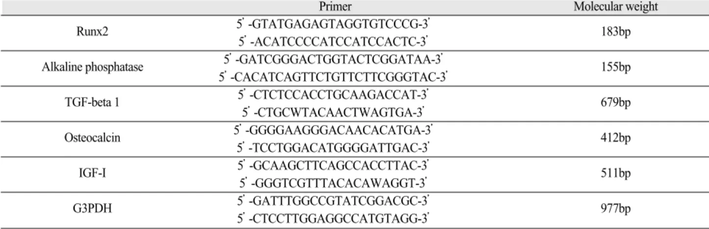
The osteoblastic differentiation of E1 cells was evaluated by RT-PCR examination of Runx2, alkaline phosphatase, osteocalcin, IGF-1, TGF beta, and G3PDH. The cells were seeded at a density of 1×10⁵ cells/ml. Cells were incubated for 24 hours. Total RNA extraction was performed with RNeasy mini kit (Qiagen, Chatsworth, CA, USA). The extracted total RNA samples were converted to cDNA. The amplifications were performed using AmpliTaq DNA polymerase (Amersham Pharmacia Biotech, Piscataway, NJ, USA). PCR products were fractionated by 1.5%
agarose gel electrophoresis and visualized by ethidium bromide staining. The intensity of the bands was quantified under UV transillumination (Eagle eye II, Stratgene, La Jolla, CA, USA). The sequences of the specific primers
Table I. Gene-specific primer sequences used in RT-PCR of MC3T3-E1 cells
Primer Molecular weight
Runx2 5’-GTATGAGAGTAGGTGTCCCG-3’ 183bp
5’-ACATCCCCATCCATCCACTC-3’
Alkaline phosphatase 5’-GATCGGGACTGGTACTCGGATAA-3’ 155bp
5’-CACATCAGTTCTGTTCTTCGGGTAC-3’
TGF-beta 1 5’-CTCTCCACCTGCAAGACCAT-3’ 679bp
5’-CTGCWTACAACTWAGTGA-3’
Osteocalcin 5’-GGGGAAGGGACAACACATGA-3’ 412bp
5’-TCCTGGACATGGGGATTGAC-3’
IGF-I 5’-GCAAGCTTCAGCCACCTTAC-3’ 511bp
5’-GGGTCGTTTACACAWAGGT-3’
G3PDH 5’-GATTTGGCCGTATCGGACGC-3’ 977bp
5’-CTCCTTGGAGGCCATGTAGG-3’
used are listed on Table I.
6. Statistical analysis
Test mean values and standard deviations (SD) were computed for MTT test and the analysis of variance (ANOVA) was used to assess the significance level of the differences between the experimental groups. All statistical analyses were performed using SPSS software (Version 12.0, SPSS Inc., Chicago, IL, USA). Differences were considered significant at P< 0.05.
RESULT
1. Cellular proliferation
The density of MC3T3-E1 cells were measured over 2 different time-periods; 24 hours and 48 hours by using the MTT assay. Figure 1 shows the MTT assay results of MC3T3-E1 cells. After 24 hours of adhesion, the osteoblast-like cell density on titanium surfaces and zirconia surfaces showed no significant difference. Grooves of zirconia had no significant effect on the proliferation of osteoblast-like cells. But after 48 hours of adhesion of MC3T3-E1 cells, the optical density of smooth zirconia did not increase significantly, but the optical density of titanium group and grooved zirconia significantly increased (P<0.05). Therefore, it is suggested that the microgrooves of the titanium disks proved to be as effective as grooves of
the zirconia on the proliferation rate and both groups increased compared to the smooth zirconia group after 48 hours of cell incubation. Overall, the osteoblast-like cells seeded onto titanium and zirconia showed similar vitality and proliferation rate.
2. Cellular attachment and morphology
The general shape and growth pattern of the osteoblast- like cells were observed using scanning electron microscopy for each group. Figure 2, 3, 4 and 5 show representative scanning electron micrographs of MC3T3-E1 cells cultured for 4 hours and 24 hours on the T group, ZS group, and ZG group. Orientation of osteoblasts of T group and ZG group were observed to be parallel to the direction of the microgrooves, whereas the cells in ZS group were observed to be oriented in random directions. Majority of the cells were found inside the microgrooves with increased formation of filopodia.
On machined titanium, SEM images show that the cells were irregularly triangular or elongated in shape. They were primarily oriented along the grooves and appeared flattened, with some long protoplasmic processes that were well attached to the substrate. However, cells cultured on zirconia disks showed higher initial adhesion properties compared to the titanium discs in the first 4 hours. After 24 hours of cell culture, osteoblast-like cells both showed more increased formation of filopodia and the cells showed more contact with each other and firm adhesion to the surface of the specimen.
3. Cellular differentiation
After incubation of E1 cells for 24 hours, the mRNA expression of alkaline phosphatase, osteocalcin, IGF-1, TGF-β, Runx2, and G3PDH on the titanium and grooved zirconia group showed similar activity (Fig. 6). Factors related to the quality of calcification; alkaline phophatase, osteocalcin, IGF-1, and TGF-βincreased only very slightly on the smooth zirconia group compared to titanium and grooved zirconia group. Overall, the gene expression analysis of E1 cells cultured showed no significant difference between the three groups.
Fig. 1. Evaluation of cellular viability by using MTT assay dur- ing 24 hours and 48 hours of MC3TC-E1 cells on titanium group, smooth zirconia group and grooved zirconia group.
(A) (B) (C)
Fig. 2. SEM images of cultured osteoblast-like cells after 4 hours (×300).
(A) titanium group, (B) smooth zirconia group, (C) grooved zirconia group
Osteoblasts of T group and ZG group were observed to be oriented parallel to the direction of the grooves.
(A) (B) (C)
Fig. 3. SEM images of cultured osteoblast-like cells after 4 hours (×1000).
(A) titanium group, (B) smooth zirconia group, (C) grooved zirconia group
Osteoblasts of T group and ZG group were found inside the microgrooves with increased formation of filopodia.
(A) (B) (C)
Fig. 4. SEM images of cultured osteoblast-like cells after 24 hours (×300).
(A) titanium group, (B) smooth zirconia group, (C) grooved zirconia group
Osteoblasts of T group and ZG group were observed to be oriented parallel to the direction of the grooves.
(A) (B) (C)
Fig. 5. SEM images of cultured osteoblast-like cells after 24 hours (×1000).
(A) titanium group, (B) smooth zirconia group, (C) grooved zirconia group
Osteoblasts after 24 hours of culture showed more contact with each other and appeared more flattened.
DISCUSSION
Alteration in surface morphology can be used to influence cell and tissue responses to implants. Surface morphology and biomaterial affects the osteoblastic-specific gene expression. Also, these surface characteristics determine how biological molecules will adsorb on the surface. Cell- material interaction occurs in two phases, the first phase involves the attachment, adhesion and spreading of the cells and it is the quality of this phase that influences the second phase, the capacity of the cell to proliferate and differentiate itself on contact with the implant.31
Smooth surfaces are considered to not favor cell adhesion, whereas micromachined surfaces inhibit epithelial downgrowth. Other investigations report that smooth surfaces favor human oral fibroblast attachment and soft tissue growth,34-36whereas rough surfaces favor osteoblast attachment and ingrowth of bone.2,37,38From the results of this study, we can suggest that grooves can favor the proliferation of osteoblasts compared to the smooth surface.
In this study, we examined the difference in cellular attachment and proliferation between titanium and zirconia.
From the results of SEM, after 4 hours of cell culture, we
could observe that titanium group showed the lowest cell adhesion. The machining of Ti6Al4V alloys has previously been shown to induce the formation of a concentration of aluminum oxides on the outermost surface.39 This phenomenon can be explained by the concentrations of Al on the machined implant surfaces since they constitute a potential risk of Al dissolution in the biological fluids surrounding alloyed Ti surgical implants. In this study, the Al dissolution from the surface may also explain the lower cell adhesion at 4 hours of cell culture.
Cell proliferation was comparable between the two materials and grooves proved to have similar cell proliferation effect as smooth surfaced material after a short-term cell culture period. However, osteoblast-like cells of the grooved zirconia group showed to be more flattened and spread evenly over the disks. Cells in a rounded configuration divide at a lower rate than those flattened and well spread on a substratum. Consequently, cells which attach to materials but spread little will show lower proliferative rates than those materials which allow greater spreading.40Cell morphology, as well as cell numbers, also affects the degree of cell attachment. Aligned cells are said to demonstrate more favorable adhesion behavior than a Fig. 6. Expression of alkaline phophatase, osteocalcin, Runx2, IGF-1, G3PDH, TGF-β.
spherically shaped cell.41 In consequence, surface topography, such as grooves as in this study, may influence the cell spread and growth especially in the early phase of cellular proliferation.
Runx2 is known to be factors for cell differentiation, alkaline phosphatase is an enzyme related to calcification.
Osteocalcin is a protein which absorbs calcium on bone surfaces and is specifically synthesized by differentiated osteoblasts. IGF-1 is known to have stimulatory effect on osteoblast proliferation. TGF-βis a well-known bone growth factor and G3PDH is a transcription factor which regulates RNA formation.42
Osteoblast differentiation generally implies alkaline phosphatase activity (ALP) and specific protein expression like osteocalcin, osteopontin, type I collagen and in vitro mineralization capacity. In vitro mechanical stimulation has shown various effects on ALP activity of cells.31The results show that no significant difference in the expression levels of Runx2, G3PDH was observed between the titanium group and zirconia group. In addition, grooves of zirconia surface have no effect on the mRNA expression of the osteoblast-like cells or HGFs. However, factors related to the quality of calcification; alkaline phophatase, osteocalcin, IGF-1, and TGF-βincreased very slightly on the smooth zirconia group compared to titanium and grooved zirconia group. This suggests that zirconia might have effects on enhancement of mineralization capacity of osteoblastic cells after a long-term cell incubation.
Recent studies of surface roughness have focused on cell attachment of titanium surfaces and showed better attachment on rough surfaces compared to smooth surfaces.43 On the contrary, it was found that on titanium disks with various degrees of roughness, proliferation and alkaline phosphatase activity was reduced when roughness increased.31In an experiment with the beagle dog, the different surface characteristics of abutment made of c.p.
titanium; rough or smooth surface failed to influence soft tissue reactions.44
To further improve the esthetic aspect for dental implants, efforts are undertaken to develop systems with tooth- colored implants and tooth-colored abutments that are biocompatible and able to withstand masticatory forces.
Zirconia implants are in clinical experiment and available on the market because of the demand for more esthetic
results.45Sollazzo et al. reported a study in which implants treated with zirconium oxide coating showed significantly higher bone-implant contact percentage than in untreated titanium.46According to a finite element analysis47and animal experiments,23,24zirconia implants seem to be able to withstand occlusal forces for a long period. Zirconia implants with rough surface can achieve higher stability in bone than zirconia implants with machined surface.
Roughening the turned zirconia implants enhances bone apposition and has a beneficial effect on the removal torque values.48However, in a recent study of Y-TZP with different surface topographies, cell attachment and cell proliferation proved to be independent of the surface treatments and even machined Y-TZP disks showed to be rough enough to enable the cells to fix onto the biomaterial.9
The results of this study show that the overall cell response to c.p. titanium and zirconia material was comparable. Further investigation is needed to identify the influence of depth and thickness of grooves on the zirconia surface. In general, zirconium oxide can be suitable for implant materials, but more clinical and mechanical trials are necessary for complete understanding of behavior of zirconia as implant materials throughout a long-time period.
CONCLUSION
The present in vitro study showed that surface topography and material of implant abutments can play an important role in expression of osteoblast phenotype markers. We evaluated the initial osteoblast-like cell response to titanium and zirconia ceramic material.
1. Zirconia ceramic showed comparable biological responses of osteoblast-like cells with titanium during a short-time cell culture period.
2. Grooves of implant material can be more effective on the cellular proliferation of osteoblast-like cells compared to the smooth surface after 48 hours of cell incubation.
3. Machined titanium surface, smooth zirconia ceramic, and grooved zirconia ceramic showed comparable osteoblast-specific gene expression However, expression of factors related to the quality of calcification; alkaline phophatase, osteocalcin, IGF-1, and TGF-βof the E1 cells increased only very slightly
on the smooth zirconia group compared to titanium and grooved zirconia group.
4. Grooves influence cell spreading and guide the cells to be aligned parallel within surface grooves.
REFERENCE
1. Kasemo B, Lausmaa J. Biomaterial and implant sur- faces: A surface science approach. Int J Oral Maxillofac Implants 1988;3:247-59.
2. Carlsson L, Rostlund T, Albrektsson B, Albrektsson T, Bra¨nemark PI. Osseointegration of titanium implants.
Acta Orthop Scand 1986;57:285-9.
3. Anselme K, Bigerelle M, Noel B, Iost A, Hardouin P.
Effect of grooved titanium substratum on human os- teoblastic cell growth. J Biomed Mater Res 2002;60:529-40.
4. Li LH, Kong YM, Kim HW, Kim YW, Kim HE, Heo SJ. Improved biological performance of Ti implants due to surface modification by micro-arc oxidation.
Biomaterials 2004;25;2867-75.
5. Brunette DM. Effects of surface topography of implant materials on cell behavior in vitro and in vivo. In: Hoch HC, editor. Nanofabrication and biosystems.
Cambridge, UK: Cambridge University Press; 1996.
6. Mustafa K, Silva Lopez B, Hultenby K, Wennerberg A, Arvidson K. Attachment and proliferation of human oral fibroblasts to titanium surfaces blasted with TiO2 particles. A scanning electron microscopic and histo- morphometric analysis. Clin Oral Impl Res 1998;9:195-207.
7. Yoshinari M, Matsuzaka K, Inoue T, Oda Y, Shimono M. Effects of multigrooved surfaces on fibroblasts be- havior. J Biomed Mater Res 2003;65A:359-68.
8. Anselme K, Linez P, Bigerelle M, Le Maguer D, Le Maguer A, Hardouin P, Hildebrand HF, Iost A, Leroy JM. The relative influence of the topography and chem- istry of Ti Al6V4 surfaces on osteoblastic cell behavior.
Biomater 2000;21:1567-77.
9. Ba¨chle M, Butz F, Hu¨bner U, Bakalinis E, Kohal RJ.
Behavior of CAL72 osteoblast-like cells cultured on zirconia ceramics with different surface topographies.
Clin Oral Impl Res 2007;18:53-9.
10. Heydecke G, Kohal R, Galser R. Optimal esthetics in single-tooth replacement with the Re-Implant system:
A case report. Int J Prosthodont 1999;12:184-9.
11. Piconi C, Maccauro G. Zirconia as a ceramic biomater- ial. Biomaterials 1999;20:1-25.
12. Garvie RC, Hannink RH, Pascoe RT. Ceramic steel?
Nature 1975;258:703-4.
13. Manicone PF, Iommetti PR, Raffaelli L. An overview of zirconia ceramics: Basic properties and clinical ap-
plications. J Dent 2007;35:819-26.
14. Ichikawa Y, Akagawa Y, Nikai H, Tsuru H. Tissue compatiblity and stability of a new zirconia ceramic in vivo. J Prosthet Dent 1992;68:322-6.
15. Josset Y, Oum’Hamed Z, Zarrinpour A, Lorenzato M, Adnet JJ, Laurent Maquin D. In vitro reactions of hu- man osteoblasts in culture with zirconia and alumina ceramics. J Biomed Mater Res 1999;47:481-93.
16. Scarano A, Piatelli M, Caputi S, Favero GA, Piatelli A.
Bacterial adhesion on commercially pure titanium and zirconium oxide disks: and in vivo human study. J Periodontol 2004;75:292-6.
17. Rimondini L, Cerroni L, Carrassi A, Torricelli P.
Bacterial colonization of zirconia ceramic surfaces: an invitro and in vivo study. Int J Oral Maxillofac Implants 2002;17:793-8.
18. Degidi M, Artese L, Scarano A, Perrotti V, Gehrke P, Piatelli A. Inflammatory infiltrate, microvessel density, nitric oxide synthase expression, vascular endothelial growth factor expression, and proliferative activity in peri-implant soft tissues around titanium and zirconium oxide healing caps. J Periodontol 2006;77:73-80.
19. Carinci F, Pezzetti F, Volinia S, Francioso F, Arcelli D, Farina, Farina E, Piatelli A. Zirconium oxide: analysis of MG63 osteoblast-like cell response by means of mi- croarray technology. Biomaterials 2004;25:215-28.
20. Lalor PA, Revell PA, Gray AB, Wright S, Railton GT, Freeman MA. Sensitivity to titanium. A cause of im- plant failure? The Journal of Bone and Joint Surgery.
British volume 1991;73:25-8.
21. Yamauchi R, Morita A, Tsuji T. Pacemaker dermatitis from titanium. Contact Dermatitis 2000;42:52-3.
22. Albrektsson T, Hansson HA, Ivarsson B. Interface his- tology of unloaded and early loaded partially stabilized zirconia endosseous implant in initial bone healing. J Prosthet Dent 1993;69:599-604.
23. Akagawa Y, Ichikawa Y, Nikai H, Tsuru H. Interface histology of unloaded and early loaded partially stabi- lized zirconia endosseous implants in initial bone heal- ing. J Prosthet Dent 1993;69:599-604.
24. Akagawa Y, Hosokawa R, Sato Y, Kamayama K.
Comparison between free-standing and tooth-connect- ed partially stabilized zirconia implants after two years’function in monkeys: A clinical and histologic study. J Prosthet Dent 1998;80:551-8.
25. Kohal RJ, Weng D, Ba¨chle M, Strub J. Loaded custom- made zirconia and titanium implants show similar os- seointegration. J Periodontol 2004;75:1262-8.
26. Kohal RJ, Hu¨rzeler MB, Mota LF, Mota LF, Klaus G, Caffesse RG, Strub JR. Custom-made root analogue tita- nium implants placed into extraction sockets. An ex- perimental study in monkeys. Clin Oral Implants Res 1997;8:386-92.
27. Sennerby L, Dasmah A, Larsson B, Iverhed M. Bone tissue responses to surface-modified zirconia implants:
a histomorphometric and removal torque study in the rabbit. Clin Implant Dent Relat Res 2005;7(suppl 1):
s13-s20.
28. Ko HC, Han JS, Ba¨chle M, Jang JH, Shin SW, Kim DJ. Initial osteoblast-like cell response to pure titanium and zirconia/alumina ceramics. Dental Materials 2007;23:1349-55.
29. Weiss P. Experiments on cell and axon orientation in vitro, the role of colloidal exudates in tissue organiza- tion. J Exp Zool 1945;100:353-86.
30. Matsuzaka K, Walboomers XF, Yoshinari M, Inoue T, Jansen JA. The attachment and growth behavior of os- teoblast-like cells on microtextured surfaces. Biomater 2003;24:2711-19.
31. Anselme K. Osteoblast adhesion on biomaterials.
Biomater 2000;21:667-81.
32. Mustafa K, Wennerberg A, Wroblewski J, Hultenby K, Lopez BS, Arvidson K. Determining optimal surface roughness of TiO2blasted titanium implant material for attachment, proliferation and differentiation of cells de- rived from human mandibular alveolar bone. Clin Oral Imp Res 2001;12:515-25.
33. Mustafa K, Oden A, Wennerberg A, Hultenby K, Arvidson K. The influence of surface topography of ce- ramic abutments on the attachment and proliferation of human oral fibroblasts. Biomater 2005;26:373-81.
34. Hormia M, Ko¨no¨nen M, Kivilahti J, Virtanen I.
Immunolocalization of proteins specific for adherents junctions in human gingival epithelial cells grown on differently processed titanium surfaces. J Perio Res 1991;26:491-7.
35. Guy SC, McQuade MJ, Scheidt MJ, McPherson JC, Rossmann JA, Van Dyke TE. In vitro study of attach- ment of human gingival fibroblasts to endosseous im- plant materials. J Periodontol 1993;64:542-6.
36. Cochran DL, Simpson J, Weber HP, Buser D.
Attachment and growth of periodontal cells on smooth and rough titanium. Int J Oral Maxillofac Implants 1988;3:21-4.
37. Bowers KT, Keller JC, Randolph BA, Wick DG, Michaels CM. Optimization of surface micromorpholo- gy for enhanced osteoblast responses in vitro. Int J Oral Maxillofac Implants 1992;7:302-10.
38. Gotfredsen K, Nimb L, Hjorting-Hansen E, Jensen JS, Holmen A. Histomorphometric and removal torque analysis for TiO2-blasted titanium implants. An experi-
mental study on dogs. Clin Oral Imp Res 1992;2:77-84.
39. Ask M, Lausmaa J, Kasemo B. Preparation and surface spectroscopic characterization of oxide films on Ti6Al4V. Appl Surf Sci 1988;35:283-301.
40. Hunter A, Archer CW, Walker PS, Blunn GW.
Attachments and proliferation of osteoblasts and fi- broblasts on biomaterials for orthopedic use.
Biomater1995;16:287-95.
41. Ismail M, Rohanizadeh R, Atwa S, Mason R, Ruys A, Martin P, Bendavid A. The influence of surface chem- istry and topography on the contact guidance of MG63 osteoblast cells. J Mater Sci Mater Med 2007; 18:705- 14.
42. Marinucci L, Balloni S, Becchetti E, Belcastro S, Gurerra M, Calvitti M, Cinzia L, Calvi EM, Locci P.
Effect of surface roughness on human osteoblast prolif- eration and gene expression in vitro. Int J Oral Maxillofac Implants 2006;21:719-25.
43. Martin JY, Schwartz Z, Hummert TW, Schraub DM, Simpson J, Lankford JJ, Dean DD, Cochran DL, Boyan BD. Effect of titanium surface roughness on proliferation, differentiation, and protein synthesis of human osteoblast-like cells (MG63). J Biomed Materi Res 1995;29:389-401.
44. Zitzmann NU, Abrahamsson I, Berglundh T, Lindhe J.
Soft tissue reactions of plaque formation at implant abutments with different surface topography. An exper- imental study in dogs. J Clin Periodontol 2002;29:456- 61.
45. Oliva J, Oliva X, Olive JD. One-year follow-up of first consecutive 100 zirconia dental implants in humans: a comparison of 2 different rough surfaces. Int J Oral Maxillofac Implants 2007;22:430-35.
46. Sollazzo V, Pezzetti F, Scarano A, Piattelli A, Bignozzi CA, Massari L, Brunelli G, Carinci F. Zirconium oxide coating improves implant osseointegration in vivo.
Dental Materials 2007;17:(Epub ahead of print).
47. Kohal RJ, Papavasiliou G, Kamposiora P, Tripodakis A, Strub JR. Three-dimensional computerized stress analysis of commercially pure titanium and yttrium- partially stabilized zirconia implants. Int J Prosthodont 2002;15:189-94.
48. Gahert M, Gudehus T, Eichhorn S, Steinhauser E, Kniha H, Erhardt W. Biomechanical and histomorpho- metric comparison between zirconia implants with varying surface textures and a titanium implant in the maxilla of miniature pigs. Clin Oral Imp Res 2007;18:662-8.
CELLULAR ATTACHMENT AND GENE EXPRESSION OF OSTEOBLAST-LIKE CELLS ON ZIRCONIA CERAMIC SURFACES
Ahran Pae1*, DMD, MSD, PhD, Heesu Lee2, DMD, MSD, PhD, Hyeong-Seob Kim3, DMD, MSD, PhD, Jin Baik4, DMD, MSD, PhD, Yi-Hyung Woo5, DMD, MSD, PhD
1Assistant Professor, Department of Dentistry, School of Medicine, Ewha Womans University
2Assistant Professor, Department of Oral Anatomy, School of Dentistry, Kangnung National University
3Associate Professor, Department of Prosthodontics, School of Dentistry, Kyung-Hee University
4Assistant Professor, Department of Prosthodontics, School of Dentistry, Kyung-Hee University
5Professor, Director, Department of Prosthodontics, School of Dentistry, Kyung-Hee University
STATEMENT OF PROBLEM: Zirconium oxide can be a substitute to titanium as implant materials to solve the esthetic problems of dark color in the gingival portion of implant restorations. PURPOSE: This study was performed to define attachment and growth behavior of os- teoblast-like cells cultured on grooved surfaces of zirconium oxide and evaluate the genetic effect of zirconium oxide surfaces using the re- verse transcriptase-polymerase chain reaction (RT-PCR). MATERIAL AND METHODS: MC3T3-E1 cells were cultured on (1) commer- cially pure titanium discs with smooth surface (T group), (2) yttrium-stabilized tetragonal zirconia polycrystal (Y-TZP) with machined sur- face (ZS group), and (3) Y-TZP with 100μm grooves (ZG group). Cell proliferation activity was evaluated through MTT assay and cell mor- phology was examined by SEM. The mRNA expression of Runx2, alkaline phosphatase, osteocalcin, TGF-β1, IGF-1, G3PDH in E1 cells were evaluated by RT-PCR. RESULTS: From the MTT assay, after 48 hours of adhesion of MC3T3-E1 cells, the mean optical density val- ue of T group and ZG group significantly increased compared to the ZS group. SEM images of osteoblast-like cells showed that significant- ly more cells were observed to attach to the grooves and appeared to follow the direction of the grooves. After 24 hours of cell adhesion, more spreading and flattening of cells with active filopodia formation occurred. Results of RT-PCR suggest that T group, ZS group, and ZG group showed comparable osteoblast-specific gene expression after 24 hours of cell incubation. CONCLUSION: Surface topography and material of implants can play an important role in expression of osteoblast phenotype markers. Zirconia ceramic showed comparable biolog- ical responses of osteoblast-like cells with titanium during a short-time cell culture period. Also, grooves influence cell spreading and guide the cells to be aligned within surface grooves.
KEY WORDS: Zirconia ceramic, MC3T3-E1 cells, Contact guidance, Cell proliferation, Gene expression
Corresponding Author: Ahran Pae
Department of Dentistry, School of Medicine, Ewha Womans University
911-1 Mokdong, Yangcheon-Ku, Seoul, 158-710, Korea +82 2 2650 2797: e-mail, [email protected] Received January 21, 2008 Last Revison February 4, 2008 Accepted June 20, 2008.