https://doi.org/10.12671/jkfs.2016.29.4.276
276
Copyright ⓒ 2016 The Korean Fracture Society. All rights reserved.
This is an Open Access article distributed under the terms of the Creative Commons Attribution Non-Commercial License (http://creativecommons.org/licenses/
by-nc/4.0) which permits unrestricted non-commercial use, distribution, and reproduction in any medium, provided the original work is properly cited.
Address reprint requests to: Hak Jun Kim, M.D.
Department of Orthopaedic Surgery, Korea University Guro Hospital, 148 Gurodong-ro, Guro-gu, Seoul 08308, Korea
Tel: 82-2-2626-3090ㆍFax: 82-2-2626-1164 E-mail: dakjul@korea.ac.kr
Financial support: None. Conflict of interest: None.
족근골 골절
박영환⋅김학준 ⋅김수현
고려대학교 구로병원 정형외과
Fractures of the Tarsal Bone
Young Hwan Park, M.D., Hak Jun Kim, M.D. , Soo Hyun Kim, M.D.
Department of Orthopaedic Surgery, Korea University Guro Hospital, Seoul, Korea
Fractures of the tarsal bone, such as the navicular, cuboid, and cuneiform, are very rare. These injuries can lead to serious walking difficulties due to pain and deformity of the foot with delayed diagnosis of tarsal bone fractures during an injury to multiple lower extremities. The diagnosis can be done on simple radiographs. Sometime weight bearing radiographs or stress radiographs may be needed for further evaluation. Computed tomography is the most widely available diagnostic tool. Navicular and cuneiform account for the medial column of the foot, whereas cuboid for the lateral column. The treat- ment of tarsal bone fractures is primarily conservative management, but operative treatment is recommended for intra- articular displacement, dislocation, or shortening of the medial or lateral column of the foot. The operative treatments include screw fixation, plate fixation, or external fixation. Complications include malunion, nonunion, posttraumatic arthritis, avascular necrosis, and deformity of the foot. Tarsal bone fracture has to be evaluated carefully to prevent serious complications.
Key Words: Tarsal bone, Navicular, Cuboid, Cuneiform, Fracture
서 론
종골 및 거골을 제외한 족근골(tarsal bone)은 중족부에 해당하는 부위로 주상골(navicula), 입방골(cuboid), 세 개 의 설상골(cuneiform)로 이루어져 있으며, 중족부에 발생하 는 골절은 종골이나 거골 골절보다는 상대적으로 덜 알려 져 있는 골절이다. 최근의 연구 결과에 의하면 다발성 손 상 환자에서 흔히 간과되기 쉬운 골절로 알려져 있어 진단 및 치료에 주의를 기울여야 한다.1,2) 중족부를 구성하는 족
근골은 전족부와 후족부를 이어 주는 구조물로 다양한 족 저 인대가 부착되어 있어 그 움직임이 대단히 제한적이며 족저궁(plantar arch)을 형성한다.3) 중족부 골절은 대부분 고 에너지 손상에 의해 일어나며 단독으로 발생하는 경우 보다는 족부의 다발성 골절, 특히 리스프랑 관절 손상과 동반되어 발생하기 쉽다.4-7) 하지에 발생하는 다발성 손상 이 있는 경우에는 간과하기 쉬우므로 낙상이나 교통사고의 경우 그 손상 기전에 주목하여 족근골 골절에 대한 주의를 기울여야 하며 적극적인 치료를 통해 기능적 회복을 도모 하여야 한다.1,2,4) 또한, 족근골은 해부학적 형태가 복잡하 여 치료 과정에서 정상적인 해부학적 형태에 대한 이해가 선행되어야 하며 해부학적 정복뿐만 아니라 기능적인 회복 도 매우 중요한 치료 방침 중에 하나이다.5,6,8)
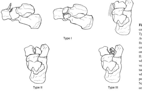
Fig. 2. Sangeorzan’s classifi- cation of navicular fracture.
Type I: Dorsal or tuberosity Avulsion fracture, the primary fracture line traverse in the coronal plane, which is seen on a lateral radiograph. Type II: Navicular body fracture, in which the fracture line is tra- verse from dorsolateral to plan- tarmedial across the body, which is seen on an antero- posterior radiograph. Type III:
Navicular body fracture with central or lateral comminution.
Fig. 1. Simple radiograph revealed the accessory navicular bone (white arrows).
주상골 골절(navicular fracture)
주상골은 근위부로 거골과 관절면을 이루고 후족부의 관 절 운동을 중족부의 설상골로 전달하는 역할을 하며 내측 세로 족저궁의 가장 높은 위치에 존재하여 족부의 초석 (keystone)으로 알려져 있고 형태가 배의 모양처럼 생겼다.
주상골 내측에는 골성 융기가 관찰되는 이를 주상골 결절 (navicular tuberosity)이라고 부르며 후경골건이 부착되는 곳이다. 주상골 결절 부위에는 제2 골화 중심이 있어서 성 인의 약 25%에서 주상골 부골(accessory navicular)이 나타
날 수 있으므로 골절과의 감별 진단이 필요하다(Fig. 1).
주상골의 골절은 체부(body)에 발생하는 골절과 결절에 발생하는 골절이 있으며, 골절의 발생 기전은 직접적인 외 력 손상이나 간접적인 손상이 있다. 직접 손상은 견열 골 절(avulsion fracture)이나 압궤 손상의 형태이고 간접 손상 은 회전력이나 족부의 축성 부하에 의해 발생하므로 주상 골 주위의 동반 손상에 대한 주의가 필요하다. 가장 흔한 손상 기전으로는 내번 또는 외번에 의한 비틀림 손상으로 족배측 인대나 관절낭에 의한 견열 골절이며 단순 족관절 염좌와 혼동하기 쉽다. 주로 달리기를 많이 하는 사람에서 반복적인 스트레스에 의한 주상골 피로 골절이 발생할 수 있는데 체부의 중앙 1/3에 주로 발생하며 단순 방사선 사 진에서 진단이 어려운 경우가 있어 골주사 검사나 자기공 명영상(magnetic resonance imaging, MRI) 촬영이 진단에 이용된다.9)
주상골 골절의 가장 흔한 임상 증상은 보행 시 주상골 부위의 통증 및 압통이다. 족부의 전후면, 측면, 내측 사면 위, 외측 사면위 사진이 가장 기본적인 단순 방사선 사진 이며 주변 관절과의 연관성 확인을 위해 전산화 단층촬영 (computed tomography, CT)이 필요하며, 부주상골과 주상 골 결절 골절과의 감별 진단을 위해서는 MRI 촬영이 필요 하기도 한다.
주상골 골절의 분류는 일반적으로 Sangeorzan 등10)이 제 안한 분류를 많이 사용한다(Fig. 2).
주상골 골절의 치료는 골절의 형태에 따라서 치료하지 않고 족부 내측주(medial column)의 길이 및 거주상 관절
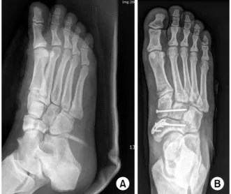
Fig. 3. (A) Type II navicular fracture and medial cuneiform subluxation were seen on an oblique radiograph. (B) Navi- cular fracture fragment and medial cuneiform were reduced and fixed with plate and screws.
(talonavicular joint)의 상합성(congruency)에 따라서 결정 한다. 즉, 족부 내측주의 길이가 유지되고 거주상 관절의 상합성이 유지되는 경우에는 단하지 석고 고정을 이용하여 6-8주간 비체중 부하를 시행한다. 운동 선수에서 주로 발 생하는 피로 골절의 경우는 진단이 어렵지만 진단이 되는 경우에는 석고 고정 없이 6주간의 비체중 부하가 권장된 다.11) 중족부의 불안정성이 동반된 주상골의 골절, 내측주 의 길이가 짧아진 골절 및 거주상 관절의 상합성이 일치하 지 않는 골절은 수술적인 치료가 권장되며 관절면의 손상 정도가 심한 골절의 경우는 일차적인 관절 유합술을 시행 하기도 한다.7,10,12,13) 수술 시기는 주상골 탈구가 동반되지 않은 경우에는 족부의 부종이 가라앉은 후에 시행하여야 연부조직 손상을 최소화할 수 있다. 주상골 골절의 수술적 인 방법은 일차적으로 정확한 관절면의 회복을 위해 관혈 적 정복술 및 내고정술이 권장되지만(Fig. 3) 분쇄가 심한 경우는 외고정 장치를 이용하여 내측주의 길이를 회복시키 는 방법이 있다.8,9)
주상골 골절의 합병증으로는 불유합, 관절의 외상후성 관절염, 무혈성 괴사 및 골 흡수에 의한 내측주의 붕괴가
있다.9,12,14) 합병증과 동반되어 발생하는 내측주의 붕괴가
가장 문제가 되므로 이러한 경우에는 내측주 정렬의 회복 및 변형 교정을 동시해 시행하여야 한다.
주상골의 탈구는 일반적으로 신경병성 족부 질환과 동반 되어 흔히 일어날 수 있다. 전족부의 과도한 족저 굴곡 상 태에서 축성 부하가 가해지면 외상에 의한 주상골 탈구가 발생할 수 있다.13,15,16) 주상골 탈구의 증상은 족부 변형과
동반된 골성 골성 돌출을 관찰할 수 있으며 주상골 탈구의 치료는 중족부의 정렬 상태를 회복하기 위해 수술적인 치 료를 시행한다.5,13,15,17,18)
입방골 골절(cuboid fracture)
입방골의 골절은 흔하지는 않으나 치료가 적절히 이루어 지지 않으면 심각한 장해가 발생할 수 있는 골절로 단독으 로 일어나기도 하지만 주로 리스프랑 관절 손상이나 거주 상 관절의 손상과 동반되어 발생한다.19-21) 입방골은 종골 원위부에 위치하고 외측주를 구성하는 피라미드 모양의 골 성 구조물로 손상 시 전후면 단순 방사선 사진에서 주상골 외측에 골편이 관찰되거나 측면 방사선 사진에서 족배부의 골편이 관찰되면 입방골 손상의 징후로 생각할 수 있으므 로 CT 등의 추가적인 검사가 필요하다.3,22) 입방골 단독 손 상은 흔하지 않으므로 족관절 염좌로 오인하여 치료 시기 를 놓치고 외측주의 단축에 의한 족부 변형이 유발될 수 있으므로 주의를 기울여야 한다.
입방골 골절은 족관절 염좌와 같이 저 에너지 손상에서 부터 다발성 골절을 일으키는 고 에너지 손상까지 다양한 손상 기전이 있으며 강력한 족저 굴곡 및 외전력에 의해 입방골에 축성 부하가 가해지면서 발생하는 입방골의 분쇄 골절은 호두까기 골절(nutcracker fracture)이라고 불린다.
증상은 외측 족배부의 통증, 압통, 부종과 피하 출혈이 있는 경우는 입방골 손상을 의심하여야 하며, 특히 중족부 골절 및 리스프랑 관절의 손상이 있는 경우는 입방골 손상 을 반드시 확인하여야 한다. 입방골은 내측 사면위(medial oblique view)에서 가장 잘 관찰할 수 있다. 입방골 주의 의 관절 불안정성이 있는 경우에는 체중 부하 방사선 사진 이 진단에 도움이 된다.
입방골 골절에 대한 확립된 분류는 없으나 AO-OTA 그 룹에서 단순 골절과 복잡 골절로 분류하지만 견열 골절도 발생할 수 있다. 골절 이외에도 드물지만 입방골 아탈구 혹은 탈구가 발생할 수 있다. 증상이 있는 입방골 탈구를
‘입방골 증후군(cuboid syndrome)’이라고 하며 중족부의 회내(pronation) 상태에서 과도한 후족부의 압력과 비틀림 에 의해 종입방 관절의 비상합성(incongruency)이 생겨서 증상이 발생한다. 입방골을 족저 방향으로 통증이 유발하 게 되는데 주로 발레 무용수에 잘 발생하며 도수 정복 후 단하지 석고 고정으로 치료한다.23,24) 입방골의 완전 탈구는 거의 일어나지 않지만 탈구는 항상 내측 족저 방향으로 일 어난다.
치료는 관절내 전위가 적거나 단순 방사선 사진이나 체 중 부하 방사선 사진에서 입방골의 탈구 및 아탈구가 없는 경우, 외측주의 단축이 없는 경우에는 6에서 8주간의 단하
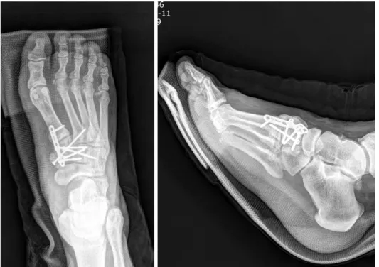
Fig. 5. Medial cuneiform frac- ture with Lisfranc injury was seen on simple radiograph and computed tomography.
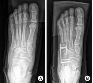
Fig. 4. (A) Nutcracker fracture of cuboid was seen on an anteroposterior radiograph. (B) Bridging plate fixation was performed from the calcaneus to the fifth metatarsal bone to restore the length of the lateral column.
지 석고 고정을 시행한다. 입방골 골절의 수술적인 치료는 관절면을 정복하고 외측주의 길이를 회복하기 위해 나사못, 금속판 및 외고정 장치를 이용한다(Fig. 4).3,17,21,22)
합병증으로 입방골 주위의 불안정성, 외상후성 관절염,
외측주의 소실이 발생할 수 있으며, 외측주의 소실 등이 발생하면 골 이식을 통한 종입방 관절 유합술을 시행할 수 있으며 입방-중족 관절의 관절염 발생 시 인대 감입 관절 성형술(interpositional arthroplasty)을 시행해볼 수 있다.3,22)
설상골 골절(cuneiform fracture)
설상골은 쐐기 모양으로 내측, 중간 및 외측 설상골로 이루어져 있으며 중족골과 관절면을 이루어 리스프랑 관절 을 형성하는 구조물로 단독 손상은 매우 드물지만 리스프 랑 관절 손상과 동반되어 자주 일어난다. 설상골의 손상 기전은 족저 굴곡 상태에서는 축성 부하에 의해 주상골 손 상과 동반되어 족배부 탈구가 발생하며, 족배 굴곡 상태에 서는 분쇄 골절 및 단축 손상이 발생한다.17,25-27) 내측 설상 골은 해부학적 특성으로 인해 불안정성이 있으므로 작은 외력에 의해서도 쉽게 탈구 등의 손상이 발생할 수 있다.
매우 드물지만 리스프랑 관절의 손상과 동반되어 전경골건 에 과도한 견인에 의한 견열 골절이 발생할 수 있다.28)
입방골의 골절은 단순 골절이나 복잡 골절로 분류할 수 있으며 탈구나 단축 등이 없는 경우에는 6에서 8주간의 단 하지 석고 고정을 이용한 보존적인 치료를 시행할 수 있 다. 수술적인 치료는 반드시 리스프랑 관절 손상 등의 동 반 손상 치료가 우선시되어야 하며 내측주의 단축을 회복 하고 탈구 및 아탈구에 대한 정복 및 고정이 필요하다(Fig.
Fig. 6. Fracture of the me- dial cuneiform and Lisfranc joint were reduced and fix- ated with plate and screws.
5, 6).17,26,27,29,30)
합병증으로는 불유합, 외상후성 관절염 및 중족부 불안 정성이 발생할 수 있으며 치료는 관절 유합술이 필요할 수 있다.
결 론
족근골에 발생한 골절은 매우 드물지만 쉽게 간과될 수 있는 골절이고 진단이 늦어져서 발생하는 합병증으로 인해 족부 변형 및 통증으로 인한 삶의 질이 저하되므로 항상 주의를 기울여야 한다. 리스프랑 관절 손상, 거골 골절 및 종골 골절 등이 있는 경우나 고 에너지로 발생한 다발성 하지 골절이 있는 경우는 반드시 족근골 골절에 대한 관심 을 가져야 한다.
References
1) Tadros AM, Eid HO, Abu-Zidan FM: Epidemiology of foot injury in a high-income developing country. Injury, 41: 137-140, 2010.
2) Wei CJ, Tsai WC, Tiu CM, Wu HT, Chiou HJ, Chang CY: Systematic analysis of missed extremity fractures in emergency radiology. Acta Radiol, 47: 710-717, 2006.
3) Yang GH, Park KC, Yoo GH, et al: Fractures and dis- locations of the midfoot and forefoot. In: Yang GH ed.
Principles of fracture management. 1st ed. Seoul, Korean Fracture Society: 837-839, 2013.
4) Lau S, Bozin M, Thillainadesan T: Lisfranc fracture dis- location: a review of a commonly missed injury of the midfoot. Emerg Med J, doi: 10.1136/emermed-2015-205317, 2016 [epub].
5) Richter M, Wippermann B, Krettek C, Schratt HE, Hufner T, Therman H: Fractures and fracture disloca- tions of the midfoot: occurrence, causes and long-term results. Foot Ankle Int, 22: 392-398, 2001.
6) Myerson MS, McGarvey WC, Henderson MR, Hakim J: Morbidity after crush injuries to the foot. J Orthop Trauma, 8: 343-349, 1994.
7) Thordarson DB: Detecting and treating common fractures of the foot and ankle part 2: the midfoot and forefoot.
Phys Sportsmed, 24: 58-64, 1996.
8) Kadow TR, Siska PA, Evans AR, Sands SS, Tarkin IS:
Staged treatment of high energy midfoot fracture disloca- tions. Foot Ankle Int, 35: 1287-1291, 2014.
9) DiGiovanni CW: Fractures of the navicular. Foot Ankle Clin, 9: 25-63, 2004.
10) Sangeorzan BJ, Benirschke SK, Mosca V, Mayo KA, Hansen ST Jr: Displaced intra-articular fractures of the tarsal navicular. J Bone Joint Surg Am, 71: 1504-1510, 1989.
11) Torg JS, Moyer J, Gaughan JP, Boden BP: Manage- ment of tarsal navicular stress fractures: conservative ver- sus surgical treatment: a meta-analysis. Am J Sports Med, 38: 1048-1053, 2010.
12) Rosenbaum AJ, Uhl RL, DiPreta JA: Acute fractures of the tarsal navicular. Orthopedics, 37: 541-546, 2014.
13) Simon JP, Van Delm I, Fabry G: Fracture dislocation of the tarsal navicular. Acta Orthop Belg, 59: 222-224, 1993.
14) Andersen RC, Neiderer K, Martin B, Dancho J: Open reduction-internal fixation of a navicular body fracture with dorsal displacement of the first and second cunei- forms: a case report. J Am Podiatr Med Assoc, 103: 246- 249, 2013.
15) Kim BS, Cho SD, Cho YS, Park TW, So CS: Isolated fracture dislocation of the tarsal navicular: case report. J Korean Soc Fract, 10: 925-928, 1997.
16) Chu IT, Yoo KH: Isolated dislocation of Tarsal Navicular bone: a case report. J Korean Soc Fract, 10: 86-90, 1997.
17) Grivas TB, Vasiliadis ED, Koufopoulos G, Polyzois VD, Polyzois DG: Midfoot fractures. Clin Podiatr Med Surg, 23: 323-341, vi, 2006.
18) Cheng Y, Yang H, Sun Z, Ni L, Zhang H: A rare mid- foot injury pattern: navicular-cuneiform and calcaneal-cu- boid fracture-dislocation. J Int Med Res, 40: 824-831, 2012.
19) Hunter JC, Sangeorzan BJ: A nutcracker fracture: cu- boid fracture with an associated avulsion fracture of the tarsal navicular. AJR Am J Roentgenol, 166: 888, 1996.
20) Macciocchi B: Etiology of tarsal cuboid bone fractures.
Minerva Chir, 4: 620-622, 1949.
21) Sangeorzan BJ, Swiontkowski MF: Displaced fractures of the cuboid. J Bone Joint Surg Br, 72: 376-378, 1990.
22) Borrelli J Jr, De S, VanPelt M: Fracture of the cuboid.
J Am Acad Orthop Surg, 20: 472-477, 2012.
23) Newell SG, Woodle A: Cuboid syndrome. Phys Sportsmed, 9: 71-76, 1981.
24) Patterson SM: Cuboid syndrome: a review of the litera- ture. J Sports Sci Med, 5: 597-606, 2006.
25) Patterson RH, Petersen D, Cunningham R: Isolated fracture of the medial cuneiform. J Orthop Trauma, 7: 94-95, 1993.
26) Miersch D, Wild M, Jungbluth P, Betsch M, Windolf J, Hakimi M: A transcuneiform fracture-dislocation of the midfoot. Foot (Edinb), 21: 45-47, 2011.
27) Lee BH, Shin DM: Treatment of fracture - dislocation of Tarsometatarsal joint. J Korean Soc Fract, 8: 606-614, 1995.
28) Ahn SH, Kim HC, Kim KY, Yoon HJ, Kim IY:
Avulsion fracture of medial cuneiform by Tibialis anterior tendon (a case report). J Korean Foot Ankle Soc, 14:
194-196, 2010.
29) Chung HJ, Park JG, Kam MC: Diagnosis and treatment of occult Lisfranc injury. J Korean Foot Ankle Soc, 17:
34-39, 2013.
30) Khatri Chhetri KM, Acharya P, Rokaya Chhetri DR:
Combined fracture dislocation of the navicular bone along with cuboid, cuneiform and longitudinal split fracture of the lateral malleolus: a rare combination of fractures. Chin J Traumatol, 17: 358-360, 2014.
Copyright ⓒ 2016 The Korean Fracture Society. All rights reserved.
This is an Open Access article distributed under the terms of the Creative Commons Attribution Non-Commercial License (http://creativecommons.org/licenses/
by-nc/4.0) which permits unrestricted non-commercial use, distribution, and reproduction in any medium, provided the original work is properly cited.
https://doi.org/10.12671/jkfs.2016.29.4.276
족근골 골절
박영환⋅김학준 ⋅김수현
고려대학교 구로병원 정형외과
주상골, 입방골 및 설상골에 발생하는 족근골(tarsal bone) 골절은 매우 드문 골절로 알려져 있으나 다발성 하지 골절 환자에서 는 쉽게 간과되어 치료가 늦어져서 발생하는 족부 변형 및 통증으로 인하여 보행에 상당한 장해를 유발할 수 있으므로 손상 발생 시 진단에 항상 주의를 기울여야 한다. 진단은 단순 방사선 사진으로 가능한 경우가 있지만 때로는 체중 부하 방사선 사진이나 스트레스 부하 방사선 사진이 필요한 경우가 있으며 전산화 단층촬영이 진단에 많은 도움이 된다. 주상골과 설상골은 족부의 내측주를 담당하고 입방골은 외측주를 담당한다. 치료는 관절 내의 전이가 없는 경우, 탈구가 없는 경우나 내측주 혹은 외측주의 단축이 없는 경우는 단하지 석고 고정을 이용한 보존적인 치료가 가능하나 관절내 전위, 탈구 및 단축이 있는 경우는 나사못, 금속판 및 외고정 장치를 이용한 수술적인 처치가 필요하다. 부정 유합, 불유합, 외상후성 관절염, 무혈성 괴사 및 족부의 변형 등의 합병증이 발생할 수 있으므로 족근골 골절의 진단에는 세심한 주의를 기울여야 진단이 늦어져서 발생하는 심각한 합병증을 예방할 수 있다.
색인 단어: 족근골, 주상골, 입방골, 설상골, 골절
교신저자 김학준
08308, 서울시 구로구 구로동로 148, 고려대학교 구로병원 정형외과 Tel 02-2626-3090, Fax 02-2626-1164, E-mail dakjul@korea.ac.kr
282