The Effects of Breath-Counting Meditation and Deep Breathing on Heart Rate Variability
Ji-Hwan Kim, Hyo-Sang Bae, Seong-Sik Park
Department of Sasang Constitutional Medicine, College of Korean Medicine, Dongguk University, Republic of Korea
Original Article
⋅Received:12 May 2016 ⋅Revised:16 June 2016 ⋅Accepted:16 June 2016
⋅Correspondence to:Seong-Sik Park
Dept. of Sasang Constitutional Medicine, Dongguk university Bundang hospital of Korean Medicine 268, Buljeong-ro, Bundang-gu, Seongnam-si, Gyeonggi-do, Rep. of Korea
Tel:+82-10-3359-8421, E-mail:[email protected]
Objectives: This study aimed to evaluate the effects of breath-counting meditation (BCM) and deep breathing (DB) on heart rate variability (HRV). These breathing techniques have the characteristics of non-paced and self-controlled breathings, resulting in less increase of HRV. We also compared BCM and DB with usual breathing (UB) or relaxing breathing (RB) which can reveal the characteristics of those.
Methods: 83 healthy volunteers sitting in chairs performed non-paced breathing; UB, RB, BCM, and DB each for 5 minutes. One minute of relaxation was permitted between breathings. Participants surfed the internet sitting in front of a computer during UB, while for RB, they remained steady with eyes closed. For BCM, they breathed inwardly counting from 1 to 10 repetitively, while they took a deep breath during DB. Physiological indices were simultaneously recorded with a biofeedback system.
Results: Respiration rate, thoracic amplitude, and mean heart rate decreased in RB compared with UB, but there was no change in HRV. Respiration rate in BCM and DB was lower than that in UB or RB, and the amplitude of thorax or abdomen, and HRV all increased (p<0.05). However, mean heart rate and skin conductance decreased in BCM compared with UB (p<0.05), whereas those were no different between DB and UB.
Conclusion: BCM, just concentrating mentally on breathing with counting each breath, can increase HRV with less sympathetic activation, while DB, actively moving thorax and abdomen for achieving the deepest respiration rate, can greatly raise HRV with the maintenance of mean vagal or sympathetic tone.
Key Words : Usual breathing, Relaxing breathing, Breath-counting meditation, Deep breathing, Heart rate variability, Biofeedback
Introduction
Respiration is naturally controlled by a pacemaker in the brain stem that receives signals from the autonomic nervous system, chemoreflexes, and baroreflexes generated by exercise, stress, and temperature changes. However, it can also be controlled consciously; individuals can control respiration rate, volume of each breath, ratio of
inspiration and expiration, etc. [1]. Consciously controlled breathing affects mind and body, and acts as an important component of meditations such as breathing awareness meditation [2], yoga [3], Dantian breathing [4], and mindfulness meditation [5].
Some previous studies used the direction of listening [6] or sight [7] for controlling participants’
respiration rate. This method has an advantage in
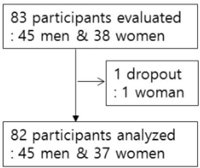
Fig. 1. Participants’ flow
Forty five men and 38 women were recruited. One woman did not complete measurements, and dropped out of the study. Therefore, total 82 participants (45 men and 37 women) were statistically analyzed.
that the respiration rates of participants can be matched as fixed pace. However, paced deep breathing caused stress in 60% of study participants [8], and autonomic nervous input heightened by this stress could lower heart rate variability (HRV) [9].
The occurrence of physical problems such as functional digestive disorders [10], inflammation [11], and hypertension [12] may correspond with a lower HRV. It is worthy to find how HRV can be increased effectively by breathing technique.
Therefore, we chose breath-counting meditation (BCM) and deep breathing (DB) among non-paced and self-controlled breathing techniques. BCM is a type of Zen meditation called Su-soku; it is performed by concentrating mentally on breathing while counting each breath [13]. On the other hand, in DB, participants concentrate physically on breathing while participants consciously maximize inspiration and expiration [14]. In order to observe the effect of these on HRV, we evaluated various physiological indices including respiration rate, the amplitude of thorax and abdomen, mean heart rate, skin conductance (SC) and body temperature with HRV while participants performed BCM and DB.
We also compared usual breathing (UB) and relaxing breathing (RB) with those in order to reveal the characteristics of BCM and DB.
Materials and Methods
1. Participants
83 young men and women attending Dongguk University were enrolled in this study (45 males, 38 females, average age 26.07±4.06). All participants were given a full explanation of the study and completed an informed consent form confirming participation of their own free will. Participants responded to questions regarding their health status, past disease history, and medication use. Participants had no previous experience with yoga or meditation,
and did not have any respiratory, cardiovascular or autonomic diseases. Participants were allowed to eat breakfast 2 hours before the beginning of the study.
However, all substances that may cause physiological changes, including coffee, were prohibited 12 hours prior to the study. One woman did not complete measurements of HRV, and dropped out of the study. Therefore, a total of 82 participants completed the study (Figure 1).
2. Outcome Measures
All measurements were digitized and recorded
with the ProComp biofeedback system (Korea
medical technology, http://www.kmtec.com, Republic
of Korea). Respiration rate, the amplitude of thorax
and abdomen were measured using respiratory
inductive plethysmography(RIP) belts strapped to the
thorax and abdomen. Mean heart rate, standard
deviation of heart rate, standard deviation of normal
intervals (SDNN), low frequency(LF; 0.04~0.15Hz),
and high frequency (HF; 0.15~0.4Hz) were measured
using an infrared photo plethysmography machine
attached to the left middle finger. SC and body
temperature were measured with sensors on the left
index and ring finger.
3. Procedures
Testing was performed in a quiet room between 9 a.m. and 11 a.m. Before the study, the participants were educated on the characteristics of each breathing method and practiced the methods with an investigator trained in advance. Afterwards, when the participants were resting while sitting in chairs, the investigator attached the measuring tools to participants’ bodies. The participants then performed UB, RB, BCM, and DB for 5 minutes each at the direction of the investigator. 1 minute of relaxation was permitted between each method. For UB, the participant was told to surf the internet using a mouse with right hand while sitting in front of a computer monitor. For RB, the participant with eyes closed breathed naturally while trying not to think of anything in particular. For BCM, the participant with eyes closed counted from 1 to 10 repetitively during expiration. For DB, the participant took a deep breath and concentrate just on physical act consciously for controlling respiration. All procedures were performed under the approval of the Institutional Review Board (IRB) of Dongguk University.
4. Statistical Analyses
Data was analyzed with SPSS (IBM SPSS Inc.
version 20). The results of LF and HF were skewed;
therefore, they were analyzed after natural log-transformation (lnLF and lnHF, respectively).
Repeated measures- analysis of variance (RM-ANOVA) was performed in order to compare breathing methods. When the sphericity assumption was not satisfied, significance was tested after correcting for the number of degrees of freedom using Greenhouse-Geisser’sε-corrected degrees of freedom. Post-hoc analysis was performed using Fisher's Least Significant Difference (LSD) method.
Results were judged to be statistically significant if the α value was less than 0.05 (p<0.05), and are shown as mean±S.D.
Results
We performed RM-ANOVA for the measured physiological indices. Post-hoc analyses of the statistically significant indices are presented in this section and summarized in Table 1.
Respiration rate significantly decreased in the following order: UB, RB, BCM, and DB (p<0.05).
Respiration rate was lowest in DB (Figure 1).
Thoracic amplitude was significantly lower during RB compared with UB and BCM (p<0.05), and was the highest during DB (p<0.05). However, there was no statistically significant difference between UB and BCM. Abdominal amplitude was significantly greater in BCM compared with RB (p<0.05), and was the highest in DB (p<0.05). However, abdominal amplitude was not significantly different between UB and either RB or BCM. Mean heart rate significantly decreased in the following order: UB, RB, and BCM (p<0.05). The mean heart rate in DB was higher than that of RB or BCM (p<0.05), and showed no difference compared with UB (Figure 2). The standard deviation of heart rate was significantly higher for DB compared with all other conditions (p<0.05). SDNN, lnLF, and lnHF were all significantly increased in BCM and DB compared with UB or RB (p<0.05). Those in DB was especially increased compared with BCM (p<0.05).
However, there was no statistically significant difference between UB and RB (Figure 3). SC was higher in UB and DB compared with RB or BCM (p<0.05). However there was no significant difference in SC between UB and DB, or between RB and BCM (Figure 4). Body temperature significantly increased in the following order: UB, RB, and BCM (p<0.05). However, there was no difference between DB and UB.
Discussion
The purpose of this study was to evaluate the
Table 1. Physiological Indices Obtained from the Results of Four Breathing States
Measure (Mean±S.D.)
Usual breath (UB)
Relaxing Breath
(RB)
Breath-counting meditation
(BCM)
Deep breathing
(DB)
RM-ANOVA Post hoc
(p<0.05)
Respiration Rate (breaths/min)
15.72
±1.59d
14.34
±2.47c
13.53
±3.00b
8.51
±2.61a
F(3,808.98)=166.95 p=0.000
UB-RB UB-BCM
UB-DB RB-BCM
RB-DB BCM-DB
Thoracic amplitude (cm)
1.81
±0.79b
1.56
±0.79a
1.86
±1.27b
7.07
±3.78c
F(1.20,1454.51)=171.30 p=0.000 ε=0.400
UB-RB UB-DB RB-BCM
RB-DB BCM-DB
Abdominal amplitude (cm)
2.88
±1.27ab
2.68
±1.23a
3.23
±2.06b
10.18
±5.41c
F(1.27,2557.11)=159.04 p=0.000 ε=0.423
UB-DB RB-BCM
RB-DB BCM-DB
Mean Heart rate (beats/min)
75.06
±9.64c
73.31
±9.62b
71.98
±9.39a
75.41
±8.58c
F(1.89,332.41)=18.49 p=0.000 ε=0.628
UB-RB UB-BCM RB-BCM RB-DB BCM-DB Standard deviation
of heart rate (beats/min)
5.54
±3.45a
5.04
±1.84a
5.24
±2.38a
9.10
±4.86b
F(2.11,432.49)=44.60 p=0.000 ε=0.702
UB-DB RB-DB BCM-DB Standard Deviation of
N-N Interval (ms)
55.07
±26.53a
56.15
±23.16a
60.13
±27.97b
96.44
±50.60c
F(1.52,63342.53)=56.22 p=0.000
ε=0.507 UB-BCM
UB-DB RB-BCM
RB-DB BCM-DB ln LF (ms2) 5.03
±0.84a
4.92
±0.89a
5.20
±0.97b
6.34
±1.20c
F(2.21,47.66)=75.71 p=0.000 ε=0.736 ln HF (ms2) 4.74
±1.06a
4.76
±0.94a
4.98
±1.02b
5.58
±1.29c
F(2.37,15.79)=22.78 p=0.000 ε=0.789
Skin conductance (μV)
0.95
±1.06b
0.76
±0.80a
0.73
±0.95a
1.09
±1.54b
F(1.65,4.22)=6.61 p=0.003 ε=0.548
UB-RB UB-BCM
RB-DB BCM-DB
Body temperature (℃)
32.96
±1.89a
33.34
±1.39b
33.50
±1.16c
33.03
±1.31a
F(1.46,10.77)=8.56 p=0.001 ε=0.487
UB-RB UB-BCM RB-BCM RB-DB BCM-DB ln : Natural logarithm
LF: Low-frequency component (0.04Hz to 0.15Hz) HF: High-frequency component (0.15Hz to 0.40 Hz)
a, b, c, d: A statistical significance is not present between the figures sharing the same alphabet subscript in the line of each index.
effect of BCM and DB on HRV using a non-paced and self-controlled breathing method. UB and RB
could reveal the characteristics of those by comparison.
In our study, in RB compared with UB, mean
Fig. 2. Changes of mean heart rate and respiration rate according to four breathing states
The mean heart rate became statistically and significantly lower as UB progressed toward RB and BCM (p<0.05). It was statistically and significantly higher for DB compared with RB and BCM (p<0.05), but was not significantly different regarding UB. Contrarily, the respiration rate became statistically, significantly, and gradually lower as UB progressed toward RB, BCM, and DB (p<0.05).
UB: Usual breathing; RB: Relaxing breathing; BCM: Breath-counting meditation; DB: Deep breathing
*p<0.05
Fig. 3. Changes of standard deviation of N-N interval (SDNN) and low frequency (LF) component of Heart rate variability (HRV) according to four breathing states
a) In the SDNN, there was no statistically significant difference for both UB and RB, but it showed a statistically significant increase pattern for BCM (p<0.05), and these indices tended to increase more for DB compared with BCM (p<0.05).
b) The changes of lnLFalsoshowed the same tendency as SDNN.This tendency was the same for lnHF(Figure not shown) SDNN: Standard deviation of N-N interval
ln: Natural logarithm
LF: Low frequency(0.04~0.15Hz)component of Heart rate variability (HRV) HF: High frequency(0.15~0.4Hz)component of Heart rate variability (HRV)
UB: Usual breathing; RB: Relaxing breathing; BCM: Breath-counting meditation; DB: Deep breathing
*p<0.05
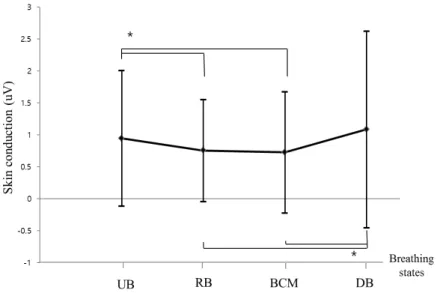
Fig. 4. Changes of skin conductance according to four breathing states
In skin conductance, UB was statistically significant compared with RB and BCM (p<0.05). DB was also statistically significant compared with RB and BCM (p<0.05). However, there was no statistically significant difference between UB and DB and between RB and BCM.
UB: Usual breathing; RB: Relaxing breathing; BCM: Breath-counting meditation; DB: Deep breathing
*p<0.05