INTRODUCTION
As the role of dental implants in the oral rehabilitation has become more important, reducing the failure rate of implant treatment has become one of the most concerned problems.
Osseointegration has been thought to be the most important pre- requisite for the long-term success of the dental implant.
Implant stability at the time of implant fixture placement, known as the primary stability, has been suggested as the crucial fac- tor for achieving successful osseointegration.1,2It has been thought that primary stability largely depends on the three major fac- tors: (1) the implant bed condition such as bone quantity and quality, (2) the mechanical shape of the fixture placed in the bone, and (3) the procedure how the fixture has been inserted in the bone.3
A number of test methods to assess the primary stability have been suggested: histology and histomorphometry, insertion torque, removal torque, push-through and pull-through, radiograph-
ic assessment, Periotest ultrasonic method, and resonance frequency analysis.1Among these test methods, resonance fre- quency analysis has been revealed and widely used as the most effective method to evaluate primary stability because of its easiness, accuracy, and non-invasiveness.4-11
A device called Osstell (Integration Diagnostic Ltd., Goteborg, Sweden) was invented to measure the resonance fre- quency value of the implant fixture through the transducer which is mounted directly to the fixture with a screw. The instrument measures the resonance frequency and display the result as the implant stability quotient (ISQ) value on a scale from 1 to 100.
Higher ISQ value means higher resonance frequency value, which means more primary stability. Finite element analysis also has been recently used to analyze the mechanical and vibration behav- ior of the three dimensionally designed structure.12-15
The aim of this study was to investigate the influence of bone quality and surgical technique on the ISQ value. Moreover, by means of the finite element analysis, a three dimensional
Effect of bone quality and implant surgical technique on implant stability quotient (ISQ) value
Hong-Gi Yoon1, Seong-Joo Heo2, DDS, MSD, PhD, Jai-Young Koak2, DDS, MSD, PhD, Seong-Kyun Kim2, DDS, MSD, PhD, Su-Young Lee2*, DDS, MSD, PhD
1Department of Dentistry, School of Dentistry, Seoul National University
2Department of Prosthodontics and Dental Research Institute, Seoul National University Dental Hospital, School of Dentistry, Seoul National University, Seoul, Korea
PURPOSE. This study investigated the influence of bone quality and surgical technique on the implant stability quotient (ISQ) value. In addi- tion, the influence of interfacial bone quality, directly surrounding the implant fixture, on the resonance frequency of the structure was also eval- uated by the finite element analysis. MATERIALS AND METHODS. Two different types of bone (type 1 and type 2) were extracted and trimmed from pig rib bone. In each type of bone, the same implants were installed in three different ways: (1) Compaction, (2) Self-tapping, and (3) Tapping.
The ISQ value was measured and analyzed to evaluate the influence of bone quality and surgical technique on the implant primary stability.
For finite element analysis, a three dimensional implant fixture-bone structure was designed and the fundamental resonance frequency of the structure was measured with three different density of interfacial bone surrounding the implant fixture. RESULTS. In each group, the ISQ val- ues were higher in type 1 bone than those in type 2 bone. Among three different insertion methods, the Tapping group showed the lowest ISQ value in both type 1 and type 2 bones. In both bone types, the Compaction groups showed slightly higher mean ISQ values than the Self-tap- ping groups, but the differences were not statistically significant. Increased interfacial bone density raised the resonance frequency value in the finite element analysis. CONCLUSION. Both bone quality and surgical technique have influence on the implant primary stability, and res- onance frequency has a positive relation with the density of implant fixture-surrounding bone. [J Adv Prosthodont 2011;3:10-5]
KEY WORDS. Compaction, Self-tapping, Tapping, Implant Stability Quotient (ISQ) Resonance frequency analysis, Finite element analysis
Corresponding author: Su-Young Lee
Department of Prosthodontics & Dental Research Institute, School of Dentistry, Seoul National University
275-1 Yeongun-dong, Jongno-gu, Seoul, 110-768, South Korea Tel. 82 2 2072 3816: e-mail, [email protected]
Received January 14, 2011 / Last Revison January 25, 2011 / Accepted February 17, 2011
ⓒ 2011 The Korean Academy of Prosthodontics
This is an Open Access article distributed under the terms of the Creative Commons Attribution Non-Commercial License (http://creativecommons.org/licenses/by- nc/3.0) which permits unrestricted non-commercial use, distribution, and reproduction in any medium, provided the original work is properly cited.
implant fixture-bone structure was also designed to evaluate the correlation between density of bone surrounding the implant fixture and resonance frequency value of the structure.
MATERIALS AND METHODS
Implant
Six Bra�nemark - type parallel implants with anodized oxi- dation surfaces (3.75 × 7 mm, Warantec, Seoul, Korea) were prepared for this study. Each implant was used nine times so that the total number of implant installation was 54 in this experiment. After each removal of inserted implants, the fixtures were cleansed with streaming water and re-used for next installation.
Specimen
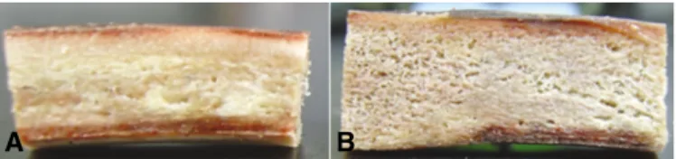
Two different types (type 1 and type 2) of bone specimens from pig rib bone were prepared for this experiment (Fig. 1).
Type 1 bones were harvested from the distal part of the pig rib with thick cortical bone and dense cancellous bone. Type 2 bone was harvested from the proximal part with less cortical bone and loose cancellous bone in comparison with the distal region. The bones were sawed to get about 4 - 5 cm long and the upper parts of cortical portions were trimmed away so that only cancellous components could be in direct contact with implant fixtures. Nine bone specimens for each type of bone were made from nine pig rib bones.
Surgical procedure
For each bone sample, three implant fixtures are installed in three different types of installation methods: (1) Compaction, (2) Self-tapping, and (3) Tapping (Fig. 2). To get rid of the poten- tial influence of implant location in bone sample on the ISQ value, each implant of different insertion technique was placed in middle for three times and in edge for six times for each bone type.
In the conventional tapping technique, the implant sites were marked first by 1.5 mm diameter round bur with drill speed of 1000 rpm, followed by twist drills of 2 mm diameter, and 3 mm diameter with 500 rpm. Finally tapping was performed in the drilled holes before the implant fixture installation. The fixtures were then inserted with an Elcomed (W&H Dentalwerk, Bu¨rmoos GmbH Austria) with engine speed of 30 rpm.
For the self-tapping installation method, round bur was used first to make a mark on the implant site, and twist drills of 2 mm/3 mm in diameter were used in sequence to make drill holes for implant fixture installation. The implant fixtures were then installed with engine speed of 30 rpm.
In the cancellous compaction technique, the implant sites were first marked by 1.5 mm diameter round bur and drilled with 2 mm wide twist drill. Then the holes were subsequently widened using varying diameters of taper-type osteotomes (2 mm/2.5 mm/3 mm) by light malleting until holes of 3 mm diam- eter were obtained. In the prepared holes, the implant fixtures were inserted with 30 rpm.
Implant stability measurement
Immediately after three implant fixtures were installed dif- ferently in each bone sample, the resonance frequencies were measured using Osstell (Integration Diagnostic Ltd., Goteborg, Sweden). The L-shaped transducer was firmly connected by hand-screw and the ISQ values were recorded in two differ- ent orientations perpendicular to the long axis of bone samples.
Statistical analysis
The data were analysed by using SPSS statistics 17.0 (SPSS Inc., Chicago, IL, USA) with 5% of significance level. The aver- age value and standard deviation of ISQ value for each six groups was calculated. Student t-test was used to compare the ISQ val- ues of two independent groups of different surgical tech- niques in each bone type. One-way ANOVA test was used for the comparison of the ISQ values of three different surgical pro- cedure groups in each bone type.
Fig. 1. Type 1 bone (A) and type 2 bone (B) specimens.
Fig. 2. A specimen with three implant fixtures inserted by three different methods.
A B
Finite element analysis
A three dimensional model for finite element analysis was created by using a finite element analysis program (ANSYS 12.1, ANSYS, Canonsburg, PA, USA) on a personal computer.
Like the implant fixtures used for RFA study, a fixture mod- el was designed to the shape of Bra�nemark type straight implant fixture with thread. The block bone model was com- posed of three compartments: (1) outer cortical bone, (2) inner cancellous bone, and (3) interfacial bone between implant fixture and cancellous bone (Fig. 3). To mimic the actu- al RFA experiment, the upper and the two side of cortical bone was removed. The interfacial bone layers surrounding the fix- ture were created to be 0.2 mm in thickness. All interfaces between two different compartments were assumed to be perfectly boned, and the model was medium meshed to gen- erate 23764 elements and 60365 nodes.
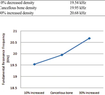
The titanium (Density of 4.5 g/cm3, Young's modulus of 117,000 MPa, and Poisson's ratio of 0.3) was chosen as the mate- rial of the model implant, and both cancellous and cortical bones were assumed to be orthotropic and linearly elastic.12,14For the interfacial bone layer, three different densities were defined : (1) normal cancellous bone density, (2) 30% increased density of the normal cancellous bone, and (3) 10% decreased density of the normal cancellous bone. All three interfacial bone layers were assumed to be orthotropic and linearly elastic, hav- ing same Poisson's ratio with cancellous bone. Each Young's modulus, however, was calculated from the equation
E = Cu0.06ρ3(where E is Young's modulus, C a constant, u the strain rate during testing, and ρthe density of bone).16All the material properties of titanium, cancellous bone, cortical bone, and three interfacial bone layers are presented in Table 1.17,18
The resonance frequency of the model could be determined in the harmonic response analysis by forcing varying fre- quencies of cyclic loads and observing the maximum dis- placements for each corresponding frequency. On the cyclic load of the resonance frequency, the maximum displacement of the model would be increased dramatically. For three dif- ferent interfacial bone layers, each resonance frequency was determined.
RESULTS
Resonance frequency analysis
The mean values and standard deviations calculated are shown in Table 2 and Fig. 4. In each type of bone, the Compaction group has the highest mean of ISQ value and the tapping group has the lowest mean of ISQ value. All surgical technique groups showed higher ISQ values in type 1 bone than in type 2 bone. The result of one-way ANOVA test showed that statistically significant difference in ISQ values between three different surgical techniques existed in each of the bone types (Table 3). The student t-test revealed statistically significant differences in ISQ value between the Self-tap-
Table 1. Material properties of bones and implant used in finite element analysis.
Material Young's moduls (MPa) Poisson's ratio Density
Ex Ey Ez vxy vyz vzx (g/cm3)
Cancellous bone 346.8 457.2 1107.1 0.05 0.01 0.322 0.55
Cortical bone 11300 13800 19400 0.274 0.237 0.237 1.94
Titanium implant 117000 0.3 4.5
10% loosed 252.8 333.3 807.1 0.05 0.01 0.322 0.495
Cancellous bone like Interfacial bone layers 346.8 457.2 1107.1 0.05 0.01 0.322 0.55
30% densed 761.9 1004.5 2432.3 0.05 0.01 0.322 0.715
Fig. 3. A three-dimensionally designed implant-bone structure.
Table 2. Mean ISQ values and standard deviations
Bone type Surgical Mean ISQ Standard
method value deviation
Type 1 Compaction 70.4 2.77
Self-Tapping 69.5 4.72
Tapping 65.1 2.53
Type 2 Compaction 63.9 3.87
Self-Tapping 62.0 4.32
Tapping 55.8 4.11
ping group and the Tapping group, and between the Compaction group and the Tapping group for each type of bone (P < .05).
However, the Compaction and the Self-tapping group were not significantly different in both type 1 and type 2 bones (Table 4).
Finite element analysis
The fundamental resonance frequencies for the three dimen- sional models with three different density of interfacial bone layers were determined and the result is presented in Table 5 and plotted in Fig 5. The result showed that the first mode of resonance frequency was increased as the interfacial bone lay- er becomes denser.
DISCUSSION
This study was to examine the influence of bone quality and surgical technique on the implant stability quotient (ISQ) value. Two different types of bone (type 1 and type 2) were extracted and the same implants were installed in three different ways.
Fig. 4. The plot of ISQ values showing the maximum value, the mean value, and the minimum value for each surgical technique groups (left for type 1 bone and right for type 2 bone).
Table 3. P-value with the one-way ANOVA test of ISQ value among different surgical techniques
Bone type Variable P value
Type 1 ISQ value vs Surgical technique .008 Type 2 ISQ value vs Surgical technique .001 The difference is statistically significant when the P-value is less than .05.
Table 4. Average difference and P-value with student t-test of ISQ value between two different surgical techniques (C, compaction; S, self- tapping; T, tapping)
Bone type Variable Average difference P value
Type 1 C vs S 0.94 .612
S vs T 4.33 .027
C vs T 5.28 .001
Type 2 C vs S 1.89 .344
S vs T 6.28 .006
C vs S 8.17 .001
The difference is statistically significant when the P-value is less than .05.
Table 5. Fundamental resonance frequency of the FEA model with different density of interfacial bone
Interfacial bone density Fundamental resonance frequency
10% decreased density 19.54 kHz
Cancellous bone density 19.95 kHz
30% increased density 20.68 kHz
Fig. 5. The plot of fundamental resonance frequency of the FEA model with three different density of interfacial bone.
A clinical instrument was recently developed to analyze res- onance frequency, which is calculated into the implant stability quotient (ISQ). Some authors4,19have shown strong correlation between ISQ value and cortical bone thickness, which suggests that cortical bone thickness plays a crucial role for implant pri- mary stability. It has been reported that no statically significant difference in ISQ value existed between different implant design types.20In the aspects of surgical technique, especial- ly between conventional drilling technique and osteotome tech- nique, some authors21 have concluded that the cancellous compaction technique increased the ISQ value than the con- ventional drilling technique. However, the other author22has shown that osteotome technique resulted in decreased ISQ val- ue, thus less primary stability, than conventional drilling technique.
In this study, only Bra�nemark type of straight implants were used so as to get rid of the influence of implant design to ISQ value. According to the quality of the cancellous bone, two types of bone were grouped: (1) type 1 bones were gained from the distal portion of the pig rib bone where cancellous bone is denser, and (2) type 2 bones were extracted from the proximal part where cancellous bone is less dense than that of distal region.
Upper cortical portions in all samples were trimmed off to exclude the influence of the cortical bone.
Comparing the results between type 1 and type 2 bones in each surgical method, the ISQ values in type 1 bones were signif- icantly higher than those in type 2, which suggests that den- sity of bone has positive relation to the implant primary sta- bility. Among three different insertion methods, the Tapping group showed the lowest ISQ value in both bone types. As a matter of fact, when removing the inserted fixture from the bone sample, especially in type 2 bone, it was so weekly anchored that even hand force would be enough to remove the implants.
This result suggests that tapping before the implant placement is not a recommended procedure if implant fixture site is absent of or with little cortical bone and not enough dense can- cellous bone.
This study showed slightly higher ISQ mean values in the Compaction group than the Self-tapping group, but the difference was not statistically significant in both bone types. In fact, the drilled hole size right before the fixture placement was 3 mm in diameter in the Self-tapping group, and the size of implant used was 3.75 mm in diameter, so during the self-tapping pro- cedure, the cancellous bone would have been compacted in some degree. This un-intended cancellous compaction from fixture itself might have raised the ISQ value approximately to the lev- el of Compaction group. If we had used bigger size of final drill for implant fixture bed preparation in Self-tapping group, the expected ISQ value would have been a little bit lower.
Through the finite element analysis, we could confirm that quality of bone directly surrounding the implant fixture plays a crucial role in determining resonance frequency. The reso-
nance frequency value of the model increased as the density of the interfacial bone surrounding the implant fixture increased.
CONCLUSION
The present study showed that both bone quality and surgical technique have influence on the implant primary stability.
Therefore, we could confirm that quality of bone directly surrounding the implant fixture plays a crucial role in deter- mining resonance frequency.
According to this experiment, the followings could be con- cluded:
1. In each three different surgical technique groups, the ISQ values were higher in type 1 bone than those in type 2 bone.
2. Among three different insertion methods, the Tapping group showed the lowest ISQ value in both type 1 and type 2 bone.
3. In both bone types, the Compaction groups showed slightly higher mean ISQ values than the Self-tapping groups, but the differences were not statistically significant.
4. Increased interfacial bone density raised the resonance fre- quency value in finite element analysis.
REFERENCES
1. Meredith N. Assessment of implant stability as a prognostic de- terminant. Int J Prosthodont 1998;11:491-501.
2. Lioubavina-Hack N, Lang NP, Karring T. Significance of pri- mary stability for osseointegration of dental implants. Clin Oral Implants Res 2006;17:244-50.
3. Sennerby L, Roos J. Surgical determinants of clinical success of osseointegrated oral implants: a review of the literature. Int J Prosthodont 1998;11:408-20.
4. Su YY, Wilmes B, Ho¨nscheid R, Drescher D. Application of a wireless resonance frequency transducer to assess primary sta- bility of orthodontic mini-implants: an in vitro study in pig il- ia. Int J Oral Maxillofac Implants 2009;24:647-54.
5. Rodrigo D, Aracil L, Martin C, Sanz M. Diagnosis of implant stability and its impact on implant survival: a prospective case series study. Clin Oral Implants Res 2010;21:255-61.
6. Zix J, Hug S, Kessler-Liechti G, Mericske-Stern R. Measurement of dental implant stability by resonance frequency analysis and damping capacity assessment: comparison of both techniques in a clinical trial. Int J Oral Maxillofac Implants 2008;23:525- 30.
7. Sim CP, Lang NP. Factors influencing resonance frequency analy- sis assessed by Osstell mentor during implant tissue integration:
I. Instrument positioning, bone structure, implant length. Clin Oral Implants Res 2010;21:598-604.
8. Sul YT, Jo¨nsson J, Yoon GS, Johansson C. Resonance frequency measurements in vivo and related surface properties of magnesium- incorporated, micropatterned and magnesium-incorporated TiUnite, Osseotite, SLA and TiOblast implants. Clin Oral Implants Res 2009;20:1146-55.
9. Bilbao A, Oliveira MH, Varela-Centelles PI, Seoane J.
Assessment of dental implant stability in osseodistraction-gen- erated bone: a resonance frequency analysis. Clin Oral Implants Res 2009;20:772-7.
10. Abrahamsson I, Linder E, Lang NP. Implant stability in relation
to osseointegration: an experimental study in the Labrador dog. Clin Oral Implants Res 2009;20:313-8.
11. Balshi SF, Wolfinger GJ, Balshi TJ. An examination of im- mediately loaded dental implant stability in the diabetic patient using resonance frequency analysis (RFA). Quintessence Int 2007;38:271-9.
12. Deng B, Tan KB, Liu GR, Lu Y. Influence of osseointegration degree and pattern on resonance frequency in the assessment of dental implant stability using finite element analysis. Int J Oral Maxillofac Implants 2008;23:1082-8.
13. Chun HJ, Cheong SY, Han JH, Heo SJ, Chung JP, Rhyu IC, Choi YC, Baik HK, Ku Y, Kim MH. Evaluation of design parameters of osseointegrated dental implants using finite element analy- sis. J Oral Rehabil 2002;29:565-74.
14. Lang LA, Kang B, Wang RF, Lang BR. Finite element analy- sis to determine implant preload. J Prosthet Dent 2003;90:539- 46.
15. Huang HM, Lee SY, Yeh CY, Lin CT. Resonance frequency as- sessment of dental implant stability with various bone qualities:
a numerical approach. Clin Oral Implants Res 2002;13:65-74.
16. Carter DR, Hayes WC. The compressive behavior of bone as a two-phase porous structure. J Bone Joint Surg Am 1977;59:954- 62.
17. Dechow PC, Nail GA, Schwartz-Dabney CL, Ashman RB.
Elastic properties of human supraorbital and mandibular bone.
Am J Phys Anthropol 1993;90:291-306.
18. Schwartz-Dabney CL, Dechow PC. Accuracy of elastic prop- erty measurement in mandibular cortical bone is improved by using cylindrical specimens. J Biomech Eng 2002;124:714- 23.
19. Roze′J, Babu S, Saffarzadeh A, Gayet-Delacroix M, Hoornaert A, Layrolle P. Correlating implant stability to bone structure. Clin Oral Implants Res 2009;20:1140-5.
20. Piao CM, Heo SJ, Koak JY, Kim SK, Han CH, Fang XH.
Effect of implant designs on insertion torque and implant stability quotient (ISQ) value. J Korean Acad Prosthodont 2006;44:325- 32.
21. Kim SK, Lee HN, Choi YC, Heo SJ, Lee CW, Choie MK. Effects of anodized oxidation or turned implants on bone healing after using conventional drilling or trabecular compaction tech- nique: histomorphometric analysis and RFA. Clin Oral Implants Res 2006;17:644-50.
22. Cehreli MC, Ko¨kat AM, Comert A, Akkocaoğlu M, Tekdemir I, Akça K. Implant stability and bone density: assessment of cor- relation in fresh cadavers using conventional and osteotome im- plant sockets. Clin Oral Implants Res 2009;20:1163-9.