ⓒ 2011 The Korean Academy of Prosthodontics
This is an Open Access article distributed under the terms of the Creative Commons Attribution Non-Commercial License (http://creativecommons.org/licenses/by- nc/3.0) which permits unrestricted non-commercial use, distribution, and reproduction in any medium, provided the original work is properly cited.
INTRODUCTION
Since introduced by Bra�nemark in Sweden, the dental implant has been remarkably developed, and has positioned as one of the methods with predictable prosthodontic treat- ment.1Accompanying with the enhancement of the dentistry, the esthetic demand of patients has been also elevated.
According to this, the ceramic restoration has been increasingly used in fabrication of the implant prostheses on the anterior region.2In addition, the recently increase of the gold prices and the development of CAD-CAM technology have been a fac- tor which has dramatically elevated the frequency of using the ceramic materials, on making the dental prosthesis.3,4
The esthetics has become more important in making anterior implant prostheses, while it is unfortunate that the esthetic restora- tions for anterior teeth have been the most difficult to gain pre- dictable results of the implant treatment. It has been caused by the variety of the healing pattern of alveolar bone and overlying gingiva according to the individual.5The predictable problems
of the anterior implant prostheses are the occurrence of the black triangle between teeth due to the loss of the interdental papil- la,6the phenomenon that the teeth have been looked long due to the gingival recession7 and the shadow effect that the underlying implant has looked grey due to the thin gingiva on the anterior region.8-10Especially, in case of the tooth loss caused by severe periodontal disease, these aforesaid problems have been more deteriorated. In this case, the effort to regenerate the damaged periodontal tissue through various way of soft or hard tissue graft has been made, and many successful results have been actually obtained.5,7,9,10However, in case of destructive loss of periodontal tissues, the effect and the application extent of these periodontal surgeries are limited. These surgeries could not be the ideal treatment for severe defects and the longer treat- ment time with the higher cost could be demerits for the regenerative surgeries.12Thus, it is essential to fabricate the implant prostheses including the artificial gingiva using the gin- giva-colored materials like pink resin or pink porcelain.
However, pink porcelain alone has several limitations as a
Evaluation of shear bond strengths of gingiva-colored composite resin to porcelain, metal and zirconia substrates
Hong-Seok An1, BS, DDS, Ji-Man Park2, DDS, PhD, Eun-Jin Park2*, DDS, PhD
1Seoul Grace Dental Clinic, 2Department of Prosthodontics, School of Medicine, Ewha Womans University, Seoul, Korea
PURPOSE. The purpose of this study is to evaluate and compare the shear bond strength of the gingiva-colored composite resin and the tooth- colored composite resin to porcelain, metal and zirconia. MATERIALS AND METHODS. Sixty cylindrical specimens were fabricated and divid- ed into the following 6 groups (Group 1-W: tooth-colored composite bonded to porcelain, Group 1-P: gingiva-colored composite bonded to porce- lain, Group 2-W: tooth-colored composite bonded to base metal, Group 2-P: gingiva-colored composite bonded to base metal, Group 3-W: tooth- colored composite bonded to zirconia, Group 3-P: gingiva-colored composite bonded to zirconia). The shear bond strength was measured with a universal testing machine after thermocycling and the failure mode was noted. All data were analyzed using the two-way analysis of variance test and the Bonferroni post-hoc test at a significance level of 0.05. RESULTS. The mean shear bond strength values in MPa were 12.39, 13.42, 8.78, 7.98, 4.64 and 3.74 for Group 1-W, 1-P, 2-W, 2-P, 3-W and 3-P, respectively. The difference between the two kinds of composite resin was not significant. The shear bond strength of Group 1 was the highest and that of Group 3 was the lowest. The differences among Group 1, 2 and 3 were all significant (P<.05). CONCLUSION. The shear bond strength of the gingiva-colored composite was not less than that of the tooth-colored composite. Thus, repairing or fabricating ceramic restorations using the gingiva-colored composite resin can be regarded as a practical method. Especially, the prognosis would be fine when applied on porcelain surfaces. [J Adv Prosthodont 2011;3:166-71]
KEY WORDS: Gingiva-colored composite resin; Shear bond strength; Ceramic repair
Corresponding author: Eun-Jin Park
Department of Prosthodontics, School of Medicine, Ewha Womans University 911-1 Mok-dong, Yangcheon-gu, Seoul, 158-710, Korea
Tel. 82 10 2984 7825: e-mail, [email protected]
Received August 30, 2011 / Last Revison September 7, 2011 / Accepted September 9, 2011
material for the artificial gingiva. First, it is hardly possible to reproduce the shade and texture of patient’s gingiva precise- ly because the porcelain artificial gingiva would be fabricat- ed only in dental laboratory. Additional chair side work is need- ed to make more well-matched and natural-looking artificial gingiva. Next, due to the contraction of porcelain during fir- ing, it is difficult to establish gapless contact between gingi- va and tissue surface of the artificial gingiva. Such disconti- nuity between the gingiva and prostheses could harm the aesthetics of the implant prosthesis. Last, the fracture on the margin of the artificial gingiva could be caused by the brittleness of ceramic. The best solution for the fracture is the replacement of prostheses. However, in many cases, immediate repair of dam- aged prostheses on chair-side is needed for aesthetical reasons, although it could not be a permanent solution. Therefore, gingiva-colored composite resin could be very useful in com- plementing these limitations. Coachman et al. has introduced the method to fabricate the natural-looking artificial gingiva by adding gingiva-colored composite resin on the existing pink porcelain of the implant prosthesis.11-13
Since the range and frequency of the use of the gingiva-col- ored composite resin is expected to increase more and more, the evaluation and understanding on the physical properties of the gingiva-colored composite resin should be preceded to use it properly in the actual clinical practieces. Especially, the under- standing of the bond strength to other materials, such as feldspathic porcelain, metal, zirconia, is important to decide the applicability of the gingiva-colored composite resin. The study for the bond strength of the composite resin to other mate- rials and the way to obtain higher bond strength has been con- ducted for a long time, and it is known that the clinically accept- able bond strength can be obtained when adding the composite resin on ceramic.14-16 Despite this, as the tooth-colored composite resin has been used in most of the previous studies, they could not provide any information for the gingiva-colored com- posite resin.
Therefore, the objectives of this study is to (1) evaluate the shear bond strength between the gingiva-colored composite resin and various base materials such as porcelain, metal and zirconia, (2) decide the clinical applicability of the gingiva-colored com- posite resin by comparing its physical properties with the one of the tooth-colored composite resin and (3) predict prognosis of using the gingiva-colored composite resin depending on the substrates used.
MATERIALS AND METHODS Fabrication of specimens
A total of sixty cylindrical specimens were fabricated according to the manufacturer’s instructions: twenty from feldspathic porcelain (VITA VM13, Vident, CA, USA) with
1.0 mm thickness and 10.0 mm diameter, twenty from nick- el-chromium base metal alloy (Bellabond Plus, BEGO, Bremen, Germany) with 1.0 mm thickness and 8.0 mm diam- eter and twenty from zirconia (ZirBlank, ACUCERA, Gyeonggi, Korea) with 1.0 mm thickness and 11.0 mm diameter.
In order to hold the specimens in place for testing, the cylindrical specimens were embedded in autopolymerizing acrylic resin (Ortho-jet, Lang, Wheeling, IL, USA) in a metal cylin- der (12 mm thickness×35 mm diameter) so that only one side of cylindrical specimens, the testing surface, was exposed. To prevent the testing surfaces from being covered with acrylic resin, the testing surfaces of the cylidirical specimens were cov- ered with gluey tape before pouring resin and the tape was removed after complete polymerization of the acrylic resin. Any resin oozing from the edge of the specimens was carefully removed with Sof-LexTMExtra Thin disk (3M, ESPE, AG, Seefeld, Germany). The specimens were cleaned using an ultra- sonic cleaner with distilled water.
Surface treatment and composite resin bonding
The surfaces of the specimens were treated with a commercially available porcelain repair system (Ceramic Repair, Ivoclar Vivadent, Schaan, Liechtenstein) according to the manufac- turer’s instruction. The detailed information on the components of Ceramic Repair is listed on Table 1. First, 50 μm alu- minum oxide were blasted on all specimens for 15 seconds with 2 bar pressure. For the purpose of cleaning the surfaces, 37% Phosphoric acid (Total Etch, Ivoclar Vivadent, Schaan, Liechtenstein) was applied for 15 seconds to the surfaces of all specimens, which were then rinsed with copious amount of water and dried completely. Then, three groups of speci- mens, such as feldspathic porcelain, base metal alloy and zirconia, were treated as follows.
�Group 1: A group of feldspathic porcelain specimens. A silane coupling agent (Monobond S, Ivoclar Vivadent, Schaan, Liechtenstein) was applied on the porcelain surface and allowed to react for 60 seconds, then air-dried.
�Group 2: A group of base metal alloy specimens. The metal surfaces were treated with a metal primer (Metal/Zirconia Primer, Ivoclar Vivadent, Schaan, Liechtenstein) for 180 seconds and dried completely. In order to mask the shade of base metal alloy, an opaque (Monopaque, Ivoclar Vivadent, Schaan, Liechtenstein) was applied to the metal surfaces and light-cured for 40 seconds.
�Group 3: A group of zirconia specimens. Zirconia sur- faces were also treated with a metal primer (Metal/Zirconia Primer, Ivoclar Vivadent, Schaan, Liechtenstein) for 180 sec- onds and dried completely.
After the surface treatments, an adhesive (Heliobond, Ivoclar Vivadent, Schaan, Liechtenstein) was applied to all kinds of specimens and air-thinned, then light-cured for 20 seconds.
The three groups of specimens were arbitrarily divided into two subgroups (P group and W group) so that each sub- group consisted of ten specimens. After the surface treat- ments, gingiva-colored composite resin (Anaxgum gingival paste, ANAXDENT, Stuttgart, Germany) and tooth-colored composite resin (Tetric N-ceram, Ivoclar Vivadent, Schaan, Liechtenstein) were bonded onto each subgroup of specimens respectively.
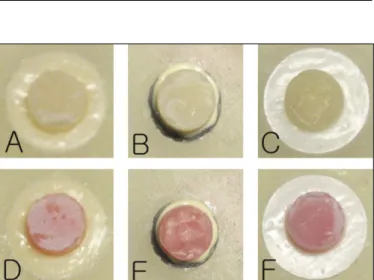
In order to obtain an equal bonding area, a glass cylindrical matrix with 6.0 mm inner-diameter and 3 mm thickness was used. The composite resin was applied in a thickness of 2.0 mm, and then cured for 20 seconds. After the glass matrix was removed, an additional 20 seconds of visible light was applied. As a result, 6 groups of specimens were fabricated as shown in Fig. 1.
Storage of specimens and measurement of shear bond strength
All specimens were stored in distilled water for 15 hours.
Then, the specimens were thermocycled between 5℃ and 55℃
for 1000 cycles with a 30 second-dwell time. After thermo- cycling, they were stored in 37℃ distilled water for an addi- tional 15 hours before being subjected to shear load. A uni- versal testing machine (Instron 3345, Instron Corp., Norwood, MA, USA) with a 10 kN load cell, a 0.5 mm/min crosshead speed and a flat-end apparatus was used to direct parallel shear- ing forces as close as possible to the resin/substrate interface.
The shear load in newtons at the point of failure was noted, and force was calculated in MPa. The mode of failure was recorded as being adhesive (failure at the substrate-resin interface), cohesive (failure within the substrate) or a combination (adhesive and cohesive). Photographs of all debonded spec- imens were taken using a digital camera (EOS 1000D, Canon, Tokyo, Japan).
Statistical analysis
Statistical analysis was performed using the SPSS statistical package (SPSS 17.0, SPSS, Chicago, IL, USA). Two-way analy- sis of variance was applied to detect any difference between groups, with shear bond strength as the dependent variable, the substrates of specimens and the types of composite resin as the independent factors. Multiple comparisons were made by Bonferroni post-hoc test to determine whether groups were sig- nificantly different. P values less than 0.05 were considered to be statistically significant in all tests.
RESULTS
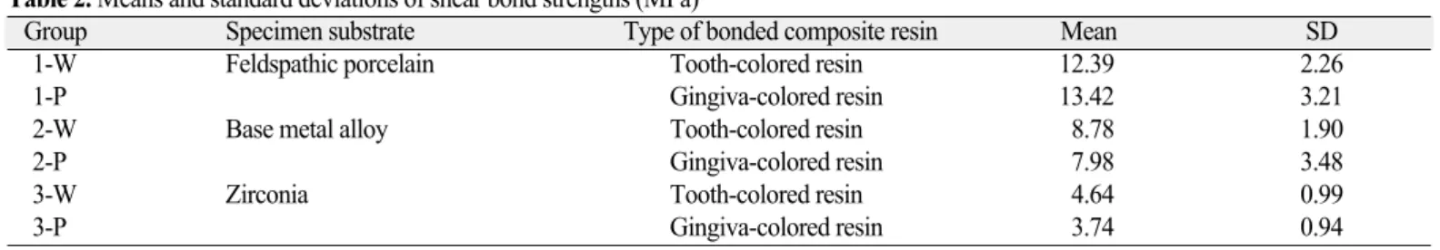
The shear bond strengths of all specimens were measured and the results of the shear bond strength tests are shown in Table 2 and Fig. 2.
The results of the two-way ANOVA test showed that the sub- strate of specimens significantly affected the shear bond strength between composite resin and the specimens regard- less of the types of composite resin (Table 3). According to the results of Bonferroni post-hoc test, the feldspathic porcelain specimens demonstrated the highest shear bond strength among all kinds of specimens and the specimens made from zirconia showed the lowest shear bond strength to composite resin (Table 4). However, for all types of specimens, there were no significant differences between the shear bond strength of tooth-colored resin and gingiva-colored resin.
In each type of specimens, both white and pink resin bond- ed specimens presented the same failure patterns. In the porcelain specimens, cohesive failures of the porcelain occurred in all specimens. In the base metal specimens, all debonded surfaces showed adhesive failures between the metal surfaces and the opaque. Lastly, all zirconia speci- mens also showed adhesive failures between the zirconia surfaces and the bonding agents (Fig. 3).
Table 1. System components of Ceramic Repair
Component Chemical composition
Total Etch 37% Phosphoric acid
Monobond S 3-methacryloxypropyl-trimthoxsilane Metal/Zirconia Primer Phosphoric acid acrylate and methacrylate
cross linking agents
Monopaque Dimethacrylates: Bis-GMA, urethane dimethacrylate and triethylene glycol dimethacrylate
Heliobond Bis-GMA, triethylene glycol dimethacrylate Tetric N-Ceram Light polymerizing hybrid composite
restorative material
Fig. 1. Prepared specimens prior to measurement of shear bond strength.
A: white resin bonded to porcelain (Group 1-W), B: white resin bonded to metal (Group 2-W), C: white resin bonded to zirconia (Group 3-W), D: pink resin bonded to porcelain (Group 1-P), E: pink resin bonded to metal (Group 2-P), F: pink resin bonded zirconia (Group 3-P).
DISCUSSION
The method to overlay the pink composite resin on the pink porcelain core has not been used for a long time since its introduction on the market, but has shown very predictable results.
When fabricating the artificial gingiva, using both the pink com- posite resin and the pink porcelain is much better than using only the pink porcelain in many aspects.11-13This hybrid tech- nique can facilitate a more biocompatible subgingival envi- ronment, and can have strong points to be easy for repair or main- tenance, as well as the excellent aesthetics.13In the event of the tooth extraction due to the severe periodontal disease, the peri- odontal tissue would remain unstable and the continuous additional bone loss would occur after extracting tooth and insert- ing the implant.7,10In this case, although the artificial gingiva has been formed with using the pink porcelain, there is still a
high possibility for the marginal integrity of the implant prostheses to be damaged due to the additional bone loss. The method of mounting the provisional restoration without aes- thetic concerns and waiting until the periodontal bone become stable also consumes too much time. In this case, the hybrid ceramic and composite artificial gingiva fabricated as the screw-retained type could be an excellent substitution.11-13It can continue the best esthetic results by repairing the implant prostheses with another add of tissue-colored composite resin, whenever needed.
Also, in case of fracture of the prostheses fabricated with the gingiva-colored porcelain, the pink composite resin can be use- ful for repairing it. Fracture and chipping of the ceramic restoration is one of the problems frequently occurred in the clinic.17Especially, the fracture on the labial surface of the implant prostheses at the anterior region has occurred frequently,18which Table 2. Means and standard deviations of shear bond strengths (MPa)
Group Specimen substrate Type of bonded composite resin Mean SD
1-W Feldspathic porcelain Tooth-colored resin 12.39 2.26
1-P Gingiva-colored resin 13.42 3.21
2-W Base metal alloy Tooth-colored resin 8.78 1.90
2-P Gingiva-colored resin 7.98 3.48
3-W Zirconia Tooth-colored resin 4.64 0.99
3-P Gingiva-colored resin 3.74 0.94
Table 3. Results of 2-way analysis of variance
Source of variation df Sum of squares Mean square F-value P value
Specimen type (A) 2 596.438 298.219 50.398 <.0001*
Resin type (B) 1 0.631 0.631 0.107 0.745
A*B 2 9.780 4.890 0.826 0.444
Error 54 266.278 5.917
Total 60 4750.616
*statistically significant P<.05
Fig. 2. Means and standard deviations of shear bond strengths (MPa) and results of Bonferroni post-hoc test. Vertical lines represent the standard deviations. *statistically significant P<.05
20 18 16 14 12 10 8 6 4 2 0
Shear bond strength (MPa)
porcelain metal zirconia Specimen type
White resin Pink resin
Fig. 3. Failure mode of specimens.
A: cohesive failure of feldspathic porcelain, B: adhesive failure in a base metal specimen, C: adhesive failure in a zirconia specimen.
Table 4. Results of Bonferroni post-hoc test for specimen type
Group P value
Porcelain vs Metal <.0001 *
Porcelain vs Zirconia <.0001 *
Metal vs Zirconia <.0001 *
*statistically significant P<.05
is caused from the brittleness of the ceramic as well as the unbear- able mechanical load due to patients’parafunctional habit or improper occlusion of the implant prostheses.17,19The gingva- colored porcelain is expected to receive relatively low mechan- ical load compared to the white porcelain because it has been mainly applied on the cervical area of implant prosthe- ses where the occlusal force is not applied directly. Thus, it is expected to cause less fracture compared to the area where the direct occlusal force is employed, like the functional cusp of the molar or the incisal edge of the incisor.20 However, mechanical stress could be transmitted from the occlusal surface to the cervical area or periodontal ligament and could cause fracture at cervical region.21,22In case of the implant pros- theses including the artificial gingiva, it could have an unde- sirable structure to endure the mechanical load as it was rel- atively lengthened, which could cause the fracture at the area of the artificial gingiva due to the similar mechanism with the cervical abfraction of the tooth. Also, the possibility that the fractured fragment includes the artificial gingiva area cannot be excluded because fracture of the ceramic restoration could occur from the occlusal to the cervical direction.19In this case, the immediate repair is required by the esthetic reason and the gingiva-colored composite resin could be very useful for it.
This study was done to evaluate the shear bond strength between the gingiva-colored composite resin and various base materials and to decide whether the shear bond strength is clinically sufficient. In order to make a meaningful decision, the clinically sufficient bond strength should be defined first.
However, as the study to determine the sufficient strength on the margin of the artificial gingiva has not been actively conducted in present, it is not easy to define the clinically suf- ficient strength. According to Behr et al., the maximum bite force in the anterior teeth is range from 150 N to 200 N. If the bond strength over 10 MPa can be obtained on the adhered sur- face, it would be regarded as the sufficient bond strength at the anterior region.23According to this standard; the pink composite resin has shown the sufficient bond strength only on the feldspathic porcelain, while it has shown insufficient on applying to the metal and zirconia. However, the conclusion that the bond strength between the pink composite and the met- al or zirconia could not have the clinically sufficient strength is not correct by the reasons shown as following. Firstly, the pink composite does not receive any direct bite forces because it is mainly applied only to the cervical area. Therefore, the mechanical stress that is applied on the adhered surfaces should be calculated by other method. Secondly, in the real clin- ical situations, it is possible to obtain the additional bond strength from the surface of the porcelain because the fractured surface includes not only the metal or zirconia but also the near porcelain in the most cases.
The feasibility of the pink composite resin is intended to be verified through the comparison to the ceramic repair method
using the tooth-colored composite resin, which is used by many clinicians and accepted as having clinically sufficient bond strength through many research works.15,16Considering this util- ity of the pink composite resin and the potential to be used expan- sively in future, the conclusions of this study that the pink com- posite has shown the similar bond strength to the white com- posite can be very meaningful. Moreover, if the adhesion surface can be enlarged by means of forming the bevel on the surface of the porcelain or preparing the retention groove on the surface, it is expected to obtain the bond strength as much as that of feldspathic porcelain.
Considering that the pink composite resin should be bond- ed on the pink porcelain in the clinical situations, the fact that the shear bond strength between the feldspathic porcelain and the pink composite resin has been shown as the highest among all types of specimens is very meaningful. On measuring the bond strength, all porcelain specimens demonstrated the cohesive failure of the feldspathic porcelain. This could be also the evidence that the bond strength between the composite resin and the feldspathic porcelain is satisfactory. In case of the met- al specimens, the major failure pattern was the adhesive fail- ure between the metal surface and the opaque. This result can be interpreted that the application of opaque weakened the adhe- sion between metal and composite resin. In case of repair of porcelain-fused-metal crowns with the white resin, the appli- cation of opaque is obligatory to prevent the reflection of the shade of underlying metal24,25 and it could result in weak- ened bond strength. However, in most clinical situation, it is possible to complement this weakened bond strength by obtaining additional adhesion from adjacent porcelain surface.
Thus, the result that the bond strength between the metal and the composite resin is a little lower does not mean that the composite resin cannot be applied when underlying metal struc- ture is exposed. In case of the ziconia specimens, the result that the shear bond strength has shown low is reasonably expect- ed. According to the results of this study, it is hardly expect- ed to obtain the sufficient bond strength in case of adhering the pink composite resin on the only-exposed surface of zirconia.
Although zirconia has been known as having less bond strength with the composite resin compared to other materi- als,23many studies has been recently conducted to overcome this weakness through various surface treatments. In this study, the surface of zirconia has been processed according to the manufacturer’s instruction, following the procedure which has been frequently used clinically, while the method to obtain the higher coherence has been introduced by the recent researches. The coherence between the zirconia and the resin is reported to be increased by the various methods including the application of phosphate monomer containing primers26,27 or silane containing primers,28,29the selective infiltration etch- ing techinique30and tribochemical silica coating.26,27,31With using these methods, it is expected to increase the bond strength
between the pink composite and zirconia.
This study has also an obvious limitation that the actual sit- uations inside the oral cavity could not be simulated as it is.
Although the 1000 cycles of thermo-cycling has been imple- mented to mimic the situations, it is not enough to simulate all the effects. Actually, if it has been exposed to the similar envi- ronment inside the oval cavity for a long time, or been through the repetitive thermo-cycling, the shear bond strength is reported to decrease.32,33Therefore, further in vivo study for a long-term basis is needed to determine the physical properies of the pink composite resin.
CONCLUSION
Within the limitation of this study, the following conclusions were drawn:
When adhered on feldspathic porcelain, metal, and zirconia, the shear bond strength of the gingiva-colored composite resin is comparable to that of the tooth-colored composite resin.
It is presumed that the best prognosis could be seen when gingival- colored composite resin is applied to feldspathic porcelain surface.
REFERENCES
1. Lesmes D, Laster Z. Innovations in dental implant design for cur- rent therapy. Oral Maxillofac Surg Clin North Am 2011;23:193-200.
2. Madan N, Pannu K. Restoration of maxillary anterior esthetics using lava all-ceramic fixed dental prostheses. Int J Comput Dent 2011;14:47-53.
3. Wall JG, Cipra DL. Alternative crown systems. Is the metal-ce- ramic crown always the restoration of choice? Dent Clin North Am 1992;36:765-82.
4. Davidowitz G, Kotick PG. The use of CAD/CAM in dentistry.
Dent Clin North Am 2011;55:559-70.
5. Cosyn J, Eghbali A, De Bruyn H, Collys K, Cleymaet R, De Rouck T. Immediate single-tooth implants in the anterior maxilla: 3-year results of a case series on hard and soft tissue response and aes- thetics. J Clin Periodontol 2011;38:746-53.
6. Chow YC, Wang HL. Factors and techniques influencing peri- implant papillae. Implant Dent 2010;19:208-19.
7. Evans CD, Chen ST. Esthetic outcomes of immediate implant placements. Clin Oral Implants Res 2008;19:73-80.
8. Miyamoto Y, Obama T. Dental cone beam computed tomography analyses of postoperative labial bone thickness in maxillary an- terior implants: comparing immediate and delayed implant placement. Int J Periodontics Restorative Dent 2011;31:215-25.
9. Kan JY, Rungcharassaeng K, Sclar A, Lozada JL. Effects of the facial osseous defect morphology on gingival dynamics after im- mediate tooth replacement and guided bone regeneration: 1-year results. J Oral Maxillofac Surg 2007;65:13-9.
10. De Rouck T, Collys K, Cosyn J. Immediate single-tooth implants in the anterior maxilla: a 1-year case cohort study on hard and soft tissue response. J Clin Periodontol 2008;35:649-57.
11. Coachman C, Salama M, Garber D, Calamita M, Salama H, Cabral G. Prosthetic gingival reconstruction in a fixed partial restora- tion. Part 1: introduction to artificial gingiva as an alternative ther- apy. Int J Periodontics Restorative Dent 2009;29:471-7.
12. Salama M, Coachman C, Garber D, Calamita M, Salama H, Cabral G. Prosthetic gingival reconstruction in the fixed partial restora- tion. Part 2: diagnosis and treatment planning. Int J Periodontics Restorative Dent 2009;29:573-81.
13. Coachman C, Salama M, Garber D, Calamita M, Salama H, Cabral G. Prosthetic gingival reconstruction in fixed partial restorations.
Part 3: laboratory procedures and maintenance. Int J Periodontics Restorative Dent 2010;30:19-29.
14. dos Santos JG, Fonseca RG, Adabo GL, dos Santos Cruz CA.
Shear bond strength of metal-ceramic repair systems. J Prosthet Dent 2006;96:165-73.
15. Haneda IG, Fonseca RG, de Almeida JG, Cruz CA, Adabo GL.
Shear bond strength of metal-ceramic repair systems. Gen Dent 2009;57:644-51.
16. Wolf DM, Powers JM, O’Keefe KL. Bond strength of composite to porcelain treated with new porcelain repair agents. Dent Mater 1992;8:158-61.
17. Ozcan M. Fracture reasons in ceramic-fused-to-metal restora- tions. J Oral Rehabil 2003;30:265-9.
18. Ozcan M, Niedermeier W. Clinical study on the reasons for and location of failures of metal-ceramic restorations and survival of repairs. Int J Prosthodont 2002;15:299-302.
19. Campos RE, Soares CJ, Quagliatto PS, Soares PV, de Oliveira OB Junior, Santos-Filho PC, Salazar-Marocho SM. In Vitro Study of Fracture Load and Fracture Pattern of Ceramic Crowns: A Finite Element and Fractography Analysis. J Prosthodont 2011;20:447-55.
20. Proos KA, Swain MV, Ironside J, Steven GP. Finite element analy- sis studies of a metal-ceramic crown on a first premolar tooth.
Int J Prosthodont 2002;15:521-7.
21. Farah JW, Craig RG. Distribution of stresses in porcelain- fused-to-metal and porcelain jacket crowns. J Dent Res 1975;54:
255-61.
22. Oyar P, Ulusoy M, Eskitascioglu G. Finite element analysis of stress distribution of 2 different tooth preparation designs in porce- lain-fused-to-metal crowns. Int J Prosthodont 2006;19:85-91.
23. Behr M, Proff P, Kolbeck C, Langrieger S, Kunze J, Handel G, Rosentritt M. The bond strength of the resin to zirconia interface using different bonding concepts. J Mech Behav Biomed Mater 2011;4:2-8.
24. Barreto MT, Bottaro BF. A practical approach to porcelain repair. J Prosthet Dent 1982;48:349-51.
25. Villarroel M, Fahl N, De Sousa AM, De Oliveira OB Jr. Direct esthetic restorations based on translucency and opacity of com- posite resins. J Esthet Restor Dent 2011;23:73-87.
26. Atsu SS, Kilicarslan MA, Kucukesmen HC, Aka PS. Effect of zirconium-oxide ceramic surface treatments on the bond strength to adhesive resin. J Prosthet Dent 2006;95:430-6.
27. May LG, Passos SP, Capelli DB, Ozcan M, Bottino MA, Valandro LF. Effect of silica coating combined to a MDP- based primer on the resin bond to Y-TZP ceramic. J Biomed Mater Res B Appl Biomater 2010;95:69-74.
28. Dias de Souza GM, Thompson VP, Braga RR. Effect of metal primers on microtensile bond strength between zirconia and resin cements. J Prosthet Dent 2011;105:296-303.
29. Kitayama S, Nikaido T, Takahashi R, Zhu L, Ikeda M, Foxton RM, Sadr A, Tagami J. Effect of primer treatment on bonding of resin cements to zirconia ceramic. Dent Mater 2010;26:426-32.
30. Aboushelib MN. Evaluation of zirconia/resin bond strength and interface quality using a new technique. J Adhes Dent 2011;13:255-60.
31. Chen L, Suh BI, Kim J, Tay FR. Evaluation of silica-coating tech- niques for zirconia bonding. Am J Dent 2011;24:79-84.
32. Blatz MB, Sadan A, Martin J, Lang B. In vitro evaluation of shear bond strengths of resin to densely-sintered high-purity zirconium- oxide ceramic after long-term storage and thermal cycling. J Prosthet Dent 2004;91:356-62.
33. Leibrock A, Degenhart M, Behr M, Rosentritt M, Handel G. In vit- ro study of the effect of thermo- and load-cycling on the bond strength of porcelain repair systems. J Oral Rehabil 1999;26:130-7.