© 2014 Korean Breast Cancer Society. All rights reserved. http://ejbc.kr | pISSN 1738-6756
INTRODUCTION
Whole-breast irradiation (WBI) has become one of the key components in breast conserving therapy. Breast cancer radio-
therapeutic methods are continuously updated as new com- prehensive therapeutic strategies for breast cancer are devel- oped. After breast conserving surgery, 80% to 90% of local re- currences occur at the site of the original tumor lesion and ad- jacent regions [1]. Therefore, some investigators have ques- tioned whether it is necessary and appropriate for all patients treated with breast conserving surgery to undergo WBI. As they have suggested, accelerated partial breast irradiation (APBI) has rapidly become the focus of clinical research.
External-beam APBI is one of the most cost-effective ther- apeutic approaches for APBI [2]. It reduces late-stage tissue fi- brosis and fat necrosis, thereby improving cosmetic outcomes compared with other irradiation modes (e.g., interstitial brachytherapy or intraoperative radiotherapy). However, ex- ternal-beam radiation may subject other healthy tissues, in- cluding breast tissue, to a moderate dose of irradiation [3].
Therefore, it is important to develop external-beam radiation techniques that are more conformal.
Accelerated Partial Breast Irradiation with Intensity-Modulated Radiotherapy Is Feasible for Chinese Breast Cancer Patients
Zhenyu He*, Sangang Wu1,*, Juan Zhou2, Fengyan Li, Jiayan Sun, Qin Lin, Huanxin Lin, Xunxing Guan
Department of Radiation Oncology, Sun Yat-Sen University Cancer Center, State Key Laboratory of Oncology in South China, Collaborative Innovation Center of Cancer Medicine, Guangzhou; Departments of 1Radiation Oncology and 2Obstetrics and Gynecology, Xiamen Cancer Center, The First Affiliated Hospital of Xiamen University, Xiamen, China
ORIGINAL ARTICLE
Purpose: Several accelerated partial breast irradiation (APBI) techniques are being investigated in patients with early-stage breast cancer. The present study evaluated the feasibility, early toxicity, initial efficacy, and cosmetic outcomes of accelerated partial breast intensity-modulated radiotherapy (IMRT) for Chi- nese female patients with early-stage breast cancer after breast- conserving surgery. Methods: A total of 38 patients met the inclu- sion criteria and an accelerated partial breast intensity-modulated radiotherapy (APBI-IMRT) plan was designed for each patient.
The prescription dose was 34 Gy in 10 fractions, 3.4 Gy per frac- tion, twice a day, in intervals of more than 6 hours. Results: Of the 38 patients, six patients did not meet the planning criteria. The remaining 32 patients received APBI-IMRT with a mean target volume conformity index of 0.67 and a dose homogeneity index of 1.06. The median follow-up time was 53 months and no local recurrence or distant metastasis was detected. The most com- mon acute toxicities observed within 3 months after radiotherapy
were erythema, breast edema, pigmentation, and pain in the ir- radiated location, among which 43.8%, 12.5%, 31.3%, and 28.1% were grade 1 toxicities, respectively. The most common late toxicities occurring after 3 months until the end of the follow- up period were breast edema, pigmentation, pain in the irradiat- ed location, and subcutaneous fibrosis, among which 6.2%, 28.1%, 21.9%, and 37.5% were grade 1 toxicities, respectively.
Thirty-one patients (96.8%) had fine or excellent cosmetic out- comes, and only one patient had a poor cosmetic outcome.
Conclusion: It is feasible for Chinese females to receive APBI- IMRT after breast conserving surgery. The radiotherapeutic tox- icity is acceptable, and both the initial efficacy and cosmetic outcomes are good.
Key Words: Breast neoplasms, Intensity-modulated radiotherapy, Segmental mastectomy
Correspondence to: Fengyan Li
Department of Radiation Oncology, Sun Yat-Sen University Cancer Center, State Key Laboratory of Oncology in South China, Collaborative Innovation Center of Cancer Medicine, Guangzhou, China
Tel: +86-20-87343543, Fax: +86-20-87343392 E-mail: [email protected]
*These authors contributed equally to this work as first authors.
This study was supported by a grant from the Sci-Tech Office of Guangdong Province (No. 2008B060600019), the Youth Foundation of the First Affiliated Hospital of Xiamen University (No. XYY2012005), the Education Scientific Research Project of Young Teachers in Fujian Province (No. JB13131) and Medical Scientific Research Foundation of Guangdong Province (No. A2010192).
Received: April 16, 2014 Accepted: July 26, 2014
Cancer
Intensity-modulated radiotherapy (IMRT) is one of the most rapidly growing therapeutic approaches in recent years.
Partial breast IMRT provides a more even and conformal dose coverage than partial breast three-dimensional confor- mal radiotherapy (3DCRT) or helical tomotherapy [4]. The volume of normal breast tissues irradiated by IMRT is smaller than that irradiated by 3DCRT [5]. Furthermore, IMRT can reduce the dose to the skin [6]. Our dosimetric comparison showed that accelerated partial breast IMRT (APBI-IMRT) could significantly reduce the cardiac radiation dosage com- pared with WBI [7]. In addition to exposure, it is important to note the cardiotoxicity of radiotherapy [8]. Therefore, patients may benefit more from IMRT as an external-beam APBI method due to its improved dose coverage and protection of normal tissues and organs. The present study focused on un- derstanding the feasibility of APBI-IMRT in Chinese female patients while investigating its initial cosmetic outcomes, tox- icity, and clinical efficacy.
METHODS
Patient selection
Chinese female patients treated with breast conserving sur- gery for early-stage breast cancer at Sun Yat-Sen University Cancer Center were recruited in this phase II study. The fol- lowing inclusion criteria were utilized for patient selection: fe- male; unilateral breast cancer; single lesion; undergoing breast conserving surgery; over 18 years of age; pathologically con- firmed as having infiltrating cancer (including duct cancer, medullary carcinoma, mucinous adenocarcinoma, and tubu- lar carcinoma); pathology-negative lymph nodes confirmed by axillary lymph node dissection or sentinel lymph node bi- opsy; American Joint Committee on Cancer (AJCC) stage I or II (pT1N0M0, pT2N0M0) with a primary tumor size ≤3 cm;
negative margins under the microscope (>1 cm); complete results including estrogen receptor (ER), progesterone recep- tor (PR), and human epidermal growth factor receptor family 2 (HER2); no small sporadic calcification in the local lesion,
as determined by ipsilateral breast postsurgical molybdenum target photography; no multicentric lesions, as determined by ipsilateral breast postsurgical magnetic resonance imaging;
and no contraindications for radiotherapy (i.e., severe heart disease, high blood pressure, rheumatic and immune diseases, or a history of other cancer). All patients and their family members provided signed informed consent, and the present study was approved by the ethics committee of Sun Yat-Sen University Cancer Center (approval number of Institutional Review Board: 2008B030301106).
Postural immobilization and computed tomography scanning Patients meeting the inclusion criteria were placed in the supine position on a postural immobilization bracket (Tho- rawedgeTM SIN-301080; Siemens, Munich, Germany). The median interval from surgery to computed tomography (CT) simulation was 4 months (range, 1–5 months). The scanning was performed using a Philips Medical Madison helix CT scanner (Philips, Amsterdam, The Netherlands) with the pa- tient breathing calmly. A metal marker was placed onto the patient’s body as a laser reference point for CT positioning.
Contiguous 5 mm CT axial images were obtained extending from the larynx to the upper abdomen, including the entire breasts and both lungs. After scanning, the imaging data were transferred into the Pinnacle 7.4f® (Philips Medical Systems, Milpitas, USA) treatment planning system for radiotherapy planning.
Delineation of the target volume and the involved organs Delineation of the surgical cavity
The surgical cavity was delineated under a CT window width of 500 and level of 0, in accordance with the following factors: the metal marker placed during operation; the residu- al seroma after operation; the interruption and density varia- tions of the breast glands in the CT images; and the surgical cavity shown by ultrasonography in the same position, includ- ing the distance between the surgical cavity or interruption of breast glands and certain fixed points on the skin; and the
A B C
Figure 1. Delineation of the surgical cavity (white arrows) based on surgical clips (A), seroma (B), and ultrasound (C).
depth, width and height of the cavity. The delineation of the surgical cavity was finally approved by three experienced ra- diation oncologists (Figure 1).
Delineation of the clinical target volume
The clinical target volume (CTV) was delineated by ex- panding the surgical cavity evenly by 10 mm with modifica- tion. The anterior boundary was located within 5 mm under the skin surface with the posterior boundary defined as the surface of the pectoral muscles.
Delineation of the planning target volume and the planning target volume for evaluation
The planning target volume (PTV) was delineated by ex- panding the CTV evenly by 10 mm. The PTV for evaluation (PTV_EVAL) was formed by modifying the PTV in accor- dance with the International Commission on Radiation Units (ICRU) 50 and ICRU62 reports, with the anterior boundary at 5 mm under the chest skin and the bottom not exceeding the thorax.
Delineation of the involved organs
The bilateral lungs, heart, and thyroid were delineated on each slice of the CT image. The ipsilateral and contralateral breasts were contoured including all of the breast tissue in the standard whole breast tangential fields from the inframam- mary fold to the clavicle in the cranial–caudal direction, with the anterior boundary at 5 mm under the skin and the bottom on the chest wall. External marker wires were placed to indi- cate the clinical expectations of the external beam tangential field borders as a guide.
Design and evaluation of the IMRT plan
The Pinnacle 7.4f ® treatment planning system was used for the design of the IMRT plan with a 6 MV photon beam. The prescription dose was 34 Gy/10 fractions/5 days (3.4 Gy/frac- tions, twice a day) with an interdose interval of more than 6 hours. The design adapted direct machine parameter optimi- zation with five coplanar or noncoplanar fields. A static multi- leaf collimator was used to modulate the intensity of the com- plete irradiation in each subfield through the step-and-shoot method. The limitation parameters were calculated and ad- justed in accordance with the actual dose to reach the desired dose coverage in the target volume while limiting the dose to the involved organs within the designated range. For each pa- tient, the IMRT dosage was evaluated using the dose-volume histogram (DVH) in accordance with the Radiation Therapy Oncology Group (RTOG)-0319 evaluation criteria [9]. The evaluation criteria for normal organs are listed as follows. For
the ipsilateral breast, <25% of the breast volume received the prescription dose, and <50% of the breast volume received
≥50% of the prescription dose. For the contralateral breast, the maximum point dose was <3% of the prescription dose.
For the ipsilateral lung, <10% of the lung tissue received 5%
of the prescription dose. For the heart, in cases of right breast cancer, <10% of the heart tissue received 5% of the prescrip- tion dose, and for cases of left breast cancer, the heart volume receiving 5% of the prescription dose was smaller than that receiving the same dose with conventional whole-breast tan- gent field irradiation. For the thyroid, the maximum point dose was <3% of the prescription dose.
Quality assurance evaluation
1) No variations (total coverage): 95% of the isodose surface covered 100% of the PTV_EVAL. All limits of the specified critical normal tissue DVH were met. 2) Minor variations (marginal coverage): 95% of the isodose surface coverage was between 95% and 100% of the PTV_EVAL. There was no portion of the PTV_EVAL receiving <93% of the prescrip- tion (isocenter) dose. All limits of the specified critical normal tissue DVH fell within 5% of the guidelines. 3) Major varia- tions (missing): 95% of the isodose surface covered <95% of the PTV_EVAL. A portion of the PTV_EVAL received <93%
of the prescription (isocenter) dose.
Plan implementation and follow-up
Treatment plans evaluated as having ‘no variations’ and ‘mi- nor variations’ were clinically implemented, and patients whose plans were evaluated as having ‘major variations’ were withdrawn and those patients were treated with WBI.
The patients were evaluated once per day during the treat- ment period for radiotherapy-induced acute toxicity. Patients continued to be evaluated at 1 week, 1 month, 2 months, and 3 months posttreatment. After 3 months, patients were evalu- ated every 3 months in the first year and every 6 months in the second year for postradiotherapy toxicity, tumor recur- rence, and metastasis. Chest radiology and breast ultrasonog- raphy or molybdenum target photography were performed periodically (generally once per year). Follow-up ended on April 1, 2014.
Calculation of the conformity index and homogeneity index The conformity index (CI)=VTref/VT×VTref/VRef, where VTref represents the target volume covered by the isodose, VT represents the target volume, and VRef represents the total vol- ume covered by 95% of the isodose. The CI range was 0–1, in which the conformity was better when the CI value was larger.
The homogeneity index (HI)=D5/D95, where D5 represents
the irradiation dose received by 5% of the PTV-EVAL volume and D95 represents the irradiation dose received by 95% of the PTV-EVAL. The closer to 1 the HI value is, the better the target uniformity will be.
Toxicity evaluation and cosmetic outcomes
The Harris criteria (1979) were used for the evaluation of breast cosmetic outcomes [10]. The National Cancer Institute’s Common Terminology Criteria for Adverse Events (CTCAE, version 3.0) were used for the evaluation of acute and late tox- icity [11]. We evaluated radiotherapy-induced toxicity that arose immediately posttreatment until the end of the last day of follow-up. The obtained values represented the most seri- ous toxicity in patients within these set periods.
RESULTS
Study accrual
A total of 38 patients meeting the inclusion criteria between October 2008 and October 2011 were selected in IMRT plan- ning. Six of the 38 patients (15.7%) were off-protocol due to planning criteria violations and were treated with WBI; the
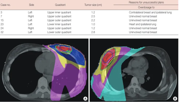
details of the dose constraints of the unsuccessful plans are shown in Table 1 and Figure 2. Therefore, 32 patients received APBI-IMRT.
Clinical characteristics and dosimetric features
The clinical pathological information is provided in Table 2.
The median age at onset was 43 years (range, 26–65 years).
Tumors arose in the upper outer quadrant in 20 of 32 patients (62.5%). Axillary lymph node dissection was performed in 23 of 32 patients (71.9%), with a median number of 15 (range, 5–32) dissected lymph nodes, and 9 of 32 (28.1%) underwent sentinel lymph node biopsy, with a median of 3 (range, 2–5) examined lymph nodes. Of the patients, 28 of 32 (87.5%) re- ceived chemotherapy with anthracycline and/or taxane regi- mens, and the median number of cycles was 6 (range, 6–8 cy- cles). All ER- and PR-positive patients received endocrine therapy. None of the HER2-positive patients were treated with trastuzumab.
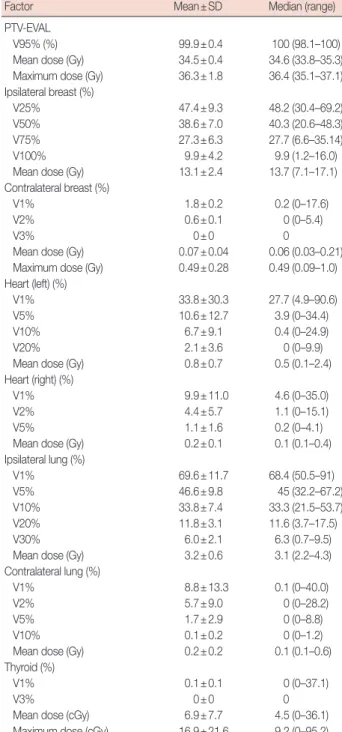
The target volume and normal tissue volume, as well as the doses for the 32 patients in this study are shown in Tables 3 and 4. Among them, 29 patients received coplanar field irra- diation, and three patients received noncoplanar field irradia- Table 1. Reasons for major variations of six patients
Case no. Side Quadrant Tumor size (cm) Reasons for unsuccessful plans
Overdosage to
3 Left Upper inner quadrant 1.2 Contralateral breast and ipsilateral lung
7 Right Upper outer quadrant 2.5 Uninvolved normal breast
15 Left Upper outer quadrant 2.2 Uninvolved normal breast
23 Left Lower inner quadrant 1.0 Heart and ipsilateral lung
24 Right Upper outer quadrant 1.2 Uninvolved normal breast
32 Left Lower outer quadrant 2.8 Uninvolved normal breast
A B
Figure 2. Dose distribution of two patients off-protocol. (A) Case 23 with tumor located in low inner quadrant, dose of heart and ipsilateral lung is in excess of the evaluation criteria of RTOG-0319. (B) Case 24 with tumor located in upper outer quadrant, dose of uninvolved normal breast is in ex- cess of the evaluation criteria of RTOG-0319.
Table 2. Clinical information on 32 patients
Factor No. (%)
Age (yr)* 43 (26–65)
Menstrual state
Premenopause 26 (81.3)
Postmenopause 6 (18.7)
Left and right side of lump
Left side 14 (43.7)
Right side 18 (56.3)
pT-staging
pT1 23 (71.9)
pT2† 9 (28.1)
Tumor location
Upper inner quadrant 7 (21.9)
Upper outer quadrant 20 (62.5)
Lower inner quadrant 0
Lower outer quadrant 5 (15.6)
Pathological classification
Infiltrating duct cancer 26 (81.3)
Medullary carcinoma 1 (3.1)
Mucinous adenocarcinoma 3 (9.4)
Tubular carcinoma 1 (3.1)
Local malignancy of intraductal papillar 1 (3.1) pN-stage
pN0 32 (100)
pN1 0
ER/PR state
Negative 3 (9.4)
Positive 29 (90.6)
HER2
Negative 27 (84.4)
Positive 5 (15.6)
Axillary lymph node treatment
Sentinel lymph node biopsy 9 (28.1)
Axillary lymph node dissection 23 (71.9) Chemotherapy
Yes 28 (87.5)
No 4 (12.5)
Endocrine therapy
Yes 29 (90.6)
No 3 (9.4)
ER=estrogen receptor; PR=progesterone receptor; HER2=human epidermal growth factor receptor 2.
*Median (range); †Tumor size ≤3 cm.
tion. The median interval from surgery to IMRT was 4 months (range, 1–5 months). The treatment planning for 18 patients was determined to have no variations, and for 14 pa- tients, it was determined to have minor variations. The aver- age target volume CI was 0.67 (range, 0.49–0.78) and the aver- Table 3. Volumes of target and normal tissues in 32 patients
Target volume and normal tissues
Volume (cm3)
Mean±SD Median (range)
Surgical cavity 4.9±3.4 5.0 (0.9–15.0)
CTV 31.3±17.0 32.6 (6.8–67.1)
PTV-EVAL 98.8±41.3 89.1 (19.3–164.3)
Ipsilateral lung 1,285.3±258.5 1,283.9 (809.8–1,779.5) Contralateral lung 1,238.5±269.7 1,255.3 (654.2–1,983.1)
Heart 498.2±57.6 500.1 (364.1–613.3)
Ipsilateral breast 496.8±258.9 425.2 (216.9–1,310.5) Contralateral breast 512.6±266.5 414.5 (251.4–1,467.6)
Thyroid 10.4±3.6 10.2 (5.2–18.6)
SD=standard deviation; CTV=clinical target volume; PTV-EVAL=planning tar- get volume for evaluation.
Table 4. Dose parameters of target volume and normal tissues in 32 patients treated with APBI-IMRT
Factor Mean±SD Median (range)
PTV-EVAL
V95% (%) 99.9±0.4 100 (98.1–100)
Mean dose (Gy) 34.5±0.4 34.6 (33.8–35.3)
Maximum dose (Gy) 36.3±1.8 36.4 (35.1–37.1) Ipsilateral breast (%)
V25% 47.4±9.3 48.2 (30.4–69.2)
V50% 38.6±7.0 40.3 (20.6–48.3)
V75% 27.3±6.3 27.7 (6.6–35.14)
V100% 9.9±4.2 9.9 (1.2–16.0)
Mean dose (Gy) 13.1±2.4 13.7 (7.1–17.1)
Contralateral breast (%)
V1% 1.8±0.2 0.2 (0–17.6)
V2% 0.6±0.1 0 (0–5.4)
V3% 0±0 0
Mean dose (Gy) 0.07±0.04 0.06 (0.03–0.21) Maximum dose (Gy) 0.49±0.28 0.49 (0.09–1.0) Heart (left) (%)
V1% 33.8±30.3 27.7 (4.9–90.6)
V5% 10.6±12.7 3.9 (0–34.4)
V10% 6.7±9.1 0.4 (0–24.9)
V20% 2.1±3.6 0 (0–9.9)
Mean dose (Gy) 0.8±0.7 0.5 (0.1–2.4)
Heart (right) (%)
V1% 9.9±11.0 4.6 (0–35.0)
V2% 4.4±5.7 1.1 (0–15.1)
V5% 1.1±1.6 0.2 (0–4.1)
Mean dose (Gy) 0.2±0.1 0.1 (0.1–0.4)
Ipsilateral lung (%)
V1% 69.6±11.7 68.4 (50.5–91)
V5% 46.6±9.8 45 (32.2–67.2)
V10% 33.8±7.4 33.3 (21.5–53.7)
V20% 11.8±3.1 11.6 (3.7–17.5)
V30% 6.0±2.1 6.3 (0.7–9.5)
Mean dose (Gy) 3.2±0.6 3.1 (2.2–4.3)
Contralateral lung (%)
V1% 8.8±13.3 0.1 (0–40.0)
V2% 5.7±9.0 0 (0–28.2)
V5% 1.7±2.9 0 (0–8.8)
V10% 0.1±0.2 0 (0–1.2)
Mean dose (Gy) 0.2±0.2 0.1 (0.1–0.6)
Thyroid (%)
V1% 0.1±0.1 0 (0–37.1)
V3% 0±0 0
Mean dose (cGy) 6.9±7.7 4.5 (0–36.1)
Maximum dose (cGy) 16.9±21.6 9.2 (0–95.2) APBI-IMRT =accelerated partial breast intensity-modulated radiotherapy;
SD=standard deviation; PTV-EVAL=planning target volume for evaluation;
Vx%=tissue volume which is ≥x% prescription dose.
gery to test the toxicity, efficacy, and cosmetic outcomes of the procedure. Our results show that APBI-IMRT achieved better target volumes and dose conformity, good cosmetic out- comes, and clinical efficacy as evidenced in the short-term follow-up, with only mild toxicity.
The cosmetic outcomes from APBI are closely correlated with the irradiation volume [12]. Women from Eastern coun- tries have significantly smaller breasts than women from Western countries [13,14]. Therefore, an irradiation mode with better conformity should improve the cosmetic outcomes in breast cancer patients receiving APBI.
In the present study, through the end of the follow-up peri- od, no local recurrences or distant metastases were found in any of the 32 patients. However, since the median follow-up period was only 53 months, it was premature to make conclu- sions regarding long-term clinical efficacy. A previous study of 199 patients receiving APBI (interstitial brachytherapy) and 199 patients receiving WBI, with a median follow-up period of 9.6 years reported local recurrence rates within 10 years of 5% among those receiving APBI and 4% among those receiv- ing WBI (p=0.48) [15]. When the median follow-up period was extended to 14.5 years, there was no difference in the local recurrence between WBI and APBI (3.8% vs. 5.0%, p=0.40) [16]. Polgár et al. [17] investigated 128 patients receiving APBI (interstitial brachytherapy or local electron beam irradiation) and 130 patients receiving WBI from 1998 to 2004, with a me- dian follow-up period of 66 months. They reported 5-year local recurrence rates of 4.7% for APBI and 3.4% for WBI (p=
0.50) and overall survival rates of 94.6% and 91.8% (p>0.05), respectively. Among these patients, 77.6% and 62.9% of the subjects had fine or excellent cosmetic outcomes (p=0.009), respectively. Taken together, these reports demonstrated the potential superior efficacy of radiotherapy and that good cos- metic outcomes could be achieved with APBI after breast conserving surgery. However, large phase III clinical trials are needed to further confirm the clinical efficacy.
In the present study, the acute toxicity and late toxicity were Table 5. Acute toxicity within 3 months and late toxicity until end of follow-up period after radiotherapy
Toxicity Grade of acute toxicity, No. (%) Grade of late toxicity, No. (%)
0 1 2 3 0 1 2 3
Erythema 18 (56.2) 14 (43.8) 0 0 30 (93.8) 2 (6.2) 0 0
Xerodermia 30 (93.8) 2 (6.2) 0 0 32 (100.0) 0 0 0
Breast edema 28 (87.5) 4 (12.5) 0 0 30 (93.8) 2 (6.2) 0 0
Skin pigmentation 22 (68.2) 10 (31.3) 0 0 23 (71.9) 9 (28.1) 0 0
Angiotelectasis 32 (100.0) 0 0 0 32 (100.0) 0 0 0
Pain in irradiated location 23 (71.9) 9 (28.1) 0 0 25 (78.1) 7 (21.9) 0 0
Radiation pneumonia 32 (100.0) 0 0 0 32 (100.0) 0 0 0
Fat necrosis 32 (100.0) 0 0 0 32 (100.0) 0 0 0
Subcutaneous fibrosis - - - - 20 (62.5) 12 (37.5) 0 0
age dose HI was 1.06 (range, 1.02–1.12). The average ratio be- tween the PTV-EVAL volume and the ipsilateral breast vol- ume was 0.20 (range, 0.07–0.45), and the ratio in six patients was >0.25 (part of the PTV_EVAL target volume was located outside of breast tissues). Three patients with a PTV-EVAL to ipsilateral breast volume ratio of >0.25 were determined to have no variations, and the other three patients were deter- mined to have minor variations.
Follow-up and efficacy
For all patients, follow-up started from the first day of sur- gery. One patient was lost to follow-up after 1 year. The overall follow-up rate was 97%, and the median period was 53 months (range, 12–64 months). Through the end of the fol- low-up period, no patients had local recurrence or distant metastasis, and both the local control rate and the overall sur- vival rate were 100%.
Radiotherapeutic toxicity and cosmetic outcomes
The most acute toxic reactions within 3 months of radio- therapy were erythema, breast edema, pigmentation, and pain in the irradiated location, among which 43.8%, 12.5%, 31.3%, and 28.1%, respectively, were grade 1 reactions. Most late tox- icities occurring by the end of the follow-up period were breast edema, pigmentation, pain in the irradiated location, and sub- cutaneous fibrosis, among which 6.2%, 28.1%, 21.9%, and 37.5%, respectively, were grade 1 reactions (Table 5).
Of the 32 patients, only one patient (3.2%) had a poor cos- metic outcome. All of the remaining 31 patients (96.8%) had a fine or excellent cosmetic outcome; of these, 22 patients (68.7%) were assessed as excellent and nine patients were (28.1%) as- sessed as fine.
DISCUSSION
In the present study, we performed APBI-IMRT in Chinese female patients who underwent with breast conserving sur-
mainly erythema, breast edema, pigmentation, pain in the ir- radiated location, and subcutaneous fibrosis; no reactions of grade 2 or higher were identified. Most patients had excellent cosmetic outcomes with a relatively short follow-up period. A study has also demonstrated that the incidence of early tox- icity (grade 1 and 2) after APBI-IMRT was significantly lesser than that after WBI (5.8% vs. 41%) [18]. Likewise, another study investigated 36 patients treated with APBI-IMRT and found that the most common acute toxicities (grade 1 and 2) were erythema, pigmentation, and pain in the breast or chest wall. No grade 3 or 4 toxicities were reported, while cosmetic outcomes were considered “excellent” or “good” by 94% of pa- tients and 97% of physicians [19]. Leonard et al. [20] investi- gated 55 patients treated with APBI-IMRT, with a median fol- low-up period of 10 months. In total, 98.2% of their subjects had fine or excellent cosmetic outcomes. Twenty patients re- ported grade 1–2 breast pain, two patients reported grade 1 breast edema, and one patient reported angiotelectasis.
Theoretically, IMRT has advantages in APBI. However, Jagsi et al. [12] reported that among 34 patients who received AP- BI-IMRT under active breathing control with 38.5 Gy/10 frac- tions and a median follow-up period of 2.5 years, seven pa- tients (21%) showed unacceptable cosmetic outcomes, which were the poorest reported outcomes in recent years. These re- sults indicated that there was strong correlation between cos- metic outcomes and normal breast irradiation volumes. The acceptable and unacceptable cosmetic outcomes for whole- breast V50 were 34.6% and 46.1% (p=0.02), and for whole- breast V100, they were 15.5% and 23.0% (p=0.02), respec- tively. Since there was a significant correlation in APBI be- tween normal breast irradiation volume and cosmetic out- comes, it is necessary to impose strict limitations on breast doses. IMRT provides more conformal protection for sur- rounding organs at risk in the target area than 3DCRT. With the help of technologies like active breathing control and im- age guide radiation therapy in the future, IMRT may be more beneficial for patients.
Currently, the indications for APBI have not been defined.
Both the American Society of Radiation Oncology (ASTRO) and the Groupe Européen de Curiethérapie and the European Society for Radiotherapy and Oncology (GEC-ESTRO) have suggested that some patients can be considered for APBI [21, 22]. Their suggestion focuses mainly on patients at a low-risk for recurrence. Indications for ABPI remain controversial in patients with early-stage breast cancer after breast conserving surgery [23]. We consider the indications suggested by ASTRO and GEC-ESTRO to be good references for screening patients to receive APBI. However, in the present study, only 3.1% (one patient >60 years of age) of patients were suitable to receive
APBI when we followed the age criteria suggested by ASTRO.
This could be because the peak age of breast cancer occurrence in Chinese females is 10 years earlier than that in Western fe- males due to the differences in the age of onset between East- ern and Western women. More studies with long-term follow- up are required to determine whether age will make any dif- ference in the prognosis of Chinese females treated with APBI.
Furthermore, the doses used in external-beam partial breast irradiation remain controversial. The reported doses in most patients were 25 to 40 Gy, and a single dose was 3.4 to 6.0 Gy [17,24-27]. However, using radiobiological calculations, the most optimized single dose should be 3.82 Gy [28]. Therefore, the recommended dose was 38.5 Gy/10 fractions, twice a day.
The dose mode was 34 Gy/10 fractions in our previous studies on partial breast 3DCRT and the primary results showed that this was a safe and effective dose [14]. Therefore, a dose of 34 Gy/10 fractions was repeatedly used in the present study. A subsequent clinical trial with stepwise dose escalation is under consideration for future studies.
In the present study, 78.1% patients had outer quadrant le- sions and the contralateral breast was exposed to a low dose of radiation; in a patient with a tumor located in the upper inner quadrant, the APBI-IMRT plan was assessed as having major variations due to overdosage to the contralateral breast. The contralateral breast dose and the incidence of secondary breast cancer following contralateral breast irradiation should be considered and compared between IMRT and convention- al whole breast irradiation; in a dosimetric study with WBI, the IMRT technique did not increase the out-of-field dose in the contralateral breast compared to conventional techniques, which should ultimately translate into a lower secondary breast cancer risk [29]. However, while breast cancer patients may live longer with the improvement of adjuvant treatment, radiotherapy-induced contralateral breast cancer still cannot be ignored. The doses of radiation to contralateral breast should be minimized when using the IMRT technique.
We recognize that there were limitations to our present study. The sample size was small, and a relatively short dura- tion of follow-up was used. In addition, since the respiratory gating system was not employed, the dosimetry might be in- fluenced by the individual’s respiration.
In conclusion, the present study demonstrates that acceler- ated partial breast IMRT is feasible for Chinese female pa- tients after breast conserving surgery with mild toxicity, good clinical efficacy, and superior cosmetic outcomes. The results from additional studies with a larger sample size and longer follow-up period are needed to further confirm its efficacy and long-term radiotherapeutic toxicity in Chinese female patients.
CONFLICT OF INTEREST
The authors declare that they have no competing interests.
REFERENCES
1. Veronesi U, Marubini E, Mariani L, Galimberti V, Luini A, Veronesi P, et al. Radiotherapy after breast-conserving surgery in small breast carci- noma: long-term results of a randomized trial. Ann Oncol 2001;12:
997-1003.
2. Sher DJ, Wittenberg E, Suh WW, Taghian AG, Punglia RS. Partial- breast irradiation versus whole-breast irradiation for early-stage breast cancer: a cost-effectiveness analysis. Int J Radiat Oncol Biol Phys 2009;74:440-6.
3. Njeh CF, Saunders MW, Langton CM. Accelerated Partial Breast Irradi- ation (APBI): a review of available techniques. Radiat Oncol 2010;5:90.
4. Oliver M, Chen J, Wong E, Van Dyk J, Perera F. A treatment planning study comparing whole breast radiation therapy against conformal, IMRT and tomotherapy for accelerated partial breast irradiation. Ra- diother Oncol 2007;82:317-23.
5. Rusthoven KE, Carter DL, Howell K, Kercher JM, Henkenberns P, Hunter KL, et al. Accelerated partial-breast intensity-modulated radio- therapy results in improved dose distribution when compared with three-dimensional treatment-planning techniques. Int J Radiat Oncol Biol Phys 2008;70:296-302.
6. Saibishkumar EP, MacKenzie MA, Severin D, Mihai A, Hanson J, Daly H, et al. Skin-sparing radiation using intensity-modulated radiotherapy after conservative surgery in early-stage breast cancer: a planning study.
Int J Radiat Oncol Biol Phys 2008;70:485-91.
7. Wu S, He Z, Guo J, Li F, Lin Q, Guan X. Dosimetric comparison of nor- mal structures associated with accelerated partial breast irradiation and whole breast irradiation delivered by intensity modulated radiotherapy for early breast cancer after breast conserving surgery. Clin Transl On- col 2014;16:69-76.
8. Darby SC, Ewertz M, McGale P, Bennet AM, Blom-Goldman U, Brøn- num D, et al. Risk of ischemic heart disease in women after radiothera- py for breast cancer. N Engl J Med 2013;368:987-98.
9. Vicini F, Winter K, Straube W, Wong J, Pass H, Rabinovitch R, et al. A phase I/II trial to evaluate three-dimensional conformal radiation ther- apy confined to the region of the lumpectomy cavity for Stage I/II breast carcinoma: initial report of feasibility and reproducibility of Radiation Therapy Oncology Group (RTOG) Study 0319. Int J Radiat Oncol Biol Phys 2005;63:1531-7.
10. Harris JR, Levene MB, Svensson G, Hellman S. Analysis of cosmetic re- sults following primary radiation therapy for stages I and II carcinoma of the breast. Int J Radiat Oncol Biol Phys 1979;5:257-61.
11. Common terminology criteria for ad verse events v3.0 (CTCAE). 2006.
National Cancer Institute. http://ctep.cancer.gov/protocolDevelopment/
electronic_applications/docs/ctcaev3.pdf. Accessed May 21st, 2008.
12. Jagsi R, Ben-David MA, Moran JM, Marsh RB, Griffith KA, Hayman JA, et al. Unacceptable cosmesis in a protocol investigating intensity- modulated radiotherapy with active breathing control for accelerated partial-breast irradiation. Int J Radiat Oncol Biol Phys 2010;76:71-8.
13. Hepel JT, Tokita M, MacAusland SG, Evans SB, Hiatt JR, Price LL, et al.
Toxicity of three-dimensional conformal radiotherapy for accelerated partial breast irradiation. Int J Radiat Oncol Biol Phys 2009;75:1290-6.
14. Li FY, He ZY, Xue M, Chen LX, Wu SG, Guan XX. Feasibility and acute toxicity of 3-dimensional conformal external-beam accelerated partial- breast irradiation for early-stage breast cancer after breast-conserving surgery in Chinese female patients. Chin Med J (Engl) 2011;124:1305-9.
15. Antonucci JV, Wallace M, Goldstein NS, Kestin L, Chen P, Benitez P, et al. Differences in patterns of failure in patients treated with accelerated partial breast irradiation versus whole-breast irradiation: a matched- pair analysis with 10-year follow-up. Int J Radiat Oncol Biol Phys 2009;74:447-52.
16. Shah C, Antonucci JV, Wilkinson JB, Wallace M, Ghilezan M, Chen P, et al. Twelve-year clinical outcomes and patterns of failure with acceler- ated partial breast irradiation versus whole-breast irradiation: results of a matched-pair analysis. Radiother Oncol 2011;100:210-4.
17. Polgár C, Fodor J, Major T, Németh G, Lövey K, Orosz Z, et al. Breast- conserving treatment with partial or whole breast irradiation for low- risk invasive breast carcinoma: 5-year results of a randomized trial. Int J Radiat Oncol Biol Phys 2007;69:694-702.
18. Livi L, Buonamici FB, Simontacchi G, Scotti V, Fambrini M, Compag- nucci A, et al. Accelerated partial breast irradiation with IMRT: new technical approach and interim analysis of acute toxicity in a phase III randomized clinical trial. Int J Radiat Oncol Biol Phys 2010;77:509-15.
19. Lewin AA, Derhagopian R, Saigal K, Panoff JE, Abitbol A, Wieczorek DJ, et al. Accelerated partial breast irradiation is safe and effective using intensity-modulated radiation therapy in selected early-stage breast cancer. Int J Radiat Oncol Biol Phys 2012;82:2104-10.
20. Leonard C, Carter D, Kercher J, Howell K, Henkenberns P, Tallhamer M, et al. Prospective trial of accelerated partial breast intensity-modulated radiotherapy. Int J Radiat Oncol Biol Phys 2007;67:1291-8.
21. Smith BD, Arthur DW, Buchholz TA, Haffty BG, Hahn CA, Harden- bergh PH, et al. Accelerated partial breast irradiation consensus state- ment from the American Society for Radiation Oncology (ASTRO).
Int J Radiat Oncol Biol Phys 2009;74:987-1001.
22. Polgár C, Van Limbergen E, Pötter R, Kovács G, Polo A, Lyczek J, et al.
Patient selection for accelerated partial-breast irradiation (APBI) after breast-conserving surgery: recommendations of the Groupe Européen de Curiethérapie-European Society for Therapeutic Radiology and Oncology (GEC-ESTRO) breast cancer working group based on clini- cal evidence (2009). Radiother Oncol 2010;94:264-73.
23. Vicini F, Arthur D, Wazer D, Chen P, Mitchell C, Wallace M, et al. Limi- tations of the American Society of Therapeutic Radiology and Oncolo- gy Consensus Panel guidelines on the use of accelerated partial breast irradiation. Int J Radiat Oncol Biol Phys 2011;79:977-84.
24. Taghian AG, Kozak KR, Doppke KP, Katz A, Smith BL, Gadd M, et al.
Initial dosimetric experience using simple three-dimensional confor- mal external-beam accelerated partial-breast irradiation. Int J Radiat Oncol Biol Phys 2006;64:1092-9.
25. Formenti SC, Rosenstein B, Skinner KA, Jozsef G. T1 stage breast can- cer: adjuvant hypofractionated conformal radiation therapy to tumor bed in selected postmenopausal breast cancer patients: pilot feasibility study. Radiology 2002;222:171-8.
26. Bourgier C, Pichenot C, Verstraet R, El Nemr M, Heymann S, Biron B, et al. Early side effects of three-dimensional conformal external beam accelerated partial breast irradiation to a total dose of 40 Gy in one week
(a phase II trial). Int J Radiat Oncol Biol Phys 2011;81:1228-35.
27. Vicini F, Winter K, Wong J, Pass H, Rabinovitch R, Chafe S, et al. Initial efficacy results of RTOG 0319: three-dimensional conformal radiation therapy (3D-CRT) confined to the region of the lumpectomy cavity for stage I/ II breast carcinoma. Int J Radiat Oncol Biol Phys 2010;77:1120-7.
28. Cuttino LW, Todor D, Pacyna L, Lin PS, Arthur DW. Three-dimension- al conformal external beam radiotherapy (3D-CRT) for accelerated partial breast irradiation (APBI): what is the correct prescription dose?
Am J Clin Oncol 2006;29:474-8.
29. Joosten A, Matzinger O, Jeanneret-Sozzi W, Bochud F, Moeckli R. Eval- uation of organ-specific peripheral doses after 2-dimensional, 3-di- mensional and hybrid intensity modulated radiation therapy for breast cancer based on Monte Carlo and convolution/superposition algo- rithms: implications for secondary cancer risk assessment. Radiother Oncol 2013;106:33-41.