DOI : 10.3341/jkos.2009.50.4.594
접수번호 : 09-22
트리암시놀론이 배양망막상피세포에서 생산된 혈관형성관련인자에 미치는 영향
이영창1⋅윤태중2⋅최광주1⋅김대현1 조선대학교 의과대학 안과학교실1, 아이안과2
목적: 트리암시놀론이 배양인체망막색소상피세포에서 생산된 혈관형성관련인자에 어떠한 영향을 미치는지 알아보고자 하였다.
대상과 방법: 트리암시놀론을 저산소환경에서 배양된 인체망막색소상피세포에 노출시킨 후 VEGF, PEDF 발현과 생산을 RT-PCR과 Western blot으로 조사하였다. 또한 배양상층액을 이용하여 맥관형성과 세포이동능력을 알아보았다.
결과: 트리암시놀론에 노출된 망막색소상피세포에서 VEGF의 유전자 발현은 감소하였고 PEDF의 유전자 발현은 변화를 보이지 않았다 (p<0.05). 또한 VEGF의 단백질 생산은 감소하였고 PEDF의 단백질 생산은 변화가 없었다(p<0.05). 맥관형성과 세포이동능력은 감소 되었다(p<0.05).
결론: 이러한 결과는 트리암시놀론이 망막색소상피의 혈관형성관련인자분비에 영향을 미쳐서 혈관신생을 억제할 수 있다는 것을 보여 준다.
<대한안과학회지 2009;50(4):594-602>
■ 접 수 일: 2008년 9월 23일 ■ 심사통과일: 2008년 11월 26일
■ 통 신 저 자: 김 대 현
광주시 동구 서석동 588 조선대학교병원 안과
Tel: 062-220-3190, Fax: 062-225-9839 E-mail: [email protected]
나이관련황반변성에서 주된 시력상실의 원인은 망막이나 망막상피세포아래에서 발생한 맥락막신생혈관으로 이로 부터 누출된 삼출물이나 혈액에 의한 직접적 망막손상과 이차적인 허혈 및 섬유성혈관조직 등에 의해 시력저하가 유발된다.1이러한 맥락막신생혈관 발생의 원인에 대해서는 산화적 스트레스, 면역반응, 염증반응, 저산소 등의 다양한 원인이 제시되고 있지만 확실히 밝혀지지 않았다.2-6 저자 들이 속해있는 조선대학교 의학연구소에서는 산화스트레스 와 저산소 환경이 배양망막상피세포에서 발생하는 혈관형 성관련인자생성에 불균형을 유도하는 것을 실험적으로 증 명하여 맥락막신생혈관의 발생기전을 제시한 바 있다.7,8
혈관형성 관련인자는 혈관신생을 촉진하는 인자와 억제 하는 인자로 분류할 수 있다. 촉진하는 인자로는 VEGF (Vascular endothelial growth factor), bFGF (Basic fibroblast growth factor), IGF-Ⅱ (insulin-like growth factor-Ⅱ) 등이 있고 억제하는 인자로는 PEDF (Pigment epithelium-derived factor), TGF–ß (Transforming growth factor–ß) Platelet factor-4, angiostatin, endostatin 등이 있다.9-14정상적으로 인체 내에서는 이들 인자간의 균형이 유지되어 불필요한 혈관신생을 막고 있으나, 저산소나 산화
스트레스 등의 환경변화에 의해 이들 인자의 균형이 깨지면 혈관신생이 유발되게 된다.7,8,15,16
Penfold et al17는 습성황반변성환자에게 유리체강내로 Triamcinolone acetonide (TA)를 투여하여 맥락막신생혈 관막(Choroidal neovascular membrane: CNVM)을 안정 화시키고 심한 시력상실을 의미 있게 줄였다고 최초로 보 고하였다. 이후 CNVM에 대한 TA의 효과가 여러 보고에 의하여 확인되고 있고, 동물실험을 통해서도 비슷한 효과 가 입증되었지만, 이의 작용기전은 정확하게 알려져 있지 않다.18-20
이에 저자들은 TA의 유리체강 내 주사가 맥락막신생혈관 치료에 영향을 주는 기전을 알아보고자 실험적인 방법으로 저산소에 노출된 망막색소상피세포주의 배양과정에 TA를 노출시켜 망막색소상피에서 분비하는 VEGF와 PEDF의 발 현의 변화를 관찰하고 더불어 이들 혈관형성관련인자들의 변화가 신생혈관형성에 어떠한 영향을 주는지를 맥관형성과 세포이동능력 검사를 통하여 확인해보고자 하였다.
대상과 방법
세포주 배양
세포 주는 인체 망막 색소 상피 세포 주(ARPE-19; ATCC CRL-2302)를 37℃, 95% air, 5% CO2의 배양기에서배양 하였다.
배양액은 56℃에서 30분간 열처리한 Fetal Bovine Serum
(FBS: Gibco BRL, Grand Island, NY, USA) 10%와 penicillin (100 U/ml), streptomycin (100 μg/ml)과 amphotericin B (1 μ g/ml)와 항진균제(antibiotic-antimycotic, Gibco BRL, NY, USA)를 함유한 DMEM (Gibco BRL. Grand Island, NY, USA)배지를 사용하였다.
Triamcinolone acetonide (TA) 준비
TA(Sigma-Aldrich, St Louis, MO, USA)를 세포배양 배지에 농도 별로 녹여 보관하여 사용하였고, 대조군은 메 탄올(Sigma-Aldrich, St. Louis, MO, USA)로 녹여 사용 하였다.
세포활성도 검사
TA에 의한 망막상피세포주의 세포 독성은 Pieters et al21의 방법을 사용하여 세포활성도를 측정하였다. 망막 상피세포주를 0.05% trypsin EDTA (Gibco, NY, USA) 로 떼어 96 well microplate (Falcon)에 0.5×10⁴/ml로 수준으로 분주하였다. 그 후 18시간 동안 배양한 뒤에 측정하고자 하는 TA를 농도별로 10 µl씩 넣고 이때 약물 대신 PBS (Gibco BRL. NY, USA)를 넣어 세포의 대조군 으로 삼고, 세포 대신 배양액만을 넣어 대조군으로 삼았다.
잘 흔든 후 2일간 CO2 배양기에서 배양한 후 모든 well에 3-(4, 5-dimethylthiazol-2-yl)-2, 5-diphenyl tetra- zolium bromide (MTT) 용액(5 mg/ml, Sigma-Aldrich, St.
Louis, MO, USA) 10 μL를 가해주고 다시 37℃, 5% CO2에 서 4~5시간 더 배양하여 MTT가 환원되도록 하였다. 각 well에서 80 μL씩 버린 다음 150 μL DMSO (Sigma- Aldrich, St. Louis, MO, USA)를 넣고 10분 동안 흔들어서 생성된 formazan 결정을 잘 녹여서 microplate reader (Bio-Tek, Cambridge, MA, USA)를 이용하여 540 nm에 서 흡광도를 측정하였다.
세포주의 저산소에 노출 후 TA의 처리
망막상피세포주가 빽빽하게 자라면 저산소 용기(혐기상 자, Y&Y Mdeipro, Gwangju, Korea)에 94% N2/5% CO2/1%
의 O2로 관류시켜 24시간 반응 후 즉시 배지를 교환하여 각 각 아무것도 처리하지 않은 대조군과 TA를 100 µM, 1 µM 수준으로 처리된 배지로 교환한 후 37℃, 95% air, 5% CO2
상태의 습윤화된 배양기 내에서 배양하였다. 배양 6시간 뒤 세포와 배지를 모아 본 실험을 시행하였다.
VEGF, PEDF의 유전자 발현측정
저산소 환경과 약물 처리된 세포를 모은 세포에서 혈관형성 관련인자의 발현측정을 위하여 TRIzol Reagent (Invitrogen.
California, USA)을 이용하여 분리 추출한 총 RNA로 RT- PCR (GeneAmp PCR system 2400, Perkin Elmer, CT, USA)을 시행하였다. First strand cDNA를 1 U/µl RNasin (Promega. Madison. USA), oligo (dT) 450 ng, 40 mM Tris-HCl (pH 8.4), 100 mM KCl, 10 mM MgCl2, 1 mM each dNTP, 10 mM DTT(Gibco BRL. Grand Island, NY, USA)와 MMLV reverse transcriptase (Invitrogen. Cali- fornia, USA) 200 U가 함유된 20 μl의 용액에서 총 RNA 1 μg으로부터 합성하였다. PCR은 1×PCR 완충액(10 mM Tris-HCl pH 8.3, 50 mM KCl, 1 mM MgCl2, 100 μg/ml gelatin, 0.05% triton X-100)에 25 ng의 RNA로부터 합성 된 cDNA, 각각 primers 10 pmole, 50 μM dNTP와 taq DNA polymerase (Perkin Elmer, CT, USA) 2.5 unit가 함 유된 25 μl의 반응액에서 시행하였다.22PCR 반응에 사용한 primer는 VEGF (sense:5'-TGCCTTGCTGCTCTACCTCC, antisense:5'-TCACCGCCTCGGCTTGTCAC), PEDF ( s e n s e : 5 ' - G G A C G C T G G A T T A G A A G G C A G , antisense:5'-TTGTATGCATTGAAACCTTACAGG), β -actin (sens:5'-GACTATGACTTAGTTGCGTTA, antisense:5'-GTTGAACTCTCTACATACTTCCG) (Bionics, Korea) 10 pM을 이용하였다.23,24 PCR조건은 VEGF의 경우 94℃에서 12분간 denaturation시키고, 94℃
30초, 62℃에서 30초, 72℃에서 1분으로 28 cycles 실시 하고 72℃에서 5분 정도 반응시켰으며, PEDF의 경우 94℃
에서 12분간 denaturation시키고, 94℃ 30초, 64℃에서 30초, 72℃에서 1분으로 31 cycles 실시하고 72℃에서 5분 정도 반응시켰다. β-actin의 경우 94℃에서 12분간 de- naturation시키고, 94℃30초, 53℃에서 30초, 72℃에서 1분으로 20 cycles 실시하고 72℃에서 5분 정도 반응시켰 다. PCR 결과물 10 μl를 1% agarous gel에서 100V로 전기 영동하여 Ethidium bromide로 염색한 뒤 gel doc system 으로 사진을 찍는다. VEGF, PEDF과 β-actin cDNA의 정량은 Fluor Chem 8900 (Alpha Inotech.)로 semi-quantification 하였으며 β-actin cDNA를 기준으로 비교하였다.24실험은 최소 2회 이상 반복하였다.
VEGF, PEDF의 생산의 측정
TA에 노출된 망막색소상피세포에서 생산된 혈관형성 관련인자를 측정하기 위하여 serum free media를 모아 실험
Figure 1.The viablity of ARPE19 cells on exposure to triamcinolone acetonide (TA). Cells viabilty is fair at lower than 100 μM TA.
에 사용하였다. 약물 처리 후 모아둔 배지는 protein assay kit (BCA assay. PIERCE, USA)를 사용하여 정량한 뒤 50 μg의 단백질을 Laemmli et al25의 방법을 이용하여 단백질 생산을 측정하였다. 12% SDS-polyacrylamide gel에100V 로 전기영동한 후 polyvinylidene fluoride transfer (PVDF) membrane (Gelman Laboatory, MI, USA)에 40 mA로 밤을 새워 전이시킨 후, membrane을 5% non fat dry milk (Gibco BRL, Grand Island, NY, USA)가 포함된 Tween-tris-buffered saline (TBS-T)로 1시간동안 실온에서 blocking한 후 10 분씩 3회 TBS-T buffer로 씻어주었다. rabbit anti-VEGF antibody (1:2000. Santa Cruz Biotechnology, Santa Cruz, California, USA)와 rabbit anti-PEDF antibody (1:2000, BD, San Diego. CA, USA)를 0.5% non fat dry milk가 든 TBS-T 로 희석시켜 2시간동안 실온에서 반응시킨 후 각각 10분씩 3회 TBS-T buffer로 씻어주었다. HRP conjugated anti- rabbit IgG antibody (Jackson ImmunoResearch, West Grove, PA, USA) 2차 항체를 1:2,000으로 희석하여 1시간 동안 실온 에서 반응시킨 후 각각 10분씩 3회 TBS-T buffer로 씻어주었 다. HRP 활성은 chemiluminescent substrate (EPD western blot detection kit, Elpis Biotec. Inc, Korea)를 사용하여 X-ray film (Fujifilm, Japan)에 노출시켜 현상한 후 VEGF와 PEDF의 발현 양상을 분석하였다.23이상의 실험은 최소 2회 이상 반복하였다.
ECV 304 cell을 이용한 맥관형성(tube formation)
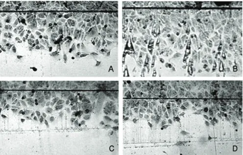
24-well plate (Iwaki, Japen)에 0.4 ml의 matrigel (Gelman Laboatory, MI, USA)을 코팅시켜 30분간 실온에서 배양하였 다. 이 well plate에 4시간 전에 serum free 상태로 배양된 ECV-304 세포에 0.05% trypsin-EDTA (Gibco BRL, NY, USA)를 처리하여 1×10⁵ cell/well 수준으로 분주였다.26,27 ECV-304 세포 배양 배지와 실험 조건에 맞게 처리한 대조군 과 저산소 환경에 노출 뒤 농도별 약물처리를 거친 망막색소 상피세포주의 conditioned medium (DMEM/F-12, 1% FBS) 을 1:1로 섞어 6시간 배양하였다.23이들 세포를 PBS로 2~3 회 세척 후 70% 에탄올로 4℃에서 30분 이상 고정한 뒤 현미 경으로 사진을 찍었다.28,29이 사진에서 무작위로 세 곳 을 선 택하여 ECV-304 세포가 만나 혈관 모양을 형성한 부분의 수 를 세어 평균값을 구해 통계 처리하였다.
세포이동능력 검사(migration assay)
60 mm2plate에 monolayer 로 ECV-304 세포와 human dermal fibroblast 1:1로 혼합해 빽빽하게 키운 후 배지를 제 거한 뒤 면도날로 60 mm2plate에 상처를 만든 뒤 한쪽의 세 포를 제거하였다.30,31PBS로 2회 세척 후 growth medium과 저산소 환경에 노출 뒤 농도별 약물처리를 거친 망막색소상 피세포주의 conditioned medium을 1:1로 섞어 24시간 배양 하였다. 이 세포를 PBS로 2~3회 세척하여 에탄올로 4℃에
A B
C D
VEGF
β -actin
Control Hypoxia Hypoxia +TA 1μm
Hypoxia +TA 100μm
PEDF
β -actin
Control Hypoxia Hypoxia +TA 1μm
Hypoxia +TA 100μm
Hypoxia TA-1μm TA-100μm VEGF
PEDF
Hypoxia TA-1μm TA-100μm 120
100 80 60 40 20 0
120 100 80 60 40 20 0
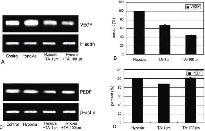
Figure 2.RT-PCR of VEGF and PEDF genes and the densitometric analysis in the ARPE-19 cells to the exposure of triamcinolone acetonide (TA). Quantitative analysis shows that expression of VEGF gene in these cells decreased after exposure to TA (A, B)(p<0.05). However, PEDF gene is not changed (C, D). VEGF=vascular endothelial growth factor; PEDF=pigment epithelium-derived factor.
서 30분 이상 고정한 뒤 현미경으로 사진을 찍었다.25이 사 진에서 무작위로 세 곳을 선택하여 상처부위에서 세포가 제 거된 부분으로 이동한 세포의 수와 가장 멀리 이동한 세포의 거리를 재어 평균을 낸 뒤 통계 처리하였다.
통계분석
실험 결과는 평균±표준편차 형태로 나타내고 통계적인 분 석은 SPSS program으로 Kruskal-wallis test를 이용하였다.
p<0.05인 경우 통계적인 유의성이 있는 것으로 간주하였다.
결 과
세포 활성도 측정을 통한 적정 TA농도의 확인
여러 농도의 TA를 배양망막색소상피세포에 노출시켜 배 양하면서 이들 농도에 따른 세포의 활성도를 조사하였다.
100 μM의 농도 이하에서 세포의 활성도가 85% 이상으로 확인되어(Fig. 1), 본 실험에서는 100 μM과 1 μM의 TA를 실험에 적합한 농도로 판단하고 사용하였다.
VEGF, PEDF의 유전자 발현측정
저산소의 환경에 24시간 노출한 후, 망막색소상피세포주 에 TA를 처치하여 RT-PCR을 이용하여 측정한 혈관형성 유도인자인 VEGF의 유전자의 발현은 저산소 상태보다 감 소하였으나 혈관형성 억제인자인 PEDF 유전자는 변화를 보이지 않았다(Fig. 2, p<0.05).
VEGF, PEDF 단백질의 생산
저산소의 환경에 24시간 노출한 후, TA를 처치한 망막 색소상피세포주의 배양액 내에서 Western blot를 통하여 측정한 혈관 형성관련인자, VEGF 단백질의 생산은 감소되 었고, 혈관 형성 억제인자인 PEDF 단백질의 생산은 변화 없었다(Fig. 3, p<0.05).
배양액의 맥관형성에 미치는 영향 확인
TA에 노출시키지 않는 배양액으로 처리한 대조군에 비 하여 세포 배양액은 ECV 304 cell의 맥관 형성을 억제하
A
B
C D
VEGF
β -tublin
Control Hypoxia Hypoxia +TA 1μm
Hypoxia +TA 100μm
PEDF
β -tublin
Control Hypoxia Hypoxia +TA 1μm
Hypoxia +TA 100μm
Hypoxia TA-1μm TA-100μm VEGF
PEDF
Hypoxia TA-1μm TA-100μm 120
100 80 60 40 20 0
120 100 80 60 40 20 0
Figure 3. Western blot on VEGF and PEDF production and the densitometric analysis in the ARPE-19 cells to the exposure of triamcinolone acetonide (TA). Quantitative analysis shows that production of VEGF protein in those cells decreased after exposure to TA (A, B)(p<0.05). However, PEDF production did not change (C, D).
VEGF=vascular endothelial growth factor; PEDF=pigment epithelium derived factor.
였다(Fig. 4, p<0.05).
배양액의 세포 이동에 미치는 영향 확인
TA로 처치하여 얻은 배양액은 TA를 처리하지 않은 배 양액에 비해서 세포 이동능력이 감소함을 알 수 있었다 (Fig. 5, p<0.05).
고 찰
인체에서 신생혈관의 발생은 혈관이 자극에 의해 발아가 일어나서 새로운 혈관이 생성되는 과정으로 먼저 혈관벽이 이완되어 투과성이 증가되고, 혈관내피세포들 간의 결합이 끊어지고 세포의 수축이 일어난다. 다음 단계로 다양한 단 백질 분해효소가 활성화되어 기저막을 분해하고 혈관내피 세포들은 혈관 벽에서 자극이 있는 방향의 혈관주변 조직 으로 이동, 증식하여 루프를 형성하며, 형성된 루프들이 분
화되어 혈관망을 생성하게 된다.32,33 혈관신생을 유도하는 저산소는 hypoxia-inducible factor-1 (HIF-1)를 증가시 켜 VEGF, bFGF, IGF-Ⅱ와 이들 수용체의 발현을 일으켜 혈관신생을 유발시킨다고 한다.30또한 PEDF는 인체망막색 소상피세포를 배양한 배지에서 처음 분리되었으며, VEGF 를 포함한 혈관형성유도인자에 강력히 대항하여 혈관형성 을 억제시키는 인자이다. PEDF는 신생혈관형성의 유도를 자극 받은 내피세포의 세포사를 일으켜 혈관신생을 억제한 다고 알려져 있다.34 정상적인 안구조직에서의 혈관형성은 VEGF와 같은 촉진인자와 PEDF 등의 억제인자 사이의 균 형에 의하여 유지되나, 저산소나 산화스트레스와 같은 자극 이 가해지면 이들 인자간의 불균형이 발생하여 혈관신생이 유발된다.34-36
맥락막모세혈관과 망막의 시세포층 사이에 위치한 망막색 소상피세포는 자체적으로 혈관형성유도인자인 VEGF와 혈 관형성억제인자인 PEDF를 생산한다. 이러한 신생혈관 유도 인자와 억제인자를 적절히 분비하여 외측으로는 맥락막모세
A B C
A=Hypoxia; B=Hypoxia+TA-1µM; C=Hypoxia+TA-100µM120
100
80
60
40
20
0
Hypoxia TA-1µM TA-100µM
D
Figure 4. Inhibition of tube formation by Triamcinolone acetonide (TA). The cells were cultured with the supernatant of HRPE cells and ECV-304 cell medium (1:1). After 6-hour culture, tube-like structure were analyzed. The supernatants of ARPE-19 cells used were collected from the APRE-19 conditioned media exposed to TA. With exposure to TA on ARPE19 cells, tube-like structures of ECV-304 cells decreased as shown in (A-D)(p<0.05).
혈관의 정상적인 기능상태를 유지하고 내측으로는 시세포층 의 무혈관상태를 유지시킨다.37그러나, 망막색소상피세포에 저산소나 산화스트레스 같은 비정상적인 환경에 노출될 때 혈관형성관련인자인 VEGF나 PEDF의 생산과 분비는 변화 되고 이러한 변화가 맥락막신생혈관의 발생을 유도한다.7,8,23 또한 이러한 변화를 정상적인 상태로 유도할 수 있는 약물이 투여된다면 신생혈관은 억제될 수 있다.17
TA는 염증성 안과질환의 치료를 위하여 오랫동안 사용 되어 온 코티코스테이로드 약제로 근래에 접어들면서 당뇨 황반부종, 폐쇄성 혈관질환, 증식유리체망막변증, 그리고 습 성나이관련 황반변성 등에 빈번히 사용되고 있다. Penfold et al17는 처음으로 습성 황반변성환자에 유리체내로 TA를 투여하여 맥락막신생혈관막을 안정화시키고 심한 시력상실 을 의미 있게 줄였다고 보고한 이후 맥락막신생혈관막에 대한 TA의 효과가 여러 보고에 의하여 확인되었고, Ciulla et al20은 동물실험을 통하여서도 비슷한 효과를 입증하였
다.18,19본 연구에서는 저산소나 산화스트레스에 노출된 배
양망막색소상피세포에서 혈관형성 관련인자들의 변화가 발 생하고 이러한 변화된 환경이 맥락막혈관신생을 유도할 수 있다는 보고들을 근거로 저산소에 노출된 망막색소상피세 포의 환경이 습성환반변성의 실험적인 상태라고 판단하고 TA을 투여한 결과 PEDF는 별다른 변화를 보이지 않았으나 VEGF의 생산과 분비가 감소되었었다.7,8,23이러한 결과는 TA가 비정상적으로 혈관형성 관련인자들이 변화된 환경에 주는 영향은 PEDF의 과발현보다는 VEGF의 저발현을 일 으킨 것으로 해석할 수 있다.
혈관신생과정은 먼저 혈관내피세포의 증식, 이동, 맥관형 성, 루프형성, 그리고 혈관형태로의 성숙과정을 거친다.31본 연구에서는 신생혈관의 발생과정에서 혈관형성의 상태가 억제되는 과정을 검증하는 방법으로 맥관형성과 세포이동 능력검사를 시행하였고 TA에 처치된 배양망막색소상피세 포의 배양액은 맥관 형성을 억제하였고 세포이동도 감소된 결과를 보여주었다. 이는 TA가 비정상적으로 혈관형성이 유도되고 있는 환경에 작용하여 혈관형성을 억제할 수 있
A B
C D
(A)=Control; (B)=Hypoxia; (C)=Hypoxia+TA-1μM; (D)=Hypoxia+TA-100μM
120 100 80 60 40 20 1
Hypoxia TA-1 µM TA-100µM Cell No.
120 100 80 60 40 20 1
Hypoxia TA-1µM TA-100 µM dlstace
Figure 5. Inhibition of cell migration by Triamcinolone acetonide (TA). Effect of the RPE-conditioned medium on human dermal microvascular endothelial cell (HDMEC) in migration assay. Conditioned medium was harvested from the culture dishes after ARPE 19 cells were exposed to TA for 6 hours. After initial wound scraping, cells were cultured for 24 hours and then observed. The degree of migration activity of HDMECs increased according to exposure to TA concentration (A-D)(p<0.05).
다는 방향으로 해석할 수 있으며 Ciulla et al20의 실험적 연 구와 동일한 결과로 생각된다. 또한 임상적으로 습성황반 변성에서 유리체내 TA의 투여로 맥락막신생혈관막을 안정 시켜 심한 시력상실이 예방되었다는 Penfold et al17의 보고 와도 부합된다.
결론적으로 TA는 저산소환경에 노출된 배양망막색소 상피에서 VEGF의 발현과 생산을 감소시켜 망막색소상피 세포가 비정상적인 환경에 노출되어 혈관신생을 유도하는 방향으로 활동할 때 혈관신생을 억제하는 방향으로 작용하 였다. 이러한 결과로 볼 때 습성황반변성환자에서 유리체 내에 TA의 투여는 VEGF의 발현을 감소시켜 혈관형성을 억제하는 방향으로 작용한다고 판단된다.
참고문헌
1) Kim JW, Kim HK, Kim HC. Photodynamic Therapy for Choroidal Neovascularization caused by age-related macular degeneration. J Korean Ophthalmol Soc 2002;43:1435-43.
2) Kosano H, Okano T, Katsura Y, et al. ProMMP-9 (92 kDa gelatinase) in vitreous fluid of patients with proliferative diabetic retinopathy. Life Sci 1999;64:2307-15.
3) Vingerling JR, Dielemans I, Hofman A, et al. The prevalence of age-related maculopathy in Rotterdam Study. Ophthalmology 1995;102:205-10.
4) Jin M, Kashiwangi K, Iizuka Y, et al. Matrix metalloproteinases in human diabetic and nondiabetic vitreous. Retina 2001;21:28-33.
5) Hanahan D, Folkman J. Patterns and emerging mechanisms of the angiogenic switch during tumorigenesis. Cell 1996;86:353-64.
6) Camochiaro PA. Retinal and choroidal neovascularization. J Cell
Physiol 2000;184:301-10.
7) Kim YD, Park YC, Choi GJ. Expression of angiogenesis-related factors in retinal pigment epithelial cells under hypoxia. J Korean Ophthalmol Soc 2006;47:629-36.
8) Kim JM, Kim JY, Lee YH, Choi GJ. Angiogenesis according to expressive change of angiogenic related factor in human RPE under oxidative stress. J Korean Ophthalmol Soc 2005;46:366-76.
9) Plate KH, Breier G, Welch HA, et al. Vascular endothelial growth factor is a potential tumor angiogenesis factor in human gliomas in vivo. Nature 1992;359:845-8.
10) Behzadian MA, Wang XL, AL-Shabrawey M, Caldwell RB. Effects of hypoxia on glial cell expression of angiogenesis-regulating factors VEGF and TGF-beta. Glia 1998;24:216-25.
11) Maione TE, Gray GS, Petro J, et al. Inhibition of angiogenesis by recombinant human platelet factor-4 and related peptides. Science 1990;247:77-9.
12) O'Reilly MS, Holmgren L, Shing Y, et al. Angiostatin: a novel angiogenesis inhibitor that mediates the suppression of metastases by a Lewis lung carcinoma. Cell 1994;79:315-28.
13) O'Reilly MS, Boehm T, Shing Y, et al. Endostatin: an endogenous inhibitor of angiogenesis and tumor growth. Cell 1997;88:277-85.
14) Yao Y, Guan M, Zhao XQ, Huang YF. Downregulation of the pigment epithelium derived factor by hypoxia and elevated glucose concentration in cultured human retinal pigment epithelial cells. Zhonghua Yi Xue Za Zhi 2003;25:1989-92.
15) Boyd SR, Zachary I, Chajravarthy U, et al. Correlation of increased vascular growth factor with neovascularization and permeability in ischemic central vein occlusion. Arch Ophthalmol 2002;120:
1644-50.
16) Ogata N, Nishikawa M, Nishimura T, et al. Unbalanced vitreous level of pigment epithelium-derived factor and vascular endothelial growth factor in diabetic retinopathy. Am J Ophthalmol 2002;
134:348-53.
17) Panfold PL, Gyory JF, Hunyor AB, Billson FA. Exudative macular degeneration and intravitreal triamcinolone: a pilot study. Aust N Z J Ophthalmol 1995;23:293-8.
18) Jonas JB, Akkoyun I, Budde WM, et al. Intravitreal reinjection of triamcinolone for exudative age-related macular degeneration.
Arch Ophthalmol 2004;122:218-22.
19) Jonas JB, Kressig I, Hugger P, et al. Intravitreal triamcinolone acetonide for exudative age-related macular degeneration. Br J Ophthalmol 2003;87:462-8.
20) Ciulla TA, Criwell MH, Danis RP, et al. Choroidal neovascular membrane inhibition in a laser treated rat model with intraocular sustained release triamcinolone acetonide microimplants. Br J Ophthalmol 2003;87:1032-7.
21) Pieters R., Huismans DR., Leyva A, Veerman AJ. Adaptation of the rapid automated tetrazolium dye based (MTT) assay for chemo- sensitivity testing in childhood leukemia. Cancer Lett 1988;41:
323-32.
22) O’Connor KA, Hansen MK, Rachal Pugh C, et al. Further characterization of high mobility group box 1 (HMGB1) as a pro- inflammatory cytokine: central nervous system effects. Cytokine 2003;24:254-65.
23) Ohno-Matsui K, Morita I, Tombran-Tink J, et al. Novel me- chanism for age-related macular degeneration: an equilibrium shift between the angiogenesis factors VEGF and PEDF. J Cell Physiol 2001;189:323-33.
24) Nakajima-Iijima S, Hamada H, Reddy P, Kakunaga T. Molecular structure of the human cytoplasmic beta-actin gene: interspecies homology of sequences in the introns. Proc Natl Acad Sci U S A 1985;82:6133-37.
25) Laemmli UK, Beguin F, Gujer-Kellenberger G. A factor preven- ting the major head protein of bacteriophage T4 from random aggregation. J Mol Biol 1970;47:69-85.
26) Matsuda S, Gomi F, Oshima Y, et al. Vascular Endothelial Growth Factor Reduced and Connective Tissue Growth Factor Induced by Triamcinolone in ARPE19 Cells under Oxidative Stress. Invest Ophthalmol Vis Sci 2005;46:1062-8.
27) Song HS, Son MJ, Lee YM, et al. Oxygen tension regulates the maturation of the blood-brain barrier. Biochem Biophys Res Commun 2002;290:325-31.
28) Heidemann J, Ogawa H, Dwinell MB, et al. Angiogenic effects of interleukin-8 (CXCL8) in human intestinal microvascular endo- thelial cells are mediated by CXCR2. J Biol Chem 2003;278:
8508-15.
29) Smith CL, Birdsey GM, Anthony S, et al. Dimethylarginine dimethylaminohydrolase activity modulates ADMA levels, VEGF expression, and cell phenotype. Biochem Biophys Res Commun 2003;308:984-9.
30) Tombran-Tink J, Chader GG, Johnson LV. PEDF: a pigment epithelium-derived factor with potent neuronal differentiative activity. Exp Eye Res 1991;53:411-4.
31) Jung S, Kim HW, Lee JH, et al. Brain tumor invasion model system using organotypic brain-slice culture as an alternative to in vivo model. J Cancer Res Clin Oncol 2002;128:469-76.
32) Yancopoulos G, Klagsbrun M, Folkman J. Vasculogenesis, angio- genesis, and growth factor: ephrins enter the fray at the border.
Cell 1998;93:661-4.
33) Risau W. Mechanisms of angiogenesis. Nature 1997;386:671-4.
34) Dawson DW, Volpert OV, Gillis P, et al. Pigment epithelium- derived factor: A potent inhibitor of angiogenesis. Science 1999;
285:245-48.
35) Bussolio F, Mantovani A, Persico G. Molecular mechanisms of blood vessel formation. Trends Biochem Sci 1997;22:251-6.
36) Raymond L, Jacobson B. Isolation and identification of stimu- latory and inhibitory cell growth factors in bovine vitreous. Exp Eye Res 1982;34:267-86.
37) Schlingemann RO. Role of growth factor and the wound healing response in age-related macular degeneration. Graefes Arch Clin Exp Ophthalmol 2004;242:91-101.
=ABSTRACT=
Effect of Triamcinolone on Angiogenesis-related Factors of Cultured Retinal Pigment Epithelial Cells
Young Chang Lee, MD1, Tae Jung Yoon, MD, PhD2, Gwang Ju Choi, MD, PhD1, Dae Hyun Kim, MD, PhD1
Department of Ophthalmology, College of Medicine, Chosun University1, Gwangju, Korea I Clinic2, Gwangju, Korea
Purpose: To examine the effects of triamcinolone on angiogenesis related factors in cultured human retinal pigment epithelial cells
Methods: Human retinal pigment epithelial cells were exposed to triamcinolone, cultured in a hypoxic environment, and expression and production of VEGF and PEDF were subsequently tested by RT-PCR and Western blot. Angiogenesis was measured via a tube formation assay using ECV 304 cells and with a migration assay using human dermal microvascular endothelial cells.
Results: Expression and production of VEGF and PEDF were tested by RT-PCR and Western blot, respectively. VEGF abundance was reduced while that of PEDF was unchanged in triamcinolone exposed retinal pigment epithelial cells cultured in hypoxic environment compared with cells with no treatment in hypoxic environment (p<0.05). Tube formation and cell migration were reduced by triamcinolone (p<0.05).
Conclusions: These results suggest that triamcinolone affects the secretion of angiogenesis-related factors and suppresses neovascularization.
J Korean Ophthalmol Soc 2009;50(4):594-602 Key Words: Angiogenesis, PDEF, Triamcinolone, VEGF
Address reprint requests to Dae Hyun Kim, MD, PhD
Department of Ophthalmology, Chosun University College of Medicine Hospital
#588 Seosuk-dong, Dong-gu, Gwangju 501-717, Korea
Tel: 82-62-220-3190, Fax: 82-62-225-9839, E-mail: [email protected]