KJTCVS
The Korean Journal of Thoracic and Cardiovascular SurgeryClinical Research
https://doi.org/10.5090/kjtcs.2020.53.1.16 pISSN: 2233-601X eISSN: 2093-6516
Korean J Thorac Cardiovasc Surg. 2020;53(1):16-21
Does Early Drain Removal Affect Postoperative Pericardial Effusion after Congenital Cardiac Surgery?
Young Eun Kim, M.D. 1 , Hanna Jung, M.D., Ph.D. 1 , Joon Yong Cho, M.D. 1 , Yeo Hyang Kim, M.D., Ph.D. 2 , Myung Chul Hyun, M.D., Ph.D. 2 , Youngok Lee, M.D., Ph.D. 1
Departments of
1Thoracic and Cardiovascular Surgery and
2Pediatrics, Kyungpook National University Hospital, School of Medicine, Kyungpook National University, Daegu, Korea
ARTICLE INFO
Received June 27, 2019 Revised August 15, 2019 Accepted August 30, 2019 Corresponding author Youngok Lee
Tel 82-53-200-5665 Fax 82-53-426-4765 E-mail yolee1210@knu.ac.kr ORCID
https://orcid.org/0000-0003-1592-4663
†
This paper was presented orally at the 50th Fall Meeting of the Korean Society for Thoracic and Cardiovascular Surgery, Seoul, Republic of Korea, October 25–27, 2018.
Background: Patients undergoing cardiac surgery require postoperative chest drainage.
However, the drain is difficult to keep in place in children with congenital heart disease.
Since 2015, at Kyungpook National University Hospital, the chest tube is removed on post- operative day 1 in patients who have undergone simple congenital cardiac surgery (i.e., closure of an atrial or ventricular septal defect). In this study, we evaluated the relationship between the duration of drain placement and the likelihood of pericardial effusion after congenital cardiac surgery.
Methods: The medical records of patients who underwent closure of an atrial or ven- tricular septal defect at our hospital between January 2014 and December 2016 were reviewed. In total, 162 patients who received follow-up echocardiography and had in- formation available on postoperative pericardial effusion after the repair procedure were enrolled.
Results: Echocardiography was performed at a median of 5 days (range, 4 to 6 days) post- operatively before discharge from the hospital. Pericardial effusion occurred in 21 patients (13.0%), of whom only 3 (1.9%) had moderate or greater pericardial effusion, regardless of the drain duration. All patients improved during outpatient follow-up without invasive management. No patient had severe complications because of pericardial effusion. The duration of drain placement did not affect the incidence of postoperative pericardial effu- sion (p=0.069). Operative survival was 100%.
Conclusion: Based on our study, we recommend removing the drain as soon as its role is complete, generally on postoperative day 1, because early removal does not increase the incidence of pericardial effusion in patients undergoing simple congenital cardiac surgery.
Keywords: Atrial heart septal defects, Congenital heart disease, Drainage, Pericardial ef- fusion, Ventricular heart septal defects
Copyright © The Korean Society for Thoracic and Cardiovascular Surgery. 2020. All right reserved.
This is an Open Access article distributed under the terms of the Creative Commons Attribution Non-Commercial License (http://creativecommons.org/licenses/
Introduction
Patients who undergo cardiac surgery routinely have 1, 2, or more chest tubes placed postoperatively to monitor postoperative bleeding or to drain pericardial effusion, which may cause hemodynamic instability because of car- diac tamponade and/or pleural effusion [1]. Surgeons hesi- tate to remove chest tubes too early because of these poten- tial postoperative complications and the possible need for an additional invasive procedure (e.g., pericardiocentesis or closed thoracostomy). However, an unnecessarily pro-
longed period of chest tube drainage is painful, immobiliz- es the patient, causes mechanical irritation to the heart and pericardium, and can be a source of infection [2-4].
Chest tube drainage is particularly problematic in chil-
dren with congenital heart disease, who lack the under-
standing and patience needed to bear the discomfort of the
chest drain. Furthermore, pediatric patients are likely to
struggle, which causes air to be sucked back into the medi-
astinum. In 2015, our congenital cardiac surgery team
started removing the chest tube on the first postoperative
day in patients undergoing simple congenital cardiac sur-
Young Eun Kim, et al. Drain Removal and Pericardial Effusion KJTCVS
gery, such as closure of an atrial septal defect (ASD) or ventricular septal defect (VSD).
The aim of this study was to evaluate the relationship be- tween the duration of drain placement and the likelihood of pericardial effusion after simple congenital cardiac sur- gery.
Methods
Patients
The study was approved by the Institutional Review Board of Kyungpook National University Hospital (IRB approval no., 2018-11-031). The requirement for informed consent was waived because of the retrospective nature of the study and the anonymity of the data. We reviewed the clinical records, echocardiograms, operative findings, and surgical outcomes in 162 patients who underwent correc- tive surgery for ASD or VSD at Kyungpook National Uni- versity Hospital between January 2014 and December 2016.
The indications for surgery were failure to thrive, conges- tive heart failure, and other standard indications for clo- sure of ASD or VSD.
Drain management
A single round 16F silicone thoracic catheter (500 mm in length; Sewoon Medical Co. Ltd., Cheonan, Korea) was placed in the retrosternum in all patients. The chest drain was connected to a disposable dry suction control chamber (OASIS Dry Suction Water Seal Chest Drain; Maquet, Ras- tatt, Germany) with 20 cmH
2O of suction. No additional manipulation (milking, stripping, or withdrawing) of the catheter was performed during the drainage period. Before 2015, the drain was kept in place for at least 24 hours post- operatively and removed when there was a decreasing trend of effusion. Starting in 2015, the drain was removed if the patient had no postoperative bleeding and stable vital
signs regardless of the amount of drainage.
Echocardiography
Echocardiography was routinely performed by pediatric cardiologists in all cases preoperatively and repeated at least once before discharge from the hospital or at the first outpatient clinic follow-up visit in the early postoperative period. The presence and amount of pericardial effusion was assessed by 2-dimensional echocardiography in the parasternal short axis, long axis, apical 4-chamber, and subcostal views. Pericardial effusion was graded according to the maximum separation between the pericardium and epicardium: less than 5 mm was considered mild, 6 to 15 mm as moderate, and more than 16 mm as severe [5].
Statistical analysis
Continuous variables are expressed as the median and interquartile range for non-normally distributed data; dif- ferences between these variables were assessed by the Mann-Whitney U-test. Categorical variables are expressed as the number and percentage; differences between these variables were evaluated using the chi-square test. Differ- ences were considered to be statistically significant when the p-value was less than 0.05. All statistical analyses were performed using IBM SPSS ver. 23.0 for Windows (IBM Corp., Armonk, NY, USA).
Results
Patient characteristics
Patients’ demographic characteristics are shown in Table 1. At the time of surgery, patients’ median age was 3 months Table 1. Patients’ characteristics (n=162)
Characteristic Value
Age (mo) 3 (1–19)
Sex
Female 78 (48.1)
Male 84 (51.9)
Weight (kg) 5.5 (3.8–10.5)
Body surface area (m
2) 0.3 (0.2–0.5) Postoperative hospital stay (day) 7 (5–9) Values are presented as median (interquartile range) or number (%).
Table 2. Intraoperative outcomes in 162 patients (n=162)
Characteristic Value
Clamp time (min) 40.5 (31.0–53.0)
Pump time (min) 64.5 (50.0–83.0)
ASD closure 32 (19.8)
MV repair 8
TV repair 11
Ventricular septal defect closure 130 (80.2)
ASD closure 102
MV repair 22
TV repair 27
Values are presented as median (interquartile range), number (%), or number.
ASD, atrial septal defect; MV, mitral valve; TV, tricuspid valve.
https://doi.org/10.5090/kjtcs.2020.53.1.16
KJTCVS
(range, 1 to 19 months), and their median body weight and surface area were 5.5 kg (range, 3.8 to 10.5 kg) and 0.3 m
2(range, 0.2 to 0.5 m
2), respectively. The median postopera- tive hospital stay was 7 days (range, 5 to 9 days).
The intraoperative outcomes are presented in Table 2.
The median clamp and pump times were 40.5 minutes (range, 31.0 to 53.0 minutes) and 64.5 minutes (range, 50.0 to 83.0 minutes), respectively. Thirty-two patients under- went ASD closure with or without mitral or tricuspid valve repair, and 130 underwent VSD closure with or without mitral or tricuspid valve repair or ASD closure.
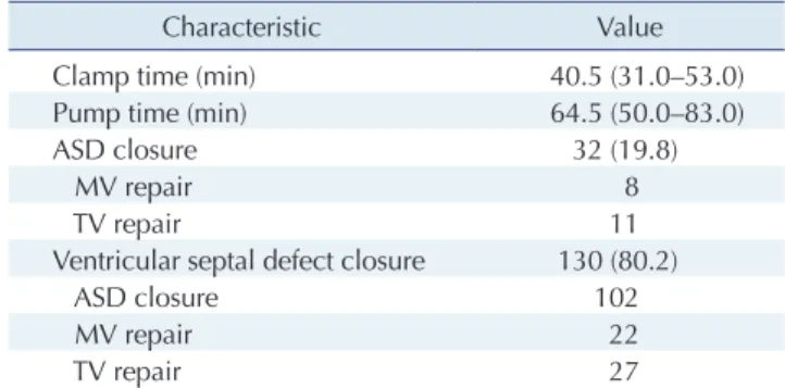
No operative complications occurred. There was no op- erative mortality or any deaths during the hospital stay or follow-up period. The distribution of the patients accord- ing to the duration of drain placement and year of surgery
is shown in Fig. 1.
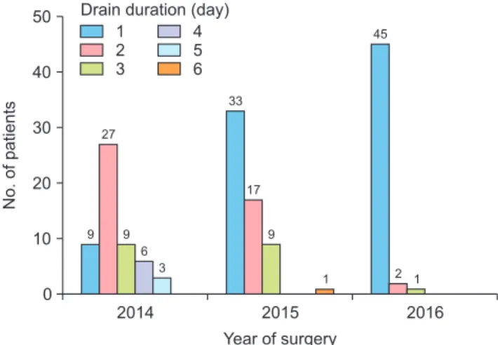
Evolution of pericardial effusion
The evolution of pericardial effusion in all 162 patients, from the postoperative echocardiogram to the second out- patient follow-up echocardiogram, is demonstrated in Fig.
2. The postoperative echocardiogram was obtained at a median of 5 days (range, 4 to 6 days) after removal of the chest tube, and the second outpatient follow-up echocar- diogram was obtained at a median of 12 months (range, 11 to 16 months) postoperatively.
The postoperative echocardiograms showed pericardial effusion in 19 patients and no pericardial effusion in 143 patients. The amount of pericardial effusion was mild in 18 cases and moderate in 1. In 18 cases, the pericardial effu- sion had resolved by the time of the first outpatient fol- low-up echocardiogram, and it eventually resolved in the remaining case (Fig. 2A). Two of the 143 patients who did not have pericardial effusion before discharge showed moderate effusion echocardiograms obtained at the time of their first outpatient clinic visit (Fig. 2B).
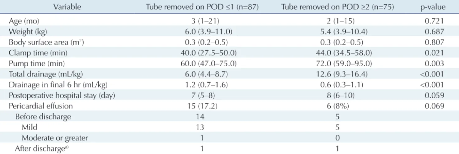
Effect of drain duration on postoperative pericardial effusion
We compared patients’ characteristics and operative out- comes according to whether the chest tube was removed on postoperative day 1 or later than postoperative day 1, as shown in Table 3. No significant difference was found in any of the patients’ characteristics or outcomes according to whether the chest tube was removed on the first postop-
50
40
30
20
10
2014
No. of patients
Year of surgery 0
2015 2016
9 27
9 6
3
33
17
9
1 2 1
1 45 2 3
Drain duration (day) 4 5 6
Fig. 1. Distribution of patients according to drain duration and year of surgery.
Echo follow-up median (IQR)
Echo follow-up median (IQR)
OPD visit 2 12 mo (11 16 mo) OPD visit 1
2 mo (2 3 mo) Postop
5 day (4 6 day) n=19
n=143
n=19
n=143
OPD visit 2 12 mo (11 16 mo) OPD visit 1
2 mo (2 3 mo) Postop
5 day (4 6 day) (+)
(+) ( )
( )
(+)
( )
(+)
( )
(+)
( )
(+)
( ) 1
18 1
2
2
A
B
Fig. 2. Frequency of pericardial ef- fusion in 162 patients after congen- ital cardiac surgery. (A) Follow- up of 19 patients who had pericardial effusion before discharge from the hospital. (B) Follow-up of 143 pa- tients who did not have pericardial effusion before discharge from the hospital. Echo, echocardiography;
IQR, interquartile range; Postop,
postoperative; OPD outpatient de-
partment.
Young Eun Kim, et al. Drain Removal and Pericardial Effusion KJTCVS
erative day (Table 3).
Predictably, however, significant differences were found in the total amount of fluid drained (p<0.001) and the amount drained during the final 6 hours of drain place- ment (p<0.001). The total volume of drainage fluid was in- evitably lower in the group with early tube removal than in the group with delayed tube removal, because the total drainage volume is smaller if the duration of drain place- ment is shorter and larger if the duration is longer. Consid- ering the need to wait for effusion to decrease, the volume drained in the 6 hours before removal of the tube in the group with delayed removal was necessarily significantly lower than that in the group with early tube removal.
However, contrary to our expectations, significant be- tween-group differences were found in clamp time (p=
0.021) and pump time (p=0.003). There are 2 possible rea- sons for these significant differences. First, even though the number of patients in these 2 groups was similar, their relative distribution changed between 2014 and 2016 (Fig.
3). Second, operator proficiency and coordination of the congenital cardiac surgery team may have improved be- tween 2014 and 2016, resulting in a significant effect on clamp and pump times (Table 4). The retrospective nature of this study meant that these factors could not be con- trolled; if the groups’ composition had been similar in terms of when the procedures were performed, there might have been no significant differences in clamp time or pump time.
Discussion
In this study, we evaluated the incidence of pericardial effusion after simple congenital cardiac surgery, (i.e., ASD or VSD closure). Pericardial effusion occurred in 21 (13.0%) of 162 patients; only 3 patients (1.9%) had moderate or greater pericardial effusion on follow-up echocardiography after surgery regardless of the drain duration. Pericardial effusion eventually improved in all patients during fol- low-up in the outpatient clinic with no need for further in- vasive management.
Pericardial effusion was detected on immediate postop- erative echocardiography in 19 patients (11.7%) and had
100
80
60
40
20
Patients (%)
Year of surgery 0
2014 2015 2016
POD < 1 POD > 2
83.3
16.7
45.0
55.0
93.8 6.2
Fig. 3. Distribution of the groups according to the timing of drain removal between 2014 and 2016. POD, postoperative day.
Table 3. Comparison of patients’ characteristics and operative outcomes according to the timing of chest tube removal
Variable Tube removed on POD ≤1 (n=87) Tube removed on POD ≥2 (n=75) p-value
Age (mo) 3 (1–21) 2 (1–15) 0.721
Weight (kg) 6.0 (3.9–11.0) 5.4 (3.9–10.4) 0.687
Body surface area (m
2) 0.3 (0.2–0.5) 0.3 (0.2–0.5) 0.807
Clamp time (min) 40.0 (27.5–50.0) 44.0 (34.5–58.0) 0.021
Pump time (min) 60.0 (47.0–75.0) 72.0 (59.0–95.0) 0.003
Total drainage (mL/kg) 6.0 (4.4–8.7) 12.6 (9.3–16.4) <0.001
Drainage in final 6 hr (mL/kg) 1.2 (0.7–1.6) 0.6 (0.3–1.1) <0.001
Postoperative hospital stay (day) 7 (5–8) 8 (6–10) 0.059
Pericardial effusion 15 (17.2) 6 (8%) 0.069
Before discharge 14 5
Mild 13 5
Moderate or greater 1 0
After discharge
a)1 1
Values are presented as median (interquartile range), number (%), or number.
POD, postoperative day.
a)