The Study of Lumbar Erector Spinea and Rectus Abdominis Activations according to the Different Gait Velocities in Young Healthy Adults
Jong-Sung Chang, PT, PhD1, Hae-Yong Lee, PT2, Mi-Young Lee, PT, PhD3
1Department of Physical Therapy, College of Health Science, Honam University, 2Department of Rehabilitation Science, Graduate School, Daegu University, 3Department of Physical Therapy, College of Health and Therapy, Daegu Haany University
Purpose: The purpose of this study was to investigate the lumbar erector spinea and rectus abdominis activations, according to the different gait velocities in young healthy adults.
Methods: We recruited 6 young male and 10 young female (mean age=21.43 years; range 19~23) in this study. We used a wireless surface electromyogram (Telemyo 2400T G2, Noraxon, USA) and a treadmill unit for the experiment. EMG activity from the lumbar erector spinea, and rectus abdominis of the dominant side was record with surface electrodes. On different day, all subjects gaited on 2.7 km/h, 4.5 km/h, and 6.3 km/h of speed in random order. They gaited at the same velocity, three times, on the treadmill unit. To reduce fatigue, sufficient rests were given between the measurements.
Results: As the gait speed increased, lumbar erector spinea and rectus abdominis activations were significantly increased (p<0.05).
Conclusion: In the current study, we found lumbar erector spinea and rectus abdominis activations were changed, according to the gait velocity. We suggested that rehabilitation intervention should be focused on the exercise velocity for the patients with problem of the trunk control.
Keywords: Trunk muscle activation, Gait velocity, Trunk control
I. Introduction
In humans, the trunk correspond to 60% of the total body mass, with its high position relative to the feet, allowing it to play a key role as an “inverted pendulum” leverage effect.
Trunk is activated by many muscles, and is highly articulated, which provides it to participate actively in the various motor task, while maintaining trunk balance.1,2 For a response to the sudden load, trunk is stabilized with strong activation of the trunk muscles. The stabilization, which occurs prior
to activation of the agonist, is able to be explained by an anticipatory postural adjustment preparing trunk with muscle contraction for posture control on the postural sway.
This could be an account for a reaction force of the body segment.3,4 Increase of muscle strength, through the trunk stabilization, works by a normal mechanism of anticipatory postural adjustment, so that it affects the balance and helps to prevent the risk of sport injury.5,6 Therefore, many previous studies have reported that improvement of balance and performance by the trunk stabilization exercise for lower back pain or sport athletes.7-9
Gait is composed of quite steady coordination modes, specific phase, and frequency relations, between the head, trunk, pelvis, and cyclical movements of the extremities.10-14 In particular, coordination between the trunk and pelvic, in addition to the activation of an associated musculature, Received May 16, 2012 Revised June 2, 2012
Accepted June 7, 2012
Corresponding author Mi-Young Lee, [email protected]
Copyright © 2012 by The Korean Society of Physical Therapy
This is an Open Access article distributed under the terms of the Creative Commons Attribution Non-Commercial License (http://creativecommons.org/licenses/by-nc/3.0/) which permits unrestricted non-commercial use, distribution, and reproduction in any medium, provided the original work is properly cited.
The Journal of Korean Society of Physical Therapy Original Article
Original Article
such as erector spinae muscle, have proven to be a useful point on the human gait. When gait speed is changed, there is changed trunk-pelvis coordination and erector spinae muscle activity to preserve the stable gait pattern and perturbation.15-17 Compared to healthy persons, patients with lower back pain on the average gait speed is slow, because they impaired trunk-pelvis coordination associated with the poorly coordinated lumbar erector spinae activity.
That is, patients with lower back pain preferred a slow gait velocity, and it is associated with the instability in adapting the trunk-pelvis coordination for various velocities.16 Gait velocity, which should represent the variation of the human, is affected by environmental factors, such as lifestyle, and genetic factors, such as difference of skeletal muscle, as well as psychological nature. Individuals also affect gait velocity, according to their physical body, the degree of fatigue, and the design or function of their shoes.18,19 Thus, a variety of gait speed is affected by the gait parameters. Most previous studies investigated kinematic changes of the lower limbs and muscle activity for the different walking speeds. However, control of trunk is important for efficiency of the locomotion such as walking and running. Therefore, the purpose of this study was to examine the changes of the lumbar erector spinea and rectus abdominis activations for various gait velocities.
II. Materials and Methods
1. Subjects
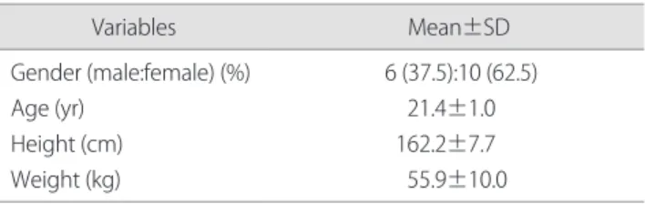
A total of 16 healthy subjects, who had no history of the neurological and musculoskeletal disorders, participated in this study. Handedness was verified by the modified Edinburg Handedness Inventory.20 All subjects gave their written informed consent before participation. The general characteristics for the subjects were as shown (Table 1).
2. Data acquisition
1) Surface electromyogram (EMG)
Muscles activations were recorded using a Telemyo G2 WiFi wireless system model (Telemyo 2400T G2, Noraxon, USA).
The frequency range of the EMG signal was set to 20~500 Hz, and all the collected data were processed via MRXP 1.07 Master editing software (Noraxon, USA) for a synchronized view of EMG signal. EMG activity, from the lumbar erector spinea and rectus abdominis of the dominant side, was recorded with surface electrodes.21,22 The ground electrode was affixed to the skin over the seventh cervical spinous process. Subjects performed maximal voluntary isometric contraction (MVIC) against manual resistance and the EMG activity was recorded from each of the two muscles.
EMG data from the MVIC were used to normalize the EMG amplitude measured during walking. The amplitude in EMG activity for each muscle was indentified from the averaged data during walking.
2) Procedure
The treadmill of the T-700ci model (T-700spirit, Apex, Korea), with fastest speed of 20 km/h and inclination of 0~16%, was used. According to the walking speed of the classification of Korean, we divided walking speed of 2.7 km/h, 4.5 km/h, and 6.3 km/h.18 Subjects were instructed to walk comfortably. Treadmill velocity was randomly assigned to the order, and the experiment assistant was setting the treadmill velocity. On different day, 2.7 km/h, 4.5 km/h, and 6.3 km/h were performed three times. Treadmill walking was conducted during 1 minute, and was performed during each warm-up and cool-down exercise, during 30-second.
To reduce fatigue, there were sufficient rests between the measurements.
3. Statistical analysis
Statistical analysis was performed to determine the difference trunk muscle activation, according to gait velocities. For the data that satisfied the normal distribution, a parametric test was used. Thus, in this study, the data were analyzed with repeated measures ANOVA. Contrast tests to examine repeated effects as within-muscle factor were used.
Table 1. The general characteristics for subjects
Variables Mean±SD
Gender (male:female) (%) Age (yr)
Height (cm) Weight (kg)
6 (37.5):10 (62.5) 21.4±1.0 162.2±7.7 55.9±10.0
Independent t-test was used to compare between lumbar erector spinea and rectus abdominis. All statistical analysis was performed using SPSS ver. 14.0 for Window (SPSS Inc., Chicago, IL, USA), and significance was set at α= 0.05.
III. Results
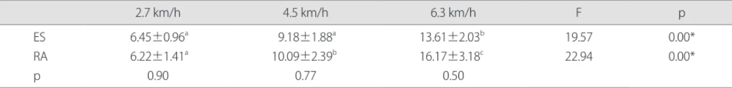
We found the difference of muscle activation according to gait velocities. In the activation of the lumbar erector spinea, there was significant difference according to the gait velocities (p<0.05). In the result of contrast test, 2.7 km/
h and 6.3 km/h, 4.5 km/h and 6.3 km/h were significantly different respectively. In addition, the activation of the rectus abdominis was also a significant difference according to the gait velocities (p<0.05). In the result of contrast test, there was a different between each velocity. However, there was no significant difference between activation of the two muscles according to the gait velocities (p>0.05) (Table 2).
IV. Discussion
In the current study, we investigated that the change of the trunk muscle (erector spinae and rectus abdominis) activation, according to the gait velocities. Gait velocities were divided with 2.7 km/h, 4.5 km/h, and 6.3 km/h, according to the classification of Korean gait velocity. In the present result, we observed that the faster the gait velocity, the activation of the trunk muscle increased more significantly. However, there was no different between activation of the two muscles according to gait velocities. Commonly, various gait velocites caused kinematic changes in the body. In a healthy gait,
interaction and coupling movement among the limbs, pelvis, trunk, and head, are relatively stable, however, these adapts flexibly to the change in velocity.10-14 That is, variable of the gait velocity causes the changes of the coordination between pelvis and trunk movement and activation of muscles in the lower extremity.
In the previous study, Lamoth et al.16 suggested that healthy persons have ability to adapt trunk-pelvis coordination to changes in velocity. In particular, lumbar-pelvic coordination was increased at higher velocities. Altered coordination reflects an attempt to stabilize the spine and prevent the occurrence of unanticipated fluctuations. In 2001, Cho and Kim23 investigated kinematics changes of the lower extremity in the gait on different speeds (3 km/h, 4 km/
h, and 5 km/h). As results, it was shown that the speed increase, consequently, increases the knee and hip joint angular velocity. In addition, Saunders et al.21 reported that amplitude of lumbo-pelvic motion changed with locomotor speed. They also demonstrated that there was association between lumbo-pelvic motion and trunk muscle activity during locomotion at different speeds. In this study, the result showed that the activation of the trunk muscle activation was significantly changed in the faster gait velocity. Based on the studies related with gait velocity, we suggested that increase of muscle activation was caused increase of angular velocity in the lower extremity and lumbar-pelvic coordination at higher gait velocity. In addition, antagonistic muscle co- activation needs for maintain spinal stability.24 In our study, we found that the activation of the between erector spinae and rectus abdominis were not significantly different. It might mean that co-activation of the trunk muscles induced stability
Table 2. The change of muscle activation according to gait velocities (unit: %MVIC)
2.7 km/h 4.5 km/h 6.3 km/h F p
ES RA p
6.45±0.96a 6.22±1.41a
0.90
9.18±1.88a 10.09±2.39b
0.77
13.61±2.03b 16.17±3.18c
0.50
19.57 22.94
0.00*
0.00*
The values (a, b, c) with different superscripts in the same column indicates the results of one factor analysis with contrast test and significance at the p<0.05 level.
ES: erector spinae, RA: rectus abdominis, MVIC: maximal voluntary isometric contraction.
*p<0.05.
during different gait velocity.
In the studies related with gait velocity, variables of the velocity varies.16,23,25 The present study was performed, according to Han’s classification18 of the Korean gait speed (2.7 km/h, 4.5 km/h, and 6.3 km/h). A 2.7 km/h is a very slow speed, which is slightly slower than the average gait speed for the people of sixty-five or over. A 4.5 km/h is an appropriate speed, which is the average gait speed for the people of the late 20 years to their early 30’s. Further, 6.3 km/h is a fast speed, which is suggested for a walk with quick steps. Using classification of the Korean gait speed, Moon investigated kinematic and EMG changes of the lower limbs for the different walking speeds - 2.7 km/h, 4.5 km/h, and 6.3 km/h.
He elucidated that muscle activation of the lower limbs was changed at different gait speeds.19 Similarly, our study showed an increase in the trunk muscles activation at different gait speeds.
In addition, the coupling of the body segments is able to elucidate the anticipatory postural adjustments (APAs), which represent changes in the muscle activation, prior to an expected mechanical perturbation. Previous studies have reported the magnitude and temporal onset time of the APAs with changes in the forthcoming dynamic task, such as arm movements at different velocities or inertial load.26,27 In 2001, Shiratori and Latash27 observed in the trunk/ leg muscles activation, prior to catching a load released. Therefore, the present finding, which is shows the change of trunk muscle activation, according to the different gait velocity, could be demonstrated by the reaction force due to the coupling of body segments.
In conclusion, the present study investigated whether the gait velocity effect the change of the lumbar erector spinea and rectus abdominis activation. We found that the more increase of gait velocity induced more increase in the change of the lumbar erector spinea and rectus abdominis activation. However, we observed that an activation of the global muscle, using a surface EMG, so the changes of the core muscle activation related to that of the trunk stabilization are unclear. For all that, the present study implies that rehabilitative intervention of patients with problem of the trunk muscle control is probably effective, when it focuses on
the exercise velocity of the lower extremity.
Author Contributions
Research design: Lee MY Acquisition of data: Lee HY
Analysis and interpretation of data: Chang JS Drafting of the manuscript: Chang JS, Lee MY
Administrative, technical, and material support: Lee HY Research supervision: Lee MY
References
1. Ceccato JC, de Sèze M, Azevedo C et al. Comparison of trunk activity during gait initiation and walking in humans. PLoS One.
2009;4(12):e8193.
2. Kumar S. Ergonomics and biology of spinal rotation. Ergonomics.
2004;47(4):370-415.
3. Cholewicki J, McGill SM. Mechanical stability of the in vivo lumbar spine: implications for injury and chronic low back pain. Clin Biomech (Bristol, Avon). 1996;11(1):1-15.
4. Hodges PW, Richardson CA. Feedforward contraction of transversus abdominis is not influenced by the direction of arm movement. Exp Brain Res. 1997;114(2):362-70.
5. Chai HK. The study for injury prevention of athleties. J Korean Soc of Saf Educ. 1998:2(2):23-35.
6. Choi BC, Kim, H. The effect of lumbar stabilization exercise on balance ability. Korean Soc Study Phys Educ. 2009;18(2):1147-56.
7. Barr KP, Griggs M, Cadby T. Lumbar stabilization: core concepts and current literature, Part 1. Am J Phys Med Rehabil. 2005;84(6):
473-80.
8. Kim GY, Ahn CS, Kim SS. The effects of 3-dimensional lumbar stabilization exercise have an effect on the improvement of pain and static or dynamic balance ability in 20’s age group with low back pain. Korean Soc Phys Med. 2011:6(2): 235-46.
9. Kim JK, Park SK, Kang JI et al. Effects of lumbar stability exercise program on trunk, lower extremity of muscle activity and balance in soccer player. J Korean Soc Phys Ther. 2010:22(5):25-31.
10. Daffertshofer A, Lamoth CJ, Meijer OG et al. PCA in studying coordination and variability: a tutorial. Clin Biomech (Bristol, Avon).
2004;19(4):415-28.
11. Donker SF, Beek PJ, Wagenaar RC et al. Coordination between arm and leg movements during locomotion. J Mot Behav.
2001;33(1):86-102.
12. Lamoth CJ, Meijer OG, Wuisman PI et al. Pelvis-thorax coordination in the transverse plane during walking in persons with nonspecific low back pain. Spine (Phila Pa 1976). 2002;27(4):E92-9.
13. Wannier T, Bastiaanse C, Colombo G, Dietz V. Arm to leg coordination in humans during walking, creeping and swimming activities. Exp Brain Res. 2001;141(3):375-9.
14. Yamasaki T, Nomura T, Sato S. Phase reset and dynamic stability during human gait. Biosystems. 2003;71(1-2):221-32.
15. de Sèze M, Falgairolle M, Viel S et al. Sequential activation of axial muscles during different forms of rhythmic behavior in man. Exp Brain Res. 2008;185(2):237-47.
16. Lamoth CJ, Daffertshofer A, Meijer OG et al. How do persons with chronic low back pain speed up and slow down? Trunk-pelvis coordination and lumbar erector spinae activity during gait. Gait Posture. 2006;23(2):230-9.
17. Lamoth CJ, Daffertshofer A, Meijer OG et al. Effects of experimentally induced pain and fear of pain on trunk coordination and back muscle activity during walking. Clin Biomech (Bristol, Avon). 2004;19(6):551-63.
18. Han SD. Eromics. Seoul, Hakmunsa, 1983.
19. Moon GS. The kinematic analysis of the ankle joint and EMG analysis of the lower limbs muscle for the different walking speed.
Korean J Sport Biomech. 2005:15(1):177-95.
20. Oldfield RC. The assessment and analysis of handedness: the Edinburgh inventory. Neuropsychologia. 1971;9(1):97-113.
21. Saunders SW, Schache A, Rath D, Hodges PW. Changes in three dimensional lumbo-pelvic kinematics and trunk muscle activity
with speed and mode of locomotion. Clin Biomech (Bristol, Avon).
2005;20(8):784-93.
22. Oh JS, Cynn HS, Won JH et al. Effects of performing an abdominal drawing-in maneuver during prone hip extension exercises on hip and back extensor muscle activity and amount of anterior pelvic tilt. J Orthop Sports Phys Ther. 2007;37(6):320-4.
23. Cho KK, Kim YS. Analysis of kinematics in gait motions on different grades and speeds of treadmill gait. Korean J Sport Biomech.
2002;12(1):175-191.
24. Granata KP, Orishimo KF. Response of trunk muscle coactivation to changes in spinal stability. J Biomech. 2001;34(9):1117-23.
25. Kwon YR, Kim JW, Kang DW et al. Changes in acceleration at the upper thigh and ankle with variations in gait speed and walkway slope. Korean J Sport Biomech. 2010:10(2): 191-96.
26. Horak FB, Esselman P, Anderson ME et al. The effects of movement velocity, mass displaced, and task certainty on associated postural adjustments made by normal and hemiplegic individuals. J Neurol Neurosurg Psychiatry. 1984;47(9):1020-8.
27. Shiratori T, Latash ML. Anticipatory postural adjustments during load catching by standing subjects. Clin Neurophysiol.
2001;112(7):1250-65.