호르몬 요법과 골다공증
순천향대학교 의과대학 순천향대학교부천병원 산부인과학교실
이 해 혁
Hormone Therapy and Osteoporosis
Hae-Hyeog Lee
Department of Obstetrics and Gynecology, College of Medicine, Soonchunhyang University, Bucheon, Korea
During the perimenopause, both the quantity and quality of bone decline rapidly, resulting in a dramatic increase in the risk of fracture in postmenopausal women. Data from the large Women's Health Initiative (WHI), in which women with an intact uterus (mean age: 63 years) were randomly assigned to estrogen plus progestin, showed that HT increased total hip BMD and reduced the risk of fractures at the hip, vertebrae, and wrist. Similarly, the estrogen-alone component of the WHI, which involved women (mean age: 64 years) with prior hysterectomy, demonstrated a reduced rate of hip fracture.
It is never too early in the menopause to evaluate women for bone loss and advise them on steps to take to prevent the declines in bone mass and quality that increase the risk of future osteoporosis and fracture. Postmenopausal women should therefore be screened for osteoporosis risk factors and have their BMD levels tested, if warranted, in accordance with current guidelines. Use of BMD assists physicians in diagnosing osteoporosis and in monitoring treatment effects.
Low-dose hormone therapy and ultralow-dose hormone therapy can provide an effective protection against the postmenopausal decrease of BMD. Estrogen depletion after menopause become involved with postmenopausal bone loss, therefore ET (estrogen therapy) and EPT (estrogen-progestogen therapy) have been considered as first-line therapy for postmenopausal osteopenia and osteoporosis.
Key Words: Hormone-therapy (HT), Osteoporosis, Menopause, Women's Health Initiative (WHI)
2) 골다공증은 골절이 발생할 때까지 많은 경우 에서 증상이 나타나지 않기 때문에 조용한 질환 (silent disease)이라고 한다1. 최근 국내에서 발표된 안성 지역사회 코호트에서 골다공증 및 척추 골절 유병률에 관한 연구에서 50세 이상 여성에서 골다
접수일: 2007년 12월 26일, 승인일: 2008년 4월 7일 책임저자: 이해혁, 순천향대학교 부천병원 산부인과
Tel: 011-273-7840, Fax: 02)6008-6874 E-mail: [email protected], [email protected]
공증 유병률은 29.6%이었으며 척추골절의 유병률 은 11.6%였다.
최근 10년 동안 폐경 여성에서 호르몬 요법 (Hormone Therapy: HT)을 사용하는 데 많은 변화가 있어 왔다.
1990년 이전까지는 호르몬 요법을 시행하는 폐경 여 성의 수가 그리 많지 않았는데, 1990년대에 들어서면 서 전 세계적으로 여러 관련학회가 창립되었고, 많은 관심과 기초 및 임상 연구가 활발하게 진행되었다.
이에 따라 호르몬 요법을 시행하는 폐경 여성의 수도
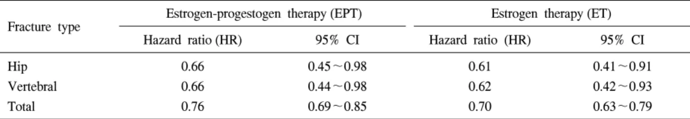
Table 1. Effect of estrogen-progestogen therapy (EPT) and estrogen therapy (ET) on clinical fracture incidence in healthy postmenopausal women2,5,6
Fracture type Estrogen-progestogen therapy (EPT) Estrogen therapy (ET)
Hazard ratio (HR) 95% CI Hazard ratio (HR) 95% CI
Hip Vertebral Total
0.66 0.66 0.76
0.45~0.98 0.44~0.98 0.69~0.85
0.61 0.62 0.70
0.41~0.91 0.42~0.93 0.63~0.79
Fig. 1. Patterns of BMD (g/cm2) of spine and femoral neck, in women treated with only 1,000 mg of calcium per day (control group), LD-HRT (1 mg estradiol+0.5 mg norethisterone acetate) or Ultra- LD-HRT (0.5 mg of 17β-estradiol and 0.25 mg of norethisterone acetate) along with 1,000 mg of calcium per day. The results are expressed as percent of variation vs. basal values. Measured at the end of the 2-year follow up study. *P<0.05 vs. corresponding control group values11.
급속히 증가되었었다. 2002년 WHI (Women's Health Initiative) 연구2가 발표되면서 호르몬 요법에 대한 위험성이 부각되기도 하였다. 그 이후로 호르몬 요 법을 중단하는 여성이 증가되었고3, 호르몬 요법을 새로이 시행하려는 폐경 여성들이 많이 감소되어 왔지만4, 최근 들어 호르몬 요법이 폐경 초기의 젊 은 여성에서는 이득이 많다는 연구들이 있어 왔으 며, 여러 관련 학회에서 폐경 여성에서 호르몬 요
법에 대한 권고 사항들이 나오고 있다. 여러 관련 학회의 호르몬 요법에 대한 권고 사항을 비롯하여 호르몬 요법에 대한 최근 동향을 기술해 보았다.
본 론
WHI 연구가 2002년 발표되면서 호르몬 요법에 대 한 많은 논란과 논의가 지금까지 지속되고 있다. 하 지만, 골다공증에 관한한 WHI의 연구 중 2002년 에 스트로겐-프로게스토겐 병합요법에 관한 연구2, 2003 년 WHI 골절 위험과 골밀도에 관한 세부 연구5와 2004년 에스트로겐 단독요법에 관한 연구6에서 골밀 도를 증가시키고, 골절을 예방한다는 결론이 지속적 으로 나오고 있다 (Table 1).
1. 골밀도 증가 및 골절 감소
WHI 연구가 호르몬 요법에 대한 일부 위험성이 부각되기는 하였지만 WHI 연구에서 호르몬 요법이 골절 방지 및 골밀도를 증가시킨다는 결론을 얻는 수확이 있었다. 대부분의 연구에서는 골다공증이 있 는 여성에서 골절 감소가 보고되었는데, WHI 연구 에서는 골감소증이 있는 여성에서도 골절 감소 효과 도 증명되었다.
일반적으로 60세 이하의 여성에서 호르몬 요법이 골다공증 예방의 일차선택제로 되어 있다. 2년간 호 르몬 요법 후 요추 및 대퇴골 골밀도의 변화를 보기 위한 연구7에서 60세 미만의 폐경 여성과 60세 이상 인 폐경 여성에서 나이에 관계없이 통계학적으로 유 의하게 모두 골밀도 증가가 있었는데, 특히, 호르몬 요법 후 6개월까지는 요추골 골밀도의 증가율은 60 세 이상의 폐경 여성에서 60세 미만의 폐경 여성에
비해 골밀도의 증가가 현저하였다 (3.6±3.7% vs. 5.7±
7.3%, P<0.05).
무작위 대조군 연구 (Randomized controlled trial:
RCT)연구와 관찰연구에서 ET/EPT를 표준 용량 사 용하였을 때 폐경 여성에서 골절 위험이 감소되었 다. WHI연구의 ET/EPT 연구에서 척추 골절, 대퇴부 골절과 총 골절에서 위약군에 비하여 현저한 감소를 보였다. 5년간 표준 용량 (CEE 0.625 mg+MPA 2.5 mg)을 투여했을 때 척추골과 대퇴골 골밀도 값이 위 약군에 비해 각각 4.5%, 3.7%로 통계학적으로 유의 하게 증가하였다.
2. 저용량 호르몬 요법
무작위 대조군 연구에서 표준 용량보다 낮은 저용 량 (경구 CEE 0.3 mg/day, 경구 micronized 17β-estradiol 0.25 mg/day, 경피 17β-estradiol 0.014 mg/day)으로 투여하였을 때 위약에 비하여 통계학적으로 유의하 게 골밀도 증가가 관찰되었다8-10.
저용량호르몬 요법과 초저용량호르몬 요법을 시 행하는 경우 폐경 여성에서 골밀도가 감소되는 것을 방지하는 효과가 있다11(Fig. 1).
골다공증이 있는 폐경 여성에서 12개월간 랄록시 펜과 저용량호르몬 요법을 사용한 비교 연구12에서 랄록시펜을 복용한 군에서 척추골 골밀도는 2.3%
증가한 반면, 저용량호르몬 요법을 시행한 군에서는 5.8% 증가를 보였고, 전신 골밀도는 랄록시펜 복용 군에서 2.9% 증가하였고, 저용량호르몬 요법을 시행 한 군에서는 4.6% 증가하였으며, 척추골 골밀도와 전신 골밀도는 각각 통계학적으로 유의한 차이가 있 었다 (P<0.01). 또한, 우리나라에는 없지만 질 삽입 링 (vaginal ring; FemringⓇ)을 통한 전신적 에스트로 겐 투여에서 위약에 비해 현저한 골밀도 증가를 관 찰할 수 있었다13.
호르몬 치료 결정은 위험을 상회하는 순 이익 여 부에 따라 고려되어야 하며, 환자의 나이와 위험 요 소에 따라 결정될 것이다. 골감소증 범위에 있는 골 밀도를 가진 폐경 여성에게 호르몬 요법을 시행하는 것을 지지하는 근거가 불충분하다는 연구14도 있었 다. 하지만, 폐경 후의 에스트로겐 결핍에 의한 골소 실이 있을 때 에스트로겐 단독요법이나 에스트로겐-
프로게스토겐 병합요법이 폐경 후 골감소증이나 골 다공증의 일차치료제로 사용이 권장되고 있다15. 호르몬 요법은 미국 식품의약청 (US Food and Drug Administration; FDA)에서 골다공증 예방 목적으로는 승인이 되었지만, 골다공증 치료 목적으로는 승인되 어 있지 않다. 이것은 미국 식품의약청에서 요구되는 골절에 관한 자료가 제출된 적이 없기 때문이다16.
3. 호르몬 요법의 중단 후 골밀도의 변화
호르몬 요법을 중단 한 후 골소실률은 폐경 초기 상태와 같이 크게 가속된다. 2년간 호르몬 요법 시 행 후 중단한 뒤 그 다음 1년간 요추골 골밀도는 4.5%까지, 대퇴골 골밀도는 3.3%까지 감소하는 결과 가 나왔다17,18.
중요한 것은 최근 5년 이내 호르몬 요법을 중단했던 폐경 여성이 이전에 한 번도 호르몬 치료를 받지 않았 던 여성에 비하여 고관절 골절률이 오히려 65% 더 높 았다는 연구19도 있었다. 그러므로 호르몬 요법을 중단 하는 폐경 여성은 골다공증 선별검사를 시행해야 하 며, 골절을 감소시키기 위하여 호르몬 요법 이외의 다 른 골다공증 치료제의 선택을 고려해야 한다20.
4. 호르몬 요법과 골다공증에 관한 각 학회의 권고안 1) 북미폐경학회 권고안
2006년 북미폐경학회 (The North Americal Menopause society: NAMS) 권고21에서 골다공증에 대한 효과를 위하여 호르몬 요법의 위험과 이득뿐 아니라 대체 치료 방법의 위험과 이득을 따져서 ET/EPT 사용을 고려할 수 있다고 하였다. ET/EPT를 시작하는 적절 한 시기와 치료의 적절한 기간은 아직 정해지지 않 았다고 하였다.
2) 국제폐경학회 권고안
2007년 국제폐경학회 (International Menopause Society;
IMS) 권고22에서는 호르몬 요법이 폐경과 관련된 골 소실을 예방하며, 저위험군에서도 척추골, 대퇴골을 포함한 골다공증 관련 골절의 빈도를 감소시킨다고 하였다. 호르몬 요법은 골절 위험이 증가된, 특히 60 세 이하의 폐경 여성에서 적절한 일차 치료제이며, 조기 폐경 여성에서 골손실을 방지한다. 하지만 60세
Table 2. Guidelines for the identification of patients management for postmenopausal osteoporosis National Osteoporosis
Foundation24(NOF)
The North American Menopause Society25(NAMS)
American Association of Clinical Endocrinologists26(AACE)
Indications of BMD testing
∙ All women aged ≥65 yr
∙ Younger postmenopausal women with ≥1 risk factor
∙ Postmenopausal women who present with fractures (to confirm diagnosis and determine disease severity)
∙ All women aged ≥65 yr
∙ All women with medical causes of bone loss
∙ Younger postmenopausal women with ≥1 risk factor
∙ All women aged ≥65 yr
∙ All women aged ≥40 yr who have sustained a fracture unrelated to major trauma
∙ All peri- and postmenopausal women who have risk factors for fractures or bone loss
Indications of treatment of osteoporosis
∙ Women with BMD T-scores
<–2.0 by hip DXA with no risk factors
∙ Women with BMD T-scores
<–1.5 by hip DXA with ≥1 risk factor
∙ Women with a prior vertebral or hip fracture
∙ Postmenopausal women with total hip or spine T-scores <–2.5
∙ Postmenopausal women with total hip or spine T-scores from –2.0 to –2.5 and ≥1 risk factor for fracture
∙ All postmenopausal women with osteoporotic vertebral fracture (no BMD needed)
∙ Women with low-trauma fracture and low BMD
∙ Women with BMD T-scores
≤–2.5
∙ Women with borderline-low BMD (T-score ≤–1.5) with risk factors
∙ Women in whom measures are ineffective
BMD=bone mineral density, DXA=dual x-ray absorptiometry.
Adapted from National Osteoporosis Foundation, Menopause, and Endocr Pract.
이후의 폐경 여성에서 골절 예방 한 가지 목적만을 위하여 표준 용량의 호르몬 요법을 시작하는 것은 권장되지 않는다. 에스트로겐 용량에 따라 생화학적 골표지자 치는 감소하는데 저용량에서도 좋은 영향 을 준다. 결론적으로 호르몬 요법의 안전성은 대부분 의 경우 나이에 의존을 하며, 60세 미만의 여성에서 호르몬 요법에 대한 안전성에 대하여 걱정해서는 안 된다. 또한 명확한 적응증이 있을 경우 호르몬 요법 은 이로운 점이 많다. 폐경이 지난 후 몇 년 이내에 호르몬 요법을 시작했을 경우 위험성은 거의 없다.
3) 유럽폐경학회 권고안
2004/2005년 유럽폐경학회 (European Menopause and Andropause Society; EMAS) 권고23에서는 ET와 EPT 둘 모두 척추골, 대퇴골뿐 아니라 다른 골다공증성 골절의 위험성을 감소시킨다는 증거가 있다고 하였 다. 대부분의 최근 연구에서 골다공증 위험성이 증 가된 모든 연령의 여성에서 호르몬 요법이 골절 예 방에 효과적이라고 하였다. 호르몬 요법에 대한 대 체치료방법이 가능하고, 대체 치료방법은 일반적으
로 고령여성에서 오랜 기간 골다공증 치료를 위해서 선호되지만, 증상이 있든 없든, 특히 젊은 여성에서 골다공증 예방에 가장 최선의 방법이다.
호르몬 요법 이외의 확립된 대체 치료방법은 질병 이 있다기보다는 위험성이 증가된 젊은 여성에서 호 르몬 요법과 같은 정도의 이로운 효과가 없는 것으 로 보인다. 이러한 여성에서 호르몬 요법의 연장 사 용을 고려할 수 있다.
5. 골밀도 측정의 권고 사항
북미폐경학회에서는 골밀도 측정 권고로 (1) 나이 에 관계없이, 골 손실의 내과적 원인이 있는 폐경 여성 (2) 추가적 위험인자와 관계없이 65세 이상의 폐경 여성으로 권고하고 있는데, 골절의 위험 인자 로는 1) 폐경 이후 골절 (skull, facial bone, ankle, finger, toe 이외의 부위), 2) 체중 57.7 kg 미만, 혹은 체질량지수 (BMI): 21 kg/m2 미만, 3) 부모가 고관절 골절된 가족력 4) 현재 흡연 등이 있다. 각 학회별 골밀도 측정에 대한 가이드라인은 도표와 같다24-26 (Table 2).
골밀도 측정은 골다공증을 진단하는 데 척추골과 대퇴골 부위의 DXA로 측정하는 방법이 선호되며, 골밀도 측정을 할 때 total hip, femoral neck과 poste- rior-anterior lumbar spine 부위 측정을 권하고, 3군데 골밀도 결과치 중 가장 낮은 치를 사용하라고 권하 고 있다.
일반적으로 치료를 받지 않는 폐경 여성에서 일반 적으로 매 5년마다 T-score 0.5 정도 감소한다. 치료 받지 않는 폐경 여성에서 매년 골소실률이 1∼1.5%
이므로 3∼5년 경과해야 골밀도 반복 측정의 변화를 알 수 있다. 골다공증 치료를 받는 여성에서는 치료 후 2년이 경과해야 임상적으로 유용한 정보를 얻을 수 있다. 골밀도 증가가 되지 않은 것이 치료 실패의 증거는 아니다. RCT 연구에서 알렌드로네이트, 랄록 시펜으로 치료한 첫 해 동안에 대부분의 여성에서 골밀도의 4% 이상 감소됨을 보였고, 같은 치료로 2 년과 3년에 걸쳐 골밀도 증가를 보여주었다.
결 론
호르몬 요법은 척추골, 대퇴골뿐 아니라 다른 골 다공증성 골절의 위험성을 감소시킨다는 증거가 있 으며, 골절위험이 증가된 모든 연령의 여성에서 골 절 예방에 효과적이다. 특히 60세 이하의 폐경 여성 에서 적절한 일차 선택제이다. 또한 60세 이후의 폐 경 여성에서 골절 예방 한 가지 목적만을 위하여 과 거 표준 용량의 호르몬 요법을 시작하는 것은 권장 되지는 않는다. 폐경 여성에서 호르몬 요법을 시행 하다 중단한 경우 한 번도 호르몬 요법을 받지 않은 폐경 여성에 비하여 골절 위험이 증가될 수 있으므 로, 호르몬 요법 중단 후 다른 약제를 사용을 고려해 야 한다.
참 고 문 헌
1. National Osteoporosis Foundation. Physician's Guide to Prevention and Treatment of Osteoporosis.
Available at: http://www.nof.org/_vti_bin/shtml.dll/
physguide/index.htm. Accessed April 27, 2005.
2. Writing Group for the Women's Health Initiative
Investigators. Risks and Benefits of Estrogen Plus Progestin in Healthy Postmenopausal Women. Principal Results From the Women's Health Initiative Randomized Controlled Trial. JAMA 2002;288:321-33.
3. Hersh AL, Stefanick ML, Stafford RS. National use of postmenopausal hormone therapy: annual trends and response to recent evidence. JAMA 2004;291:
47-53.
4. Ettinger B, Grady D, Tosteson ANA, Pressman A, Macer JL. Effect of the Women's Health Initiative on women's decisions to discontinue postmenopausal hormone therapy. Obstet Gynecol 2003;102:1225-32.
5. Cauley JA, Robbins J, Chen Z, Cummings SR, Jackson RD, LaCroix AZ, et al. Effects of estrogen plus progestin on risk of fracture and bone mineral density: the Women's Health Initiative randomized trial. JAMA 2003;290:1729-38.
6. Anderson GL, Limacher M, Assaf AR, Bassford T, Beresford SA, Black H, et al. Effect of conjugated equine estrogen in postmenopausal women with hysterectomy: the Women's Health Initiative rando- mized controlled trial. JAMA 2004;291: 2212-20.
7. 김우선, 윤병구, 김지영, 최두석, 이제호, 김주한 등. 호르몬 대치요법이 60세 이상의 폐경 후 여 성 골밀도에 미치는 영향. 대한폐경회지 2007;13:
114-22.
8. Lindsay R, Gallagher JC, Kleerekoper M, Pickar JH. Effect of lower doses of conjugated equine estrogens with and without medroxyprogesterone acetate on bone in early postmenopausal women.
JAMA 2002;287:2668-76.
9. Prestwood KM, Kenny AM, Kleppinger A, Kulldorff M. Ultralow-dose micronized 17beta-estradiol and bone density and bone metabolism in older women: a randomized controlled trial. JAMA 2003;290:1042-8.
10. Ettinger B, Ensrud KE, Wallace R, Johnson KC, Cummings SR, Yankov V, et al. Effect of ultralow- dose transdermal estradiol on bone mineral density:
a randomized clinical trial. Obstet Gynecol 2004;
104:443-51.
11. Gambacciani M, Cappagli B, Ciaponi M, Pepe A, Vacca F, Genazzani AR. Ultra low-dose hormone replacement therapy and bone protection in postme- nopausal women. Maturitas 2008;59:2-6. Epub 2007 Dec 3.
12. Dane C, Dane B, Cetin A, Erginbas M. Comparison of the effects of raloxifene and low-dose hormone replacement therapy on bone mineral density and bone turnover in the treatment of postmenopausal osteoporosis. Gynecol Endocrinol 2007;23:398-403.
13. Al-Azzawi F, Lees B, Thomson J, Stevenson JC.
Bone mineral density in postmenopausal women treated with a vaginal ring delivering systemic doses of estradiol acetate. Menopause 2005;12:331-9.
14. Hohenhaus MH, McGarry KA, Col NF. Hormone Therapy for the prevention of bone loss in menopausal women with osteopenia: is it a viable option? Drugs 2007;67:2311-21.
15. Taniguchi R, Higuchi T, Mizunuma H. Hormone replacement Up-to-date. effect of estrogen therapy (ET) and estrogen-progestogen therapy (EPT) on the bone metabolism. Clin Calcium 2007;17:1342-8.
16. Gass M, Dawson-Hughes B. Preventing osteoporosis- related fractures: An overview. Am J Med 2006;119:
3S-11S.
17. Greenspan SL, Emkey RD, Bone HG, Weiss SR, Bell NH, Downs RW, et al. Significant differential effects of alendronate, estrogen, or combination therapy on the rate of bone loss after discontinu- ation of treatment of postmenopausal osteoporosis:
a randomized, double-blind, placebo-controlled trial.
Ann Intern Med 2002;137:875-83.
18. Gallagher JC, Rapuri PB, Haynatzki G, Detter JR.
Effect of discontinuation of estrogen, calcitriol, and the combination of both on bone density and bone markers. J Clin Endocrinol Metab 2002;87:4914-23.
19. Yates J, Barrett-Connor E, Barlas S, Chen YT, Miller PD, Siris ES. Rapid loss of hip fracture
protection after estrogen cessation: evidence from the national osteoporosis risk assessment. Obstet Gynecol 2004;103:440-6.
20. Barrett-Connor E, Wehren LE, Siris ES, Miller P, Chen YT, Abbott TA 3rd, et al. Recency and duration of postmenopausal hormone therapy: effects on bone mineral density and fracture risk in the National Osteoporosis Risk Assessment (NORA) study. Menopause 2003;10:412-9.
21. The Board of Trustees of The North Menopause Society (NAMS). Position Statement. Management of osteoporosis in postmenopausal women: 2006 position statement of The North American Menopause Society. Menopause 2006;13:340-67.
22. Genazzani AR, Gambacciani M, Simoncini T.
Position Statement. Menopause and aging, quality of life and sexuality. Climacteric 2007;10:88-96.
23. Skouby SO, Al-Azzawi F, Barlow D, Ertungealp JCE, Graziottin A, Hudita D, et al. Climacteric medicine: European Menopause and Andropause Society (EMAS) 2004/2005 position statements on peri- and postmenopausal hormone replacement therapy. Maturitas 2005;51:8-14.
24. National Osteoporosis Foundation. Physician’s Guide to Prevention and Treatment of Osteoporosis. Available at:http://www.nof.org/_vti_bin/shtml.dll/physguide/
index.htm. Accessed April 27, 2005.
25. North American Menopause Society, Management of postmenopausal osteoporosis position statement of the North American Menopause Society. Menopause 2002;9:84-101.
26. Hodgson SF, Watts NB, Bilezikian JP, Clarke BL, Gray TK, Harris DW, et al. American Association of Clinical Endocrinologists medical guidelines for clinical practice for the prevention and treatment of postmenopausal osteoporosis 2001 edition, with selected updates for 2003. Endocr Pract 2003;9:
544-64.